

Abstract
Filariasis is a disabling parasitic disease and the prevalence of lymphatic filariasis caused by Wuchereria bancrofti is quite high in India. However, W. bancrofti presenting as a subcutaneous swelling and a demonstration of microfilariae in cytological smears from upper extremity lesions is extremely rare. We report a case of 20-year-old male who presented with a small subcutaneous swelling near medial aspect of the left cubital fossa. The wet mount preparation showed many motile microfilariae. Cytology smears revealed a large number of sheathed microfilariae with the tail tip free of nucleus, identified as W. bancrofti without significant inflammatory cell infiltrate. Indirect ELISA was highly positive for specific recombinant W. bancrofti filarial antigen (WL-L2). The role of cytology cannot be underestimated in clinically unanticipated cases of bancroftian filariasis, especially with the amicrofilaremic state. Filariasis should always be considered in the differential diagnosis during cytological evaluation of any swelling, especially in endemic areas.
INTRODUCTION
Filariasis is a disabling parasitic disease, widely distributed throughout the tropics and subtropics. Filariasis is caused by slender, thread-like nematodes that dwell in the skin and subcutaneous tissue (Onchocerca volvulus and Loa loa) or the lymphatic system (Wuchereria bancrofti and Brugia malayi) [1]. India contributes ∼40% of the global burden and accounts for ∼50% of the people at risk of infection. Nine Indian states (Andhra Pradesh, Bihar, Gujarat, Kerala, Maharashtra, Orissa, Tamil Nadu, Uttar Pradesh and West Bengal) contribute to ∼95% of total burden [2, 3]. In India, the prevalence of lymphatic filariasis is quite high (5–10%); of which, 98% of the diagnosed cases are caused by W. bancrofti [4].
The different types of microfilaria found in humans are mainly divided under two broad categories: sheathed and unsheathed. Microfilariae bancrofti, Microfilariae malayi and Microfilariae loa are the sheathed Microfilaria. Microfilariae perstans and Microfilariae ozzardi are the unsheathed variety.
Common methods of diagnosis of filariasis are by a demonstration of microfilaria in stained or unstained blood films, circulating filarial antigen detection and demonstration of organism in histopathological sections. Fluid cytology or fine-needle aspiration cytology (FNAC) is rarely applied for even routine diagnosis of clinically suspected filariasis. However, filarial organism can be detected in cytological smears from various sites of body in clinically unsuspected cases of filariasis. Incidental detection of filarial organism has been reported in cytological smears from almost any part of the body. Microfilaria is the most common form of filarial organism detected in cytological smears; however, ova of the organism and fragments of the adult worm can also be detected rarely [5]. Thus, the role of cytology in diagnosis of filariasis cannot be underestimated in clinically unanticipated cases.
The microfilaria bancrofti was detected by aspiration cytology at so many different sites like breast, thyroid, liver, lungs and lymph nodes and a small number of cases have been reported in bone marrow and body fluids, but subcutaneous nodule is an extremely rare presentation [1, 6, 7]. Contrasting W. bancrofti, L. loa commonly presents as subcutaneous nodules and usually presents with daytime motility in peripheral blood. We hereby present a case of bancroftian filariasis presenting with a small subcutaneous swelling near medial aspect of the left cubital fossa.
CASE REPORT
A 20-year-old male, resident of Saifai village of Etawah district, Uttar Pradesh, India, presented in the Surgery Out-patient Department with a small swelling near medial aspect of the left cubital fossa since 2 months. There was no history of pain, trauma, discharge or a rapid increase in size of the swelling. On local examination, a small subcutaneous, slightly elongated, soft, non-tender and cystic swelling of size 2 x 1 cm, and without any fixity to deeper tissue was observed (Fig. 1). The patient was referred to Pathology Department for FNAC with a clinical diagnosis of benign soft tissue neoplasm. One milliliter of clear fluid was aspirated. The wet mount preparation and the smears made from the fluid were studied microscopically.
Figure 1:
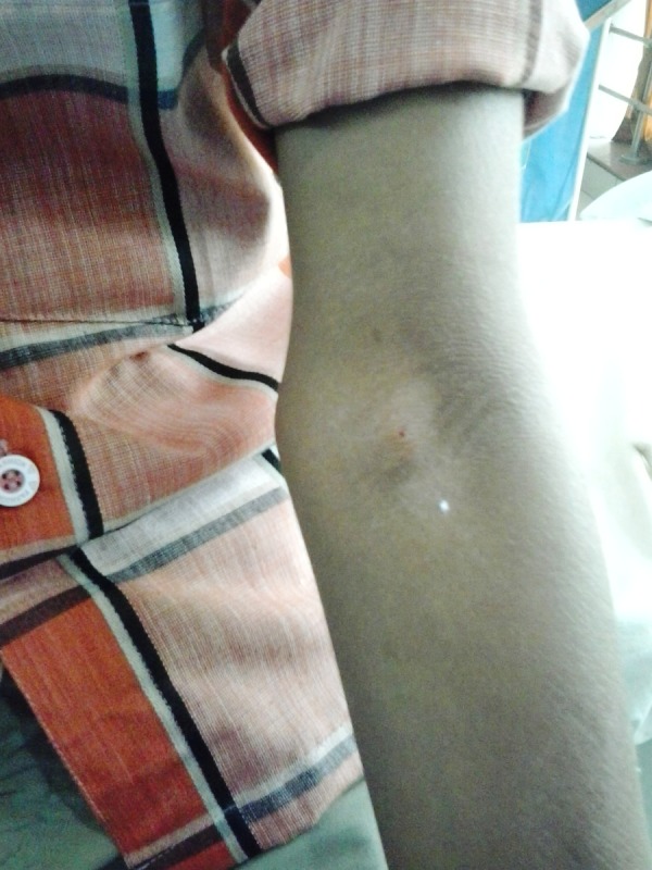
A small subcutaneous swelling near medial aspect of the left cubital fossa.
The wet mount preparation showed many motile microfilariae (Fig. 2). Cytology smears revealed a large number of microfilariae, identified as W. bancrofti because of the presence of hyaline sheath and cephalic space length : breadth ratio was 1 : 1; nuclei were almost spherical, regularly placed, appeared in regular row, well separated without any overlapping and were absent at the tip tail, without significant inflammatory cell infiltrate (Fig. 3). With the cytomorphological diagnosis of subcutaneous filariasis, the patient had nocturnal blood examination, but no microfilariae were found on three consecutive nights. Patient's routine hematological including blood eosinophil counts (3%) and biochemical investigations were also within normal limits. Indirect ELISA was performed which was highly positive for specific recombinant W. bancrofti filarial antigen (WL-L2). Swelling subsided after administration of diethylcarbamazine (100 mg three times a day) for 21 days.
Figure 2:
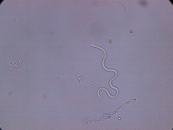
Photomicrograph of wet mount preparation of the fluid shows microfilaria of W. bancrofti.
Figure 3:
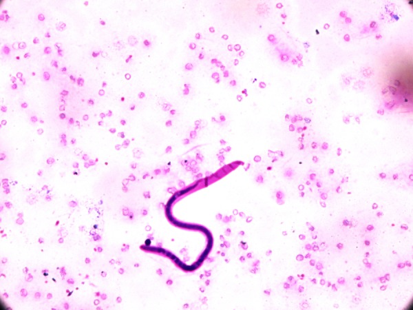
Photomicrograph of a sheathed microfilaria of W. bancrofti with a clear space free of nuclei at the caudal end (May Grunwald Giemsa, ×400).
DISCUSSION
Wuchereria bancrofti, presented as subcutaneous swelling, is a very rare presentation. Its typical presentations are elephantiasis, chronic lymphoedema, epididymitis, funiculitis and lymphadenitis. The subcutaneous filariasis is mainly caused by L. loa, O. volvulus and Mansonella streptococca; of which, L. loa is found in both peripheral blood and subcutaneous nodule; and the other two found only in the skin [1, 7, 8]. Dirofilariasis, an accidental infection in humans caused by Dirofilaria spp. (D. immitis and D. repens), affects wild and domestic animals with canines as a principal reservoir. Human dirofilariasis is uncommon and mostly presents as periorbital and subconjunctival cysts. Human ocular dirofilariasis, though uncommon, has been reported from many parts of the world including Asia [9].
The diagnosis of filarial infection in symptomatic cases with typical clinical presentation is often easy and straightforward, but demonstration of microfilariae in circulating blood is the only conventional means by which one can make definite diagnosis [10]. Unfortunately, in endemic areas, a majority of the affected individuals remain asymptomatic with continued disease transmission [11]. Regardless of high incidence of this parasite in an endemic zone, microfilaremia is often absent and presence of microfilariae in cytological smears and body fluids is an incidental finding. The absence or transient microfilaremia in these endemic zones further complicates the detection of disease.
Adult worms live in regional lymphatics and microfilariae circulate in the blood which is their natural habitat. As the parasite circulate in the lymphatic and vascular systems, appearance of filarial organism in tissue fluids and exfoliated surface material probably occurs due to conditions causing lymphovascular obstruction resulting into extravasations of blood and release of microfilariae. The lymphatic blockade and vessel obstruction are determined by local factors such as tumors, inflammation, trauma or stasis [11, 12]. Cytology has an important role in the diagnosis of subclinical filariasis and can demonstrate these extravasated larvae in tissue spaces or fluids as was evident in our case. Diagnosis of filariasis in cytological smears can also be made by the presence of fragments of adult female or male worms and ova of the filarial organism with or without simultaneous presence of microfilaria [5].
In our case, the patient came from an endemic area explaining the lack of clinical symptoms and the amicrofilaremic state. Similar to our findings, subsequent peripheral examination following cytological diagnosis did not reveal microfilariae [10, 13], suggesting that filaria can exist without microfilaremia. The majority of cases in endemic regions neither show microfilariae in blood, nor any symptom [10]. Blood eosinophil counts within the normal range, as observed in our case, were also reported by Varghese et al. [13]. However, Valand et al. [1] demonstrated eosinophilia in their case. These observations suggest that there is no consistent relationship between filarial infection and blood eosinophilia, which in turn reflects the difference in host response to parasite from person to person. Diagnosis of filarial infection by detection of antigen would obviate the problem in low level of microfilaremia.
Despite high incidence of filariasis, microfilaria in FNAC is not a very common finding. Microfilaria may present in an unusual form; consequently, a high index of suspicion is required to diagnose such unusual presentation of W. bancrofti. As a consequence, careful screening of cytological smear can bestow definitive diagnosis of early, asymptomatic and clinically unanticipated cases of bancroftian filariasis, especially with the amicrofilaremic state. A possibility of filariasis must always be kept in mind as a possible differential diagnosis while evaluating the aspirates from subcutaneous swellings, especially in endemic areas.
CONFLICT OF INTEREST STATEMENT
None declared.
REFERENCES
- 1.Valand AG, Pandya BS, Patil YV, Patel LG. Subcutaneous filariasis: an unusual case report. Indian J Dermatol 2007;52:48–49. [Google Scholar]
- 2.Basu A, Gon S, Berra S, Chakravarti S. Breast filariasis: a rare cytomorphological diagnosis. J Pak Med Stud 2013;3:103–105. [Google Scholar]
- 3.Michael E, Bundy DAP, Grenfell BT. Re-assessing the global prevalence and distribution of lymphatic filariasis. Parasitology 1996;112:409–428. [DOI] [PubMed] [Google Scholar]
- 4.Sabesan S, Palaniyandi M, Das PK, Michael E. Mapping of lymphatic filariasis in India. Ann Trop Med Parasitol 2000;94:591–606. [DOI] [PubMed] [Google Scholar]
- 5.Jain S, Sodhani P, Gupta S, Sakhuja P, Kumar N. Cytomorphology of filariasis revisited. Expansion of the morphologic spectrum and coexistent with other lesions. Acta Cytol 2001;45:186–191. [DOI] [PubMed] [Google Scholar]
- 6.Chaturvedi S, Arora VK. Soft tissue swelling: cytology comes to rescue. J Postgrad Med 2001;47:144. [PubMed] [Google Scholar]
- 7.Panicker NK, Buch AC, Vimal S, Dharwadkar AP. Cytological diagnosis of microfilariae in subcutaneous nodule. Med J DY Patil Univ 2012;5:71–72. [Google Scholar]
- 8.Dey P, Walker R. Microfilariae in a fine needle aspiration from skin nodule. Acta Cytol 1994;38:114. [PubMed] [Google Scholar]
- 9.Hoti SL. Filariasis of uncommon nature in India. Trop Parasitol 2012;2:2–3. [DOI] [PMC free article] [PubMed] [Google Scholar]
- 10.Sivakumar S. Role of fine needle aspiration cytology in detection of Microfilariae: report of 2 cases. Acta Cytol 2007;51:803–805. [DOI] [PubMed] [Google Scholar]
- 11.Phukan JP, Sinha A, Sengupta S, Bose K. Cytodiagnosis of filariasis from a swelling of arm. Trop Parasitol 2012;2:77–79. [DOI] [PMC free article] [PubMed] [Google Scholar]
- 12.Chatterjee KD. Phylum Nemathelminthes: Class Nematoda. In: Chatterjee KD, ed. Parasitology (Protozoology and Helminthology) in Relation to Clinical Medicine, 12th edn Kolkata: Chatterjee Medical Publishers, 1980, pp. 184–199. [Google Scholar]
- 13.Varghese R, Raghuveer CV, Pai MR, Bansal R. Microfilariae in cytological smears: a report of six cases. Acta Cytol 1996;40:299–301. [DOI] [PubMed] [Google Scholar]