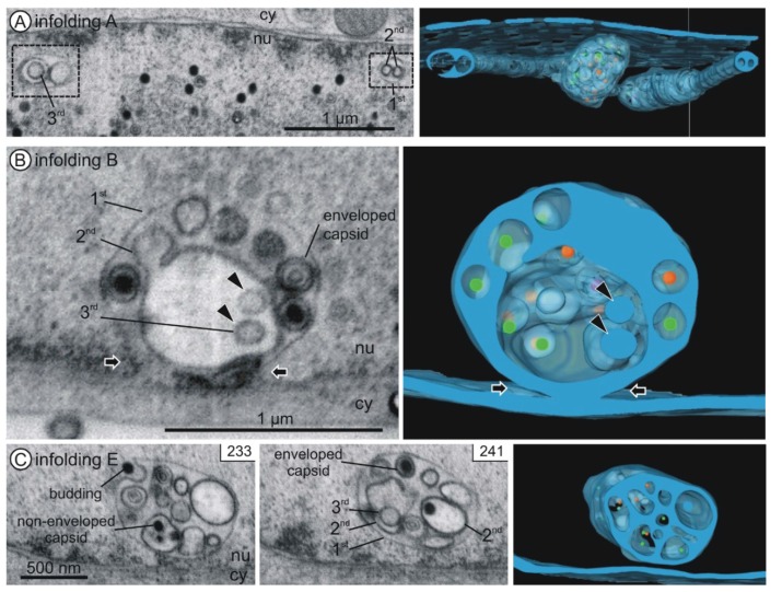
Figure 4.
Internal structure of nuclear infoldings. Left and middle panels: FIB/SEM micrographs of the reconstructed details. See also Movies S1 and S2. Right panels: Three-dimensional reconstructions. Nuclear infoldings (semi-transparent blue) and capsids inside the infoldings (A capsids purple; B capsids orange; C capsids green). (A) Infolding A. Cross sections through two tubular segments (1st order infoldings) with 2nd order infoldings and a 3rd order infolding. Three-dimensional imaging reveals the tubular shape of the 2nd order infoldings and the spherical shape of the 3rd order infolding; (B) Connection of infolding B with the inner nuclear membrane (black arrows). Vesicles (2nd order infoldings) and three enveloped capsids are visible within the 1st order infolding. The 2nd order infolding contains two 3rd order infoldings (black arrowheads); (C) Infolding E with 1st, 2nd and 3rd order infoldings, a budding capsid, enveloped capsids in 1st order infoldings and non-enveloped capsids in 2nd order infoldings. nu nucleoplasm, cy cytoplasm.

