

Abstract
BACKGROUND. Rabbit-generated antithymocyte globulins (ATGs), which target human T cells, are widely used as immunosuppressive agents during treatment of kidney allograft recipients. However, ATGs can induce immune complex diseases, including serum sickness disease (SSD). Rabbit and human IgGs have various antigenic differences, including expression of the sialic acid Neu5Gc and α-1-3-Gal (Gal), which are not synthesized by human beings. Moreover, anti-Neu5Gc antibodies have been shown to preexist and be elicited by immunization in human subjects. This study aimed to assess the effect of SSD on long-term kidney allograft outcome and to compare the immunization status of grafted patients presenting with SSD following ATG induction treatment.
METHODS. We analyzed data from a cohort of 889 first kidney graft recipients with ATG induction (86 with SSD [SSD+] and 803 without SSD [SSD–]) from the Données Informatisées et Validées en Transplantation data bank. Two subgroups of SSD+ and SSD– patients that had received ATG induction treatment were then assessed for total anti-ATG, anti-Neu5Gc, and anti-Gal antibodies using ELISA assays on sera before and after transplantation.
RESULTS. SSD was significantly associated with long-term graft loss (>10 years, P = 0.02). Moreover, SSD+ patients exhibited significantly elevated titers of anti-ATG (P = 0.043) and anti-Neu5Gc (P = 0.007) IgGs in late post-graft samples compared with SSD– recipients.
CONCLUSION. In conclusion, our data indicate that SSD is a major contributing factor of late graft loss following ATG induction and that anti-Neu5Gc antibodies increase over time in SSD+ patients.
FUNDING. This study was funded by Société d’Accélération du Transfert de Technologies Ouest Valorisation, the European FP7 “Translink” research program, the French National Agency of Research, Labex Transplantex, the Natural Science and Engineering Research Council of Canada, and the Canadian Foundation for Innovation.
Introduction
Polyclonal anti-human T lymphocyte IgGs (antithymocyte globulins [ATGs]) from various animal sources have been used since the very beginning of allotransplantation (1, 2). ATGs are administered either immediately following surgery (refereed to as “induction treatment”) to decrease early rejection (3) or as treatment for acute rejection (4), particularly in steroid-resistant cases (5). Despite being associated with a higher incidence of bacterial and viral opportunistic infections and their related complications (3, 6), ATGs are being increasingly used as induction treatment in patients exhibiting high immunological risks (3, 7, 8). ATGs are also used in other T cell–mediated diseases, such as malignancies, graft-versus-host disease (9), and aplastic anemia (10).
ATG treatment, which involves a polyclonal and foreign anti–T cell agent, has been associated with severe side effects, such as cytokine release storm (11), or immune complex disease symptoms, ranging from fever and skin rashes to serum sickness disease (SSD) (12). SSD occurs in almost all cases in which ATG is not associated with immunosuppressive drugs (13). Early biochemical characterization of the major antigenic determinants of ATG stressed the role of “heterophilic” epitopes and, particularly, of the Neu5Gc antigen, coined as the “serum sickness antigen” (12, 14–16). Humans cannot synthesize the sialic acid Neu5Gc (glycolyl form of neuraminic acid) from the acetylated form, Neu5Ac, following the mutation of the cytidine monophosphate-N-acetylneuraminic acid hydroxylase (CMAH) (17, 18). In addition, humans have preexisting anti–galactose-α(1,3)-galactose (Gal) and anti-Neu5Gc antibodies (19, 20).
While early immune complex–related clinical symptoms have mostly been considered as merely a severe discomfort, the possible long-term deleterious role of anti-ATG immune complex formation on the graft clinical outcome following induction treatment has not been yet established in allograft recipients. However, there are several theoretical possibilities supporting an eventual long-term detrimental effect of anti-xenogenic response after ATG treatment, involving the generation of soluble immune complexes during the acute stage of the disease and also, as endothelial cells (ECs) can accumulate detectable diet-derived Neu5Gc moieties (21–23), a possible direct attack on the recipient and graft vessel walls (24), following the paradigm of an “in situ immune complex” disease (25). Significantly, use of sialyl chips has provided evidence of a shift in the pattern of recognition of Neu5Gc conformational epitopes following immunization (26), making at least theoretically understandable the paradox of a deleterious effect of elicited anti-Neu5Gc compared with that of “natural” anti-Neu5Gc.
In this article, using a large and clinically homogeneous cohort of patients treated with rabbit ATG in an induction treatment strategy, we assessed long-term graft survival according to patient SSD status. Furthermore, we additionally aimed to compare the immunization statuses of two matched subgroups of patients (patients with SSD [SSD+] and without SSD [SSD–]) against several kinds of xenogenic antigens: anti-Neu5Gc, anti-Gal, and anti-global ATG antibodies.
Results
Overall characteristics of cohort A
As shown in Figure 1A, the 889 patients in cohort A were retained from an initial cohort of 1,370 first graft recipients treated with rabbit ATG induction therapy. Baseline characteristics are presented in Table 1.
Figure 1. Flow chart of the study cohorts.

(A) Cohorts A and (B) B are shown, along with the selection of the SSD+ group and the control SSD– group matched for five clinical variables. See “Patients” for details on the selection procedure.
Table 1. Population and transplantation characteristics of SSD+ (n = 86) and SSD– patients (n = 803) at baseline.
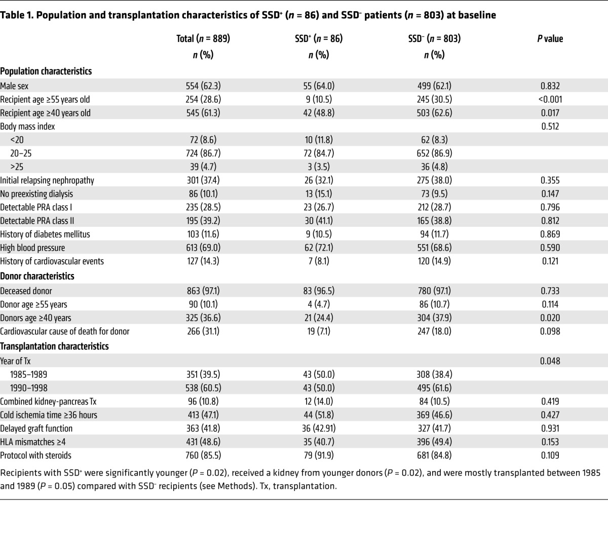
Differences between SSD+ and SSD– patients in cohort A
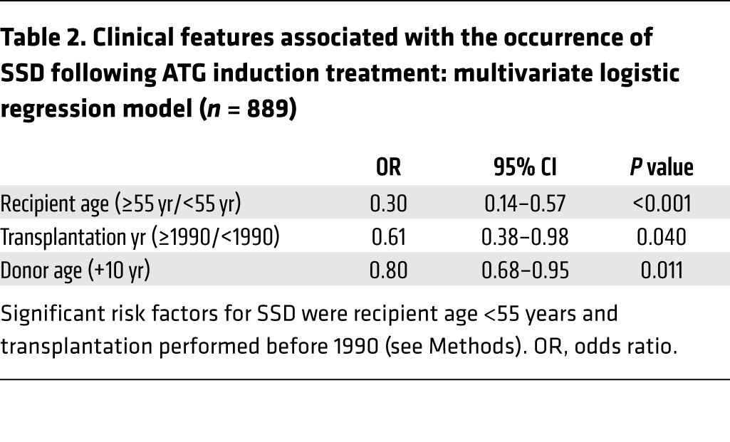
Among the 889 patients, 86 (9.7%) exhibited a SSD within the first month following induction onset. They were younger (P = 0.02) and received allografts from younger donors (P = 0.02) than SSD– patients (Table 1). Multivariate analysis identified three variables significantly associated with SSD occurrence status: young donor, recipient ages, and transplantation before 1990 (Table 2). Patients from the SSD+ group exhibited significantly more biopsy-proven acute rejection episodes (AREs, n = 39, 45.3%) compared with SSD– patients (n = 236, 29.4%, P = 0.003). AREs occurred after SSD onset with no time overlap, at a median of 4.7 months after transplant, and only one patient had an ARE diagnosed within the first month.
Table 2. Clinical features associated with the occurrence of SSD following ATG induction treatment: multivariate logistic regression model (n = 889).
Relationship between SSD occurrence and graft survival in cohort A
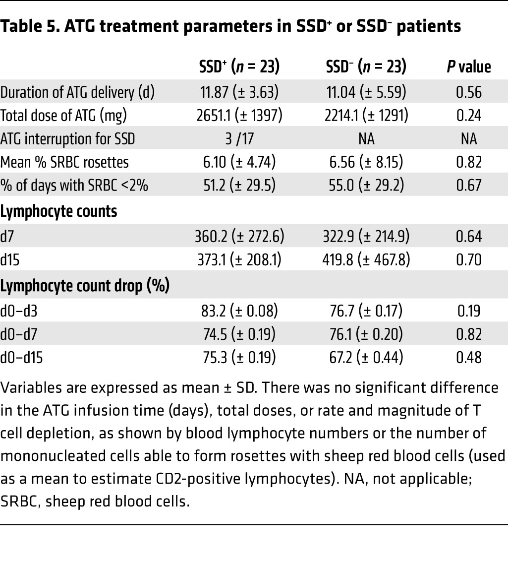
Multivariate analysis showed that SSD occurrence was significantly associated with late graft failure (>10 years), with a 1.72-fold relative risk of late graft failure (death censored) compared with SSD– patients (95% CI = 1.08–2.72), P = 0.02). Death-censored graft survival is shown in Figure 2, and survival of these patients is shown in Table 3. At 15 years posttransplantation, 352 recipients (315 SSD– and 37 SSD+) of the initial global sample returned to dialysis, 169 died (153 SSD– and 16 SSD+), and 272 remained alive with a functioning kidney (246 SSD– and 26 SSD+). In SSD– patients, the survival probabilities at 5, 10, 15, and 20 years posttransplantation were estimated to be 79.7% (95% CI = 76.9–82.7), 64.0% (95% CI = 60.5–67.7), 52.2% (95% CI = 58.4–56.3), and 42.6% (95% CI = 38.4–47.2), respectively. In SSD+ patients, the survival probabilities at the same posttransplantation times were estimated at 77.2% (95% CI = 68.5–87.1), 67.1% (95% CI = 57.2–78.7), 47.6% (95% CI = 37.0–61.3), and 32.2% (95% CI = 22.2–46.7), respectively. The other factors also associated with a higher risk of graft failure were male sex, presence of class I panel-reactive antibodies (PRAs) before engraftment (pre-graft), age of donor, prolonged cold ischemia time, and occurrence of an ARE (Table 4). Table 5 and Supplemental Figure 1 (supplemental material available online with this article; doi:10.1172/JCI82267DS1) show the differences in ATG doses and responses in the SSD+ and the SSD– matched controls (see “Patients”). There was no significant difference in the mean ATG infusion time, total doses, or rate and magnitude of T cell depletion, as recorded by blood lymphocyte numbers or the number of mononucleated cells able to form rosettes with sheep red blood cells (an estimation of CD2-positive lymphocytes; ref. 27). Length of ATG treatment was slightly truncated in only 3 patients exhibiting SSD, with no significant difference in the total dose. The mean onset time of SSD was 14.2 ± 3.7 days, suggesting that there was no suboptimal immunosuppression linked to SSD.
Figure 2. Death-censored graft survival curves estimated from cohort A, according to SSD status.

The SSD+ and SSD– populations have different rates of late graft survival (P = 0.02). The median follow-up was 9.0 years (interquartile range, from 3.4 to 16.5 years).
Table 3. Number of at-risk patients.
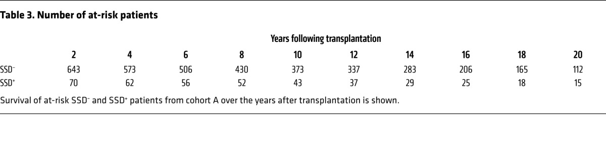
Table 4. Clinical variables associated with an increased risk of graft failure: results of the multivariate Cox model (n = 889).
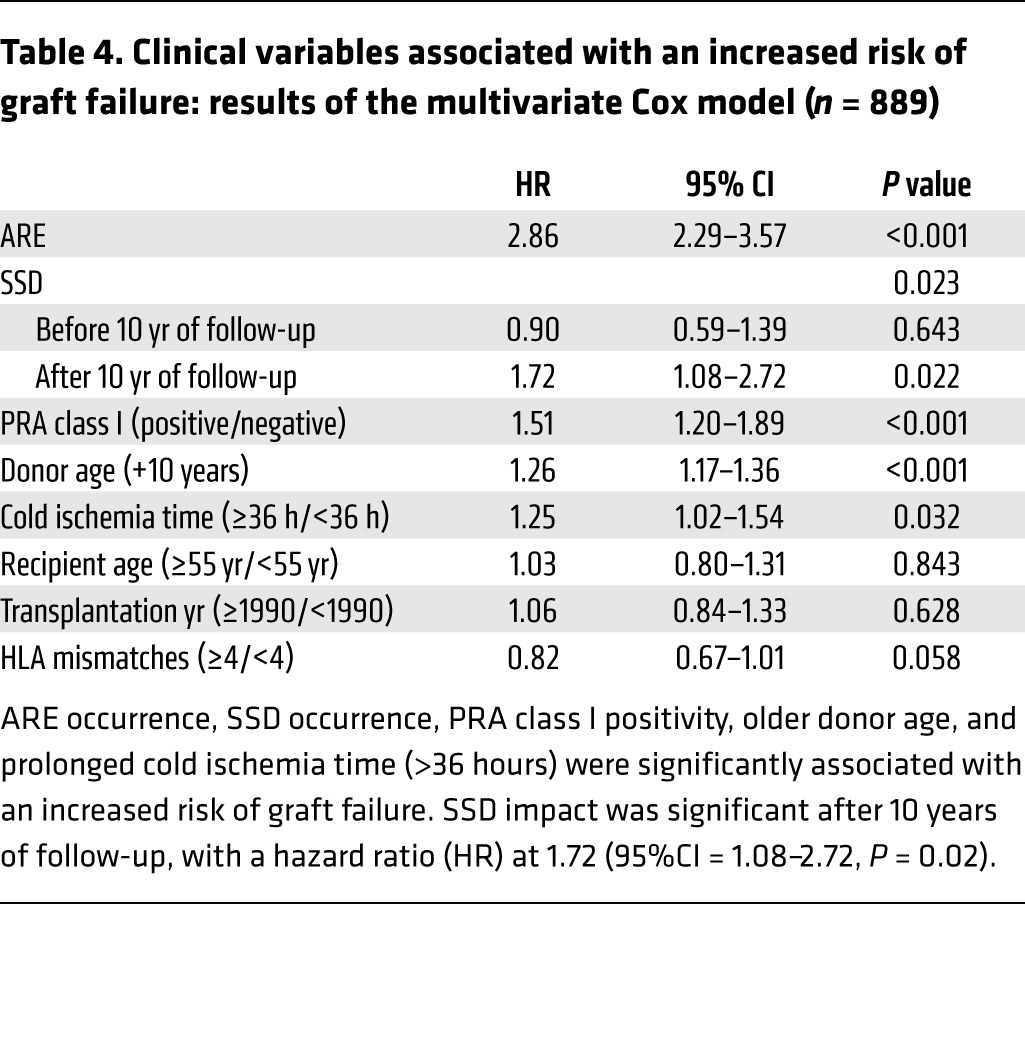
Table 5. ATG treatment parameters in SSD+ or SSD– patients.
Cross-sectional study of the immune response against total ATG, Neu5Gc, and Gal antigens in cohort B
In a secondary aim of the study, 13 SSD+ recipients of a first allograft treated with ATG induction therapy were paired with 13 SSD– patients according to five variables: recipient age, year of transplantation, PRA class I, PRA class II, and number of HLA mismatches (Figure 1B). To take into account the kinetics of antibody levels among samples taken within the first year posttransplantation (T1), those taken more than 4 years posttransplantation (T2), and those taken before transplantation (T0), SSD+ and SSD– groups were compared using ΔT1 as the difference between T1 and T0 antibody levels and ΔT2 as the difference between T2 and T0 levels.
Pre-graft sera.
Pre-graft anti-ATG levels did not differ from those of age/gender-matched healthy individuals (mean 17.5 ± 26.3 ng/μl), whatever the post-graft SSD status (Supplemental Figure 2A). In the same way, there was no difference in pre-graft anti-Neu5Gc or anti-Gal antibody levels compared with those in sera from healthy individuals, whatever the SSD status (Supplemental Figure 2, B and C). 73.1% of recipients and 83.1% of healthy individuals were found to be positive for anti-Neu5Gc antibodies, with a mean of 2.6 ng/μl (0.01% of total IgGs).
Post-graft sera comparison of T0 and T1 antibody levels.
No significant increase in total anti-ATG antibodies was observed in the two groups in the year post-graft (Figure 3A). In the same way, no significant difference was observed between paired SSD+ and SSD– patients when looking at the early evolution (ΔT1) in anti-Neu5Gc antibody levels (Figure 3B) and anti-Gal antibody levels (Figure 3C).
Figure 3. Cross-sectional analysis of anti-ATG, anti-Neu5Gc, and anti-Gal IgG serum levels in SSD+ and SSD– patients from cohort B.

Results are expressed as mean ± SEM of the ΔT1 or ΔT2 values. Comparisons between SSD+ and SSD– matched patients were performed using a Wilcoxon paired test. (A) In SSD+ patients, anti-ATG levels increased from a mean of 9.49 ± 9.02 ng/μl in T0 sera to 17.41 ± 13.74 ng/μl in T1 sera, while SSD– patients had T0 levels of 10.33 ± 4.95 ng/μl and T1 levels of 13.85 ± 11.90 ng/μl (ΔT1, NS). (B) In SSD+ patients, anti-Neu5Gc levels increased from 1.3 ± 1.87 ng/μl (T0 sera) to 2.28 ± 3.08 ng/μl in T1 sera, in comparison with SSD– patients (T0 = 12.10 ± 29.22 ng/μl vs. T1 = 6.91 ± 14.64 ng/μl, ΔT1, NS). (C) In SSD+ patients, anti-Gal levels remained stable (5.62 ± 5.24 ng/μl in T0 sera vs. 5.78 ± 7.33 ng/μl in T1 sera), in comparison with SSD– patients (T0 = 5.83 ± 3.89 ng/μl vs. T1 = 3.13 ± 2.48 ng/μl, ΔT1, NS). (D) In SSD+ patients, anti-ATG levels increased from a mean of 9.49 ± 9.02 ng/μl in T0 sera to 16.48 ± 12.99 ng/μl in T2 sera, while SSD– patients had T0 levels of 10.33 ± 4.95 ng/μl and T2 levels of 13.85 ± 19.33 ng/μl (ΔT2, P = 0.04). (E) In SSD+ patients, anti-Neu5Gc levels significantly increased from 1.3 ± 1.87 ng/μl to 9.85 ± 19.8 ng/μl in T2 sera, in comparison with SSD– patients (T0 = 12.10 ± 29.22 ng/μl vs. T2 = 4.55 ± 13.54 ng/μl, ΔT2, P = 0.007). (F) In SSD+ patients, anti-Gal levels remained stable (5.62 ± 5.24 ng/μl in T0 sera vs. 5.5 ± 5.41 ng/μl in T2 sera), in comparison with SSD– patients (T0 = 5.83 ± 3.89 ng/μl vs. T1 = 4.42 ± 3.02 ng/μl, ΔT2, NS).
Post-graft sera comparison of T0 and T2 antibody levels.
We noted a significant increase of anti-ATG antibody levels in the T2 samples (ΔT2, P = 0.04, Figure 3D) from patients exhibiting a SSD compared with paired grafted controls. Moreover, the difference in ΔT2 anti-Neu5Gc antibody levels was highly significant (P = 0.007, Figure 3E) when comparing the two groups of patients. These results suggest a long-term anti-Neu5Gc response and exposure following SSD. No difference was noted in anti-Gal antibodies at this time point (Figure 3F).
Longitudinal study of the immune response against total ATG, Neu5Gc, and Gal antigens in cohort B
In the longitudinal study, the total number of patients reached 14 SSD+ and 41 SSD– (including the 13 SSD+ and 13 SSD– patients described previously, see “Patients”). Again, no difference was noted between pre-graft antibody levels and those in healthy volunteers in this extended cohort (data not shown). In the SSD+ group, no increase in anti-ATG antibodies was noted in T1 samples (Figure 4A, P = 0.3), but a highly significant rise in anti-ATG levels was observed in T2 samples compared with pre-graft samples (Figure 4B, P = 0.001). Moreover, anti-Neu5Gc levels were increased in T1 samples (Figure 4C, P = 0.047), and a borderline statistical significance was also noted in anti-Neu5Gc IgGs in T2 samples compared with T0 samples (Figure 4D, P = 0.054). No difference was found in anti-Gal antibodies in the SSD+ group. Anti-ATG, anti-Neu5Gc, and anti-Gal IgG levels remained stable in the patients from the control SSD– group at all the follow-up times studied (data not shown).
Figure 4. Longitudinal analysis of anti-ATG and anti-Neu5Gc IgG serum levels in SSD+ patients from cohort B.

T0, T1, and T2 samples for each patient in the SSD+ group were assessed for (A and B) anti-ATG and (C and D) anti-Neu5Gc antibody levels. Antibody levels were quantified using ELISA assays, with median and interquartile range shown for each group. Comparisons between time points for one patient were performed using a Wilcoxon paired test. (A) Anti-ATG IgGs in T0 (mean 9.99 ± SD 10.96 ng/μl) and T1 (15.06 ± 12.78 ng/μl) samples (n = 9 patients at T1, NS). (B) Anti-ATGs in T0 (mean 7.04 ± SD 1.93 ng/μl) and T2 (16.89 ± 12.83 ng/μl) samples (n = 12 patients at T2, P = 0.001). (C) Anti-Neu5Gc IgGs in T0 (mean 0.74 ± SD 0.91 ng/μl) and T1 (2.46 ± 2.92 ng/μl) samples (n = 9 patients at T1, P = 0.047). (D) Anti-Neu5Gc in T0 (mean 1.41 ± SD 1.91 ng/μl) and T2 (9.78 ± 19.82 ng/μl) samples, n = 12 patients at T2, P = 0.054. Similar analysis in the SSD– group did not yield significant differences (data not shown).
Finally, there was no significant decrease of recipient total IgG serum levels among the T0, T1, and T2 samples (data not shown).
Association of anti-Neu5Gc, anti-Gal, and anti-ATG levels with clinical outcome
First, we found no significant correlation between preexisting anti-Neu5Gc, anti-Gal, or anti-ATG levels and early clinical events (SSD, ARE, delayed graft function) or graft failure (data not shown). Next, we attempted to assess a possible long-term association of exposure to high titers of anti-Neu5Gc antibodies and late graft survival, whatever the patient SSD status. Patients with sera available at 7 ± 3 years after transplantation (median of the T2 sampling time) and with a functional kidney allograft through this period were included in order to analyze homogeneous data in terms of the time interval of the post-graft assay (n = 36 patients). The third of patients with the highest levels of antibodies (n = 12) constituted the “high level” group, and the remaining patients constituted the “low level” group (n = 24). This procedure was similarly performed for anti-Neu5Gc, anti-ATG, and anti-Gal antibody levels. No significant differences were observed in the occurrence of delayed graft function, ARE, or anti-HLA antibodies between the groups (Supplemental Table 1), but patients with “high anti-Neu5Gc levels” had a lower graft survival compared with patients from the “low anti-Neu5Gc level” group (Supplemental Figure 3A, P = 0.004). In contrast, no significant association between the levels of total anti-Gal or anti-ATG and graft survival was observed (Supplemental Figure 3, B and C).
Early kinetics of anti-ATG, anti-Neu5Gc, and anti-Gal antibodies
The short-term kinetics of the anti-ATG antibody level variations were tested in 17 recipients, all SSD–, from the “early response” cohort with sera sampled at 0, 7, 14, 21, 60, and 90 days after ATG induction onset (Supplemental Figure 4). There was no decrease in circulating total anti-ATG antibodies, suggesting a moderate effect on rabbit IgG bioavailability from the recipient preexisting anti-Neu5Gc or anti-Gal antibodies (Supplemental Figure 4A). In contrast, there was a significant decrease in anti-Neu5Gc (P < 0.01 at days 14, 21, 60, and 90 (Supplemental Figure 4B) and anti-Gal IgGs (P < 0.05 at day 14, Supplemental Figure 4C) early following transplantation, compared with preexisting antibody levels. No difference was observed in total post-graft anti-ATG IgM levels (Supplemental Figure 4D). Levels of circulating thymoglobulin were also investigated in the whole cohort, with a maximum mean concentration measured at day 7 (57.5 ± 8.2 μg/ml, ranging from 6.7 to 114.6 μg/ml, Supplemental Figure 4E); however, there was no link between serum thymoglobulin concentrations and levels of anti-Neu5Gc or anti-Gal antibodies (data not shown).
Rabbit IgGs display Neu5Gc, but no Neu5Ac, epitopes and baseline levels of Gal
Supplemental Figure 5 presents the mass spectrometric confirmation of preliminary results, showing borderline positivity of Gal expression on rabbit IgGs, using the IB4 Gal-specific lectin in an ELISA assay. Only a few Galα-1,3Galβ-1,4GlcNAc branchings are observed following β galactosidase treatment. However, thymoglobulin was found to express substantial levels of Neu5Gc, without Neu5Ac residues. These data unambiguously validate the presence of xeno-sugar antigens on rabbit IgGs.
Presence of Neu5Gc on fresh human aortic ECs and activation of ECs by anti-Neu5Gc–containing sera
Sera from patients with substantial amounts of anti-Neu5Gc antibodies can activate ECs toward an inflammatory profile. TNF-α and culture media were used as positive and negative controls, respectively. Stimulated ECs were cultured for 2 weeks with Neu5Gc-containing fetal calf serum and displayed the same levels of membrane Neu5Gc as fresh aortic ECs (Supplemental Figure 6). Supplemental Figure 7 shows an accumulation of all the transcripts tested in ECs (VCAM1, ICAM1, E selectin, ADAM10, TNFA, IL1B, IL6, and IL8) following incubation of ECs for 4 hours with 3 sera containing high levels of anti-Neu5Gc (from 29.8 to 75.9 ng/μl). There was no accumulation following incubation with the 3 patient sera without detectable anti-Neu5Gc. When tested undiluted, 2 sera were positive for anti-HLA: 1 serum of 3 without anti-Neu5Gc was positive for anti-HLA class I. Reactivity disappeared at the dilution used in the EC stimulation test (1:100). This serum did not activate ECs. Another serum was positive for anti–HLA class II. The anti–class II antibodies were still detectable at a 1:100 dilution. This serum, which belongs to the group with anti-Neu5Gc, elicited only a borderline activation of ECs. The serum with the strongest stimulating effect on ECs in this group of sera with anti-Neu5Gc was negative for anti-HLA, even undiluted. These data, using several sera and particularly “immune” sera from patients primed by ATG, complements and reinforces previous data using a serum from a normal individual (28). Despite restrictions on available sera, which do not allow relevant statistical analysis, the increase of all transcript species tested and the strong transcript accumulation (for instance, for VCAM1, IL1, and IL8) suggest that anti-Neu5Gc antibodies activate ECs.
Discussion
In this study, we bring the first evidence to our knowledge that SSD occurrence is a significant risk factor for long-term allograft loss in first kidney transplant recipients who have received an induction treatment with rabbit anti-human lymphocyte IgGs. In addition, we show an increased humoral immune response against rabbit IgGs following SSD and, more specifically, against xenogeneic Neu5Gc epitopes, despite exposure to major immunosuppressive drugs.
If restricted to prospective randomized studies, the incidence of SSD following thymoglobulin treatment of kidney recipients ranges from 4% to 28% (6, 29–31). The 9.7% incidence reported in our study included only SSD with arthralgia, irrespective of other “minor” symptoms, such as skin rashes, fever, or headaches. SSD is considered as the human paradigm of the experimental “one shot” immune complex disease, as described in the rabbit following the seminal work of Dixon et al. (32). SSD occurs in all patients receiving ATG treatment without additional immunosuppressive medications (13). However, SSD in transplant recipients differs by the fact that the foreign protein is administered over roughly a week or two and with major immunosuppressive and antiinflammatory drugs given during and after the treatment with the xenoantigen. Furthermore, almost all humans have preexisting anti-ATG, anti-Gal, and anti-Neu5Gc antibodies, and, significantly, anti-Neu5Gc antibodies can affect ECs through a model of “planted immune complexes” (25), due to the existence of dietary-derived Neu5Gc on ECs, which may affect the graft tissues in the long term. Our multivariate Cox model identified SSD as a variable affecting long-term graft outcome. The etiology of this late failure following SSD remains a matter of speculation. Owing to the immune complex nature of SSD, early graft vasculature damage in the course of SSD may restrict the long-term adaptation to the nephron functional overload following transplantation (33) and can be accelerated by many factors, such as calcineurin inhibitor (CNI) toxicity, hypertension, hyperlipidemia (see ref. 34 for review), or inadequate nephronic mass (35) for instance. The patients experiencing SSD may have a strong responder phenotype. Indeed, AREs were more frequent in the SSD group than in the control group. This increased frequency is not explained by suboptimal immunosuppression (Table 5). As a severe early inflammation episode, SSD may also be responsible for an aspecific stimulation of recipient immune response, leading to an increased occurrence of rejection but also to increased production of Neu5Gc. However, anti-Gal antibody levels were not increased, arguing against this interpretation. In addition, as elicited anti-Neu5Gc antibodies recognize different patterns of Neu5Gc epitopes than “natural” ones (26) and their presence results in increased long-term exposure of Neu5Gc-positive ECs to these antibodies, they may trigger a chronic inflammation of graft vascular walls, as further discussed below. Indeed, in Cmah KO mice, an animal model unable to biosynthesize Neu5Gc, similarly to humans, it has been demonstrated that anti-Neu5Gc antibodies can stimulate chronic inflammation in dietary Neu5Gc-charged tissues (36).
A number of types of antibody specificities against rabbit IgG were explored. First of all, we confirmed that sera from healthy individuals as well as pretransplantation sera of all patients contained anti-ATG and anti-Gal antibodies and that 83% of all patient sera contained anti-Neu5Gc, a close match with studies performed in other smaller cohorts (20, 37). No significant association was noted between the preexisting titers and early post-graft clinical events. We did not detect an early increase in overall global titers of total anti-ATG, anti-Neu5Gc, or anti-Gal within the first 3 months after treatment. Instead, there was a slight decrease in these antibodies. In contrast to the sialic acid Neu5Gc, which was clearly evidenced on rabbit IgGs using an anti-Neu5Gc polyclonal IgY, Gal was not evidenced on rabbit IgGs (using the Gal-specific lectin IB4 for detection, data not shown). However, mass spectrometry unambiguously demonstrates the presence of Gal on thymoglobulin, although at a much lower level than for Neu5Gc epitopes. Of note, rabbit IgG (as well as horse IgGs) did not display Neu5Ac (Supplemental Figure 5). Immunosuppressive drugs should not influence dramatically the titers of these preformed antibodies during this early period after surgery, suggesting that part of the preexisting anti-Neu5Gc and anti-ATG antibodies is complexed by the infused ATG.
Both cross-sectional and longitudinal studies suggest that recipients who have experienced SSD have a distinct immune status compared with those who have not, in particular, a delayed increase in anti-Neu5Gc and total anti-ATG antibodies, despite sustained and major immunosuppression. It is possible that chronic exposure to immune complexes with Neu5Gc-bearing epitopes of dietary origin (21, 23, 28), which are able to activate the complement, offers optimal conditions for B cell response through a more efficient display on dendritic follicular cells in the lymph nodes (38).
Unfortunately, we were not able to study the antibody response at the time of SSD. Finally, our data, which require confirmation from a larger cohort, suggest that a higher risk of graft failure is associated with high anti-Neu5Gc levels, whatever the SSD status. In contrast, late graft failure was not significantly associated with global anti-ATG or anti-Gal antibody levels tested in a similar procedure.
Despite the fact that a possible causal link between anti-Neu5Gc antibody levels and long-term graft survival cannot be confirmed from this clinical study, we show that living, fresh, uncultured human ECs express substantial amounts of Neu5Gc on their cell surface. Furthermore, human ECs displaying surface membrane Neu5Gc, mimicking the flow cytometry pattern of fresh aortic ECs, cultured for 4 hours in the presence of patient sera with high titers of anti-Neu5Gc, exhibited an increased expression of VCAM1, ICAM1, E selectin, ADAM10, TNFA, IL1B, IL6, and IL8 transcripts (Supplemental Figure 7), a profile highly suggestive of an inflammatory phenotype of the stimulated ECs (39). Of potential interest, ADAM-10, which is not usually regulated by TNF-α (40) and involved in several cytokine “maturation processes” (41), has been found to be overexpressed by ECs (42). Despite being highly dispersed, our data, using Q-PCR, confirm a previous report on the increased cytoplasmic content of several cytokines following stimulation of Neu5Gc-positive cells (28). Our study identified a set of proinflammatory genes, including adhesion molecules, which are upregulated in ECs by anti-Neu5Gc–containing sera. This pattern suggests that anti-Neu5Gc antibodies may activate the NF-κB pathway. This pattern of regulation was not observed in response to anti-HLA antibodies, as in our previous studies. Findings from our group indicated that anti–class I (43) and anti–class II (44, 45) HLA antibodies mediate proliferative and protective effects, respectively, mostly via PI3 kinase signaling. Importantly, the sole serum with anti-HLA in the anti-Neu5Gc group induced only a low inflammatory response on ECs, whereas the serum displaying the strongest stimulation on ECs did not display anti-HLA in the Luminex assay. Further studies using affinity-purified anti-Neu5Gc antibodies are nevertheless required. However, our data differ from those in Pham’s study (28), since our sera were obtained from patients primed by ATG who have exhibited a SSD, instead of from an unaffected individual. Furthermore, our sera were diluted to 1:100 (instead of 1:2), diluting other potential antibodies, and with final anti-Neu5Gc concentration close to that of the unaffected individual’s sera. As mentioned previously, anti-Neu5Gc epitope recognition by serum IgG drastically differs between primed and unprimed individuals (26). Therefore, different patterns of Neu5Gc epitopes recognized by elicited anti-Neu5Gc antibodies in primed individuals, as in our study, in addition to exposure to increased levels of anti-Neu5Gc, may be important in understanding a potential role of such antibodies in this unique situation of a “physiological” immune complex condition.
We are conscious of some limitations of our study. Particularly, as in any study on long-term clinical outcome, the cohort, even with a homogeneous treatment, is subjected to the continuous evolution of the techniques of patient monitoring and clinical management, the differences in surgical methods, and the quality of the data dating from 1985, restricting the possible confounding factors analyzed, despite the comparison with variables-matched controls (including graft year) in the initial design of our study. We also suggest, but do not formally demonstrate, the detrimental effect of an increased exposure of ECs to elicited anti-Neu5Gc antibodies; an interaction with graft epithelial cells may also be involved.
Taken together, our data show that, despite providing valuable aid in the clinical management of allografts, rabbit anti-human lymphocyte IgGs are not without toxicity. In particular, we show for the first time to our knowledge that SSD is associated with poor long-term graft survival. Our data warrant further study on the immune response of graft recipients receiving animal-derived material and foster the development of animal-derived ATG without Neu5Gc content.
Methods
Patients
Cohort A.
First kidney (n = 793) or first combined kidney-pancreas (n = 96) recipients from the Centre Hospitalier Universitaire de Nantes between 1985 and 1998 were included (Figure 1A). Only recipients receiving ATG (Thymoglobulin, Genzyme) as induction therapy associated with CNI and mycophenolic acid (MMF) (2 g/d) were considered, whatever their status concerning prednisone (Table 1). 85.5% of the patients received a tritherapy (CNI, MMF, and prednisone), whereas 14.5% were treated with only CNI and MMF (patients were part of controlled randomized assays on corticoid avoidance). All patients received a peroperative bolus of hydrocortisone. To avoid heterogeneity of ATG indication, only patients treated before 1999 were considered, since after this time point, indication of ATG was restricted to immunologically high-risk patients at our institute. ATG was administered over 6 days at 1.5 mg/kg. Donor and recipient data were extracted from the prospectively harvested Données Informatisées et Validées en Transplantation (DIVAT) data bank (www.divat.fr, Commission nationale de l’informatique et des libertés [CNIL] reference for the French Research Ministry RC12_0452, May 2013) (46). Codes were used to ensure donor and recipient anonymity.
Cohort B.
The secondary objective of the study was to analyze the xenogenic immunization statuses in two subgroups of patients who have received ATG induction treatment: SSD+ and SSD– patients. Two types of analysis were done: cross-sectional comparisons (SSD+ and SSD– matched control group) and longitudinal comparisons (various values obtained for each patient at different times after transplantation). For this specific purpose, first graft recipients exhibiting SSD transplanted between 1995 and 2008 were included, due to poor serum availability from patients grafted prior to 1995. All biological samples from the patients from cohort B were obtained from the DIVAT data bank and were not harvested specifically for this study.
Cross-sectional study.
A paired SSD– control group was selected by taking into account five clinical variables chosen for their clinical relevance (immunization status of recipients) or for their association with poor graft survival in cohort A univariate analysis: recipient age, ± 10 years; transplantation year, ± 2 years; PRA class I positivity; PRA class II positivity; and number of HLA mismatches (≥4/<4). Each SSD+ patient (n = 13) was thus paired with one highly matched SSD– control patient (n = 13). Figure 1B shows the control cohort selection procedure. Regarding the relative heterogeneity of the times posttransplantation at which sera was sampled for each SSD+ and SSD– patient, one post-graft sample obtained within the first year posttransplantation (T1) and one late sample obtained >4 years posttransplantation (T2) were considered, in addition to the pretransplantation sera (T0). Samples from patients who returned to dialysis were excluded.
Longitudinal study.
T0, T1, and T2 samples from each patient were compared. In this serial analysis, which did not require pairing, additional patients with available sera, but which were not fully matched in the paired process described above, were included (n = 14 SSD+ and n = 41 SSD– patients, including those from the cross-sectional study). Finally, we also compared patient anti-xenoantibody values with those of 70 control healthy volunteers matched for age and sex. Healthy control serum samples were obtained from the Etablissement Français du Sang Pays de la Loire, and all donors signed a consent form.
Demographic data and clinical characteristics.
For cohorts A and B, donor parameters studied were sex, age, and cause of death. For the recipients, the variables collected at time of transplantation were sex, age, weight and body mass index, history of diabetes mellitus, initial nephropathy (classified into relapsing or not), kidney or combined pancreas-kidney allograft, preemptive grafts, HLA-A-B-DR mismatches, pretransplantation anti-HLA immunization (historical peak of class I or class II PRA determined by complement-dependent cytotoxicity and Luminex screening), and duration of cold ischemia time. Class II PRAs could not be considered in the entire cohort, as they were not part of routine screening before 1989. Delayed graft function (the need for dialysis in the first week after surgery) was also recorded. Only overt SSD, as defined by a combination of arthralgia and painful temporomandibular joint/trismus, was considered. Renal function was assessed yearly by serum creatininemia and estimated glomerular filtration rate. Biopsy-proven (according to the histological criteria of the year of diagnostic) AREs were recorded.
Supplemental “early response” cohort.
An additional group of 17 recently transplanted patients from the Institute for Clinical and Experimental Medicine, Department of Nephrology, with pre-graft and serially harvested posttransplantation sera sampled at 7, 14, 21, 30, 60, and 90 days, was used specifically and exclusively for the analysis of the early response to ATG and for measurement of thymoglobulin blood levels. None of these patients, who were treated with CNI and MMF, exhibited a SSD. All patients gave informed consent.
Test tubes were coded by personnel from outside the study, and all ELISA tests were carried out blind.
Total anti-rabbit IgG and IgM antibodies
Quantification of human serum IgGs against anti-rabbit IgGs (Thymoglobulin, Genzyme) was adapted from Prin Mathieu et al. (47). Plates (NUNC Maxisorp; NUNC AB) were coated overnight with thymoglobulin (1 μg/ml) in 50 mM sodium carbonate-bicarbonate buffer (pH 9). Wells were blocked for 2 hours at 37°C with PBS, 0.05% Tween20 (Sigma-Aldrich), and 1% Ovalbumin (PBSTO, Sigma-Aldrich). Human serum samples diluted 1:400 in PBS 0.05% Tween20 (PBST) were added and incubated for 2 hours at 37°C, before the addition of a HRP donkey anti-human IgG (H+L) (1:7,000, 709-035-149, Jackson Immunoresearch) for 1 hour at 37°C and development using TMB substrate (Sigma-Aldrich). For standard curves, wells were coated with serial dilutions of human IVIg (concentrations started at 800 ng/ml; Privigen, CSL Berhing SA).
A similar protocol was used for quantification of human serum IgM against thymoglobulin, with serum dilutions of 1:50 and 1:100 and a HRP goat anti-human IgM (μ-chain–specific) secondary antibody (A0420, Sigma-Aldrich). For the standard curve, human-purified IgM (I8260, Sigma-Aldrich) was used, with the initial working concentration of 200 ng/ml.
Anti-Neu5Gc antibodies
Anti-Neu5Gc antibodies were quantified using a modified ELISA originally described by V. Padler-Karavani et al. (26, 48). This assay uses mouse serum proteins as coating antigens in the ELISA plate, offering an exhaustive display of the major epitopes containing Neu5Gc (48). The plates were coated overnight at 4°C with wild-type C57BL/6 mouse sera (Janvier Labs) at 1 μg per well diluted in 50 μl of coating buffer. Diluted human sera were preincubated for 2 hours at 4°C with Cmah KO mouse sera (CMAH-deficient mice, B6.129X1-Cmahtm1Avrk/J adult females, The Jackson Laboratory) diluted 1:100 in PBSTO. After washing and blocking, pretreated human sera were added to the plates for 2 hours at room temperature. IgGs were detected as described previously (26). Again, serial dilutions of IVIg at concentrations starting from 200 ng/ml served as standards. Sera from KO mice were screened for the potential presence of mouse antibodies cross-reacting with human serum determinants, as described by V. Padler-Karavani et al. (48). Positive KO mouse sera were excluded. The absence of Neu5Gc antigens in KO mouse sera was checked using the Neu5Gc-specific chicken IgY antibody (clone Poly21469, Biolegend) and secondary HRP-coupled anti-IgY antibody (ab97135, Abcam).
Anti-Gal antibodies
An ELISA was adapted from Buonomano et al. (49). Plates were coated with Gal1-3Gal-polyacrylamide conjugate (5 μg/ml; PAA-Bdi, Lectinity) overnight at 4°C and then blocked with PBS 0.5% fish gelatin (Sigma-Aldrich) for 2 hours at 37°C. Human sera (at 1:100, 1:500, and 1:1,000 in PBST) were incubated for 2 hours at 37°C. A rabbit anti-human IgM+G and a HRP goat anti-rabbit antibody (1:2,000, 309-005-107, and 1:2,000, 111-035-144, respectively; Jackson ImmunoResearch) were used as secondary antibodies. A titration curve used serial dilutions of human IVIg (Privigen), with an initial working concentration of 50 ng/μl.
Blood thymoglobulin levels
Thymoglobulin circulating levels were measured using a commercial Rabbit IgG ELISA Quantitation Set (Bethyl Laboratories Inc.), and the assay was performed according to the manufacturer’s specifications.
Measurement of total IgG levels
Total IgG assays were performed on a Cobas 8000 C502 (Tina-quant IgG Gen.2. Roche Diagnostics GmbH), using an immunoturbidimetry method.
Analysis of Neu5Gc, Neu5Ac, and Gal moieties on rabbit IgGs by mass spectrometry
Rabbit thymoglobulin (50 μg) was reduced with dithiothreitol (Sigma-Aldrich), alkylated with iodoacetamide (Sigma-Aldrich), and digested with trypsin (2 μg, Promega). The digestion mixture was separated by reversed-phase HPLC. Pooled glycopeptide fractions were further digested with β-galactosidase from bovine testes (Prozyme). All MS analyses were performed on an UltrafleXtreme mass spectrometer (Bruker Daltonics) equipped with LID-LIFTTM technology for tandem MS experiments. Dihydroxy benzoic acid was used as the matrix.
EC preparation and stimulation
Human aortic ECs (HAECs) were isolated from unused aortic pieces harvested from organ donors at the Centre Hospitalier Universitaire de Nantes following the specific agreement of the national Biomedicine Agency (PFS08-017). Aortas were incubated with collagenase A (2.2 U/ml; Roche Diagnostics GmbH) for 40 minutes at 37°C, and cells harvested by mild scrapping of the lumen. ECs were selected using CD31-Dynabeads (Invitrogen, ThermoFisher Scientific) for 20 minutes at 4°C, washed 3 times, plated on plastic dishes, and grown in Endothelial Cell Basal Medium (Promocell) supplemented with 10% FCS, glutamine (200 mM), penicillin and streptomycin (100 UI/ml), ECGS (0.004 ml/ml), hEGF (0.1 ng/ml), hbFGF (1 ng/ml), hydrocortisone (1 μg/ml), and Heparin (90 μg/ml) at 37°C in a 5% CO2. Passage 5 HAECs were used for stimulation experiments.
For quantification of Neu5Gc by flow cytometry, fresh, uncultured HAECs were used immediately after the selection step. Fresh HAECs were labeled with mouse anti-CD31-PE (555446, BD Pharmingen) and chicken anti-Neu5Gc (clone Poly21469, Biolegend), revealed by donkey anti-chicken IgY-A647 (703-606-155, Jackson Immunoresearch) in a 0.5% gelatin fish buffer, read on BD FACSCanto II (BD Biosciences), and analyzed by FlowJo software.
For the stimulation experiments and PCR, HAECs cultured in FCS+ or human serum (HuS+; Sigma-Aldrich) EC medium for 15 days were incubated for 4 hours with 3 SSD+ patient sera with high anti-Neu5Gc levels (29.76 to 75.92 ng/μl) and 3 sera with negative Neu5Gc titers, diluted 10 or 100 fold. Controls consisted of EC medium only or TNF-α at 500 U/ml (R&D Systems). Total RNA was isolated using the TRIzol method (ThermoFisher Scientific). Total RNA (500 ng) was reverse transcribed with the Omniscript Reverse Transcription Kit (Qiagen) according to the manufacturer’s instructions. Quantitative realtime PCR was performed in an ViiA 7 Real-Time PCR System using labeled TaqMan probes (ThermoFisher Scientific). The following ready-to-use primers/probe mixes were used: VCAM1 (Hs01003372_m1), ICAM1 (Hs00153853_m1), E selectin (Hs00950401_m1), ADAM10 (Hs00153853_m1), TNFA (Hs01113624_g1), IL1B (Hs04555410_m1), IL8 (Hs00174103_m1), and IL6 (Hs00174131_m1). GAPDH (Hs99999905_m1) was used to normalize the amounts of RNA. Relative expression was calculated according to the 2–DDCt method, in which the reference represents 1-fold expression.
Statistics
Cohort A.
The main objective of the study was to evaluate the graft survival of the SSD+ patients compared with that of the SSD– patients. Comparisons of baseline characteristics between SSD+ and SSD– patients were based on χ2 tests for identifying potential confounding factors. The time to event was the time between the transplantation and graft failure (i.e., return to dialysis). Patient deaths were considered right censored. Survival curves were estimated using the Kaplan-Meier estimator. A multivariate Cox model (50) was used to take into consideration the previous potential confounding factors. Univariate analyses were performed for first variable selection (P < 0.20). If the log-linearity assumption was unconfirmed, the quantitative variable was categorized according to usual thresholds. Then, a step-by-step backward selection method was manually performed: no significant variable was progressively removed, taking out the less significant variable at each step (P < 0.05). To guarantee the comparability between the SSD+ and the SSD– patients, adjustments were at least performed for all of the following pretransplant confounding factors: recipient age, transplantation period (before or after 1990), HLA-A-B-DR mismatches, and pretransplantation anti-HLA immunization (historical peak of class I PRA). The proportional hazard assumption was checked by plotting log-minus-log survival curves and by using weighted residuals analysis (51). To consider nonproportional covariates, the Cox model with time-dependent regression coefficients (52) was used. Statistical analyses were performed using version 3.0.1 of the R software (53).
Cohort B.
For antibody level analyses, comparisons between the two groups (SSD+ and SSD–) and comparisons among sampling times (T0, T1, and T2) for every patient inside the groups were performed using Wilcoxon paired tests. Kidney allograft survival, according to antibody levels, was analyzed using a log-rank test.
Study approval
All patients provided informed consent, allowing use of clinical, biological, and histological information in the DIVAT cohort. This noninterventional DIVAT study complies with the guidelines for good practice of the Centre Hospitalier Universitaire de Nantes and was approved by CNIL (www.divat.fr, CNIL 891735 2nd version 2, August 2004, CNIL, Ministère de l’enseignement supérieur et de la recherche, Paris, France, dossier 13.334 for “cohort DIVAT RC12_0452”).
Author contributions
GCD participated in study design, analyzed the clinical data, and wrote the manuscript. AS participated in study design, performed and analyzed antibody measurements, and wrote the manuscript. LLB performed and analyzed antibody measurements, performed EC stimulation, and wrote the manuscript. GE participated in study design and performed and analyzed antibody measurements. OV, PH, and PV obtained sera from the short-term patient cohort. PG extracted the clinical data and sera from the database. TD performed total IgG measurements. JR performed and analyzed antibody measurements. AN manages the Cmah KO mouse colony and obtained sera from Cmah KO and WT mice. JMB reviewed the manuscript and helped acquire study funding. EA and HP performed the mass spectrometry experiments. YF and ML participated in study design and performed statistical analysis on clinical and in vitro data. SB reviewed the manuscript and helped acquire study funding. SC performed the Luminex assays. MG supervised extraction of clinical data from the DIVAT data bank. JH contributed to the assessment of anti-Neu5Gc antibody measurements and reviewed the paper. BC supervised experiments on ECs. JPS organized the study and wrote the manuscript.
Supplementary Material
Acknowledgments
The authors wish to thank Robert Floch, Odile Duvaux, and Emanuele Cozzi for scientific discussions. The authors are grateful to the Unité thérapeutique expérimentale de l’Institut de Recherche en Santé de l’Université de Nantes facility (S. Lemarchand-Mindé, M. Liabeuf) for providing blood from CMAH-KO mice. A. Salama was funded in part by the Société d’Accélération du Transfert de Technologies Ouest Valorisation. J.P. Soulillou and L. Le Berre are supported by the European FP7 “Translink” research program (603049). M. Lorent was funded by a grant from the French National Agency of Research (ANR-11-JSV1-0008-01). J.P. Soulillou, S. Brouard, and J. Harb are supported by a grant from the Labex Transplantex. H. Perreault and E. Ang are supported by the Natural Science and Engineering Research Council of Canada (RGPIN/170241) and Canadian Foundation for Innovation (22391). A. Nicot is supported by the Région Pays de la Loire.
Footnotes
Conflict of interest: Jean-Paul Soulillou and Jean-Marie Bach are cofounders of the start-up Xenothera.
Reference information:J Clin Invest. 2015;125(12):4655–4665. doi:10.1172/JCI82267.
References
- 1.Woodruff MF. Purification of antilymphocytic antibody. Nature. 1968;217(5131):821–824. doi: 10.1038/217821a0. [DOI] [PubMed] [Google Scholar]
- 2.Gaber AO, Knight RJ, Patel S, Gaber LW. A review of the evidence for use of thymoglobulin induction in renal transplantation. Transplant Proc. 2010;42(5):1395–1400. doi: 10.1016/j.transproceed.2010.04.019. [DOI] [PubMed] [Google Scholar]
- 3.Brennan DC, Daller JA, Lake KD, Cibrik D, Del Castillo D, Thymoglobulin Induction Study Group Rabbit antithymocyte globulin versus basiliximab in renal transplantation. N Engl J Med. 2006;355(19):1967–1977. doi: 10.1056/NEJMoa060068. [DOI] [PubMed] [Google Scholar]
- 4.Gaber AO, et al. Results of the double-blind, randomized, multicenter, phase III clinical trial of Thymoglobulin versus Atgam in the treatment of acute graft rejection episodes after renal transplantation. Transplantation. 1998;66(1):29–37. doi: 10.1097/00007890-199807150-00005. [DOI] [PubMed] [Google Scholar]
- 5.Gaber LW, et al. Utility of standardized histological classification in the management of acute rejection. 1995 Efficacy Endpoints Conference. Transplantation. 1998;65(3):376–380. doi: 10.1097/00007890-199802150-00013. [DOI] [PubMed] [Google Scholar]
- 6.Soulillou JP, et al. Randomized controlled trial of a monoclonal antibody against the interleukin-2 receptor (33B3.1) as compared with rabbit antithymocyte globulin for prophylaxis against rejection of renal allografts. N Engl J Med. 1990;322(17):1175–1182. doi: 10.1056/NEJM199004263221702. [DOI] [PubMed] [Google Scholar]
- 7.Thiyagarajan UM, Ponnuswamy A, Bagul A. Thymoglobulin and its use in renal transplantation: a review. Am J Nephrol. 2013;37(6):586–601. doi: 10.1159/000351643. [DOI] [PubMed] [Google Scholar]
- 8.Gharekhani A, Entezari-Maleki T, Dashti-Khavidaki S, Khalili H. A review on comparing two commonly used rabbit anti-thymocyte globulins as induction therapy in solid organ transplantation. Expert Opin Biol Ther. 2013;13(9):1299–1313. doi: 10.1517/14712598.2013.822064. [DOI] [PubMed] [Google Scholar]
- 9.Bacigalupo A, et al. Thymoglobulin prevents chronic graft-versus-host disease, chronic lung dysfunction, and late transplant-related mortality: long-term follow-up of a randomized trial in patients undergoing unrelated donor transplantation. Biol Blood Marrow Transplant. 2006;12(5):560–565. doi: 10.1016/j.bbmt.2005.12.034. [DOI] [PubMed] [Google Scholar]
- 10.Feng X, et al. In vivo effects of horse and rabbit antithymocyte globulin in patients with severe aplastic anemia. Haematologica. 2014;99(9):1433–1440. doi: 10.3324/haematol.2014.106542. [DOI] [PMC free article] [PubMed] [Google Scholar]
- 11.Nashan B. Antibody induction therapy in renal transplant patients receiving calcineurin-inhibitor immunosuppressive regimens: a comparative review. BioDrugs. 2005;19(1):39–46. doi: 10.2165/00063030-200519010-00005. [DOI] [PubMed] [Google Scholar]
- 12.Pirofsky B, Ramirez-Mateos JC, August A. “Foreign serum” heterophile antibodies in patients receiving antithymocyte antisera. Blood. 1973;42(3):385–393. [PubMed] [Google Scholar]
- 13.Gitelman SE, et al. Antithymocyte globulin treatment for patients with recent-onset type 1 diabetes: 12-month results of a randomised, placebo-controlled, phase 2 trial. Lancet Diabetes Endocrinol. 2013;1(4):306–316. doi: 10.1016/S2213-8587(13)70065-2. [DOI] [PMC free article] [PubMed] [Google Scholar]
- 14.Hanganutziu M. Hémagglutinines hétérogénétiques après injection de sérum de cheval. CR Séances Soc Biol. 1924;91:1457–1459. [Google Scholar]
- 15.Higashi H, Naiki M, Matuo S, Okouchi K. Antigen of “serum sickness” type of heterophile antibodies in human sera: identification as gangliosides with N-glycolylneuraminic acid. Biochem Biophys Res Commun. 1977;79(2):388–395. doi: 10.1016/0006-291X(77)90169-3. [DOI] [PubMed] [Google Scholar]
- 16.Merrick JM, Zadarlik K, Milgrom F. Characterization of the Hanganutziu-Deicher (serum-sickness) antigen as gangliosides containing n-glycolylneuraminic acid. Int Arch Allergy Appl Immunol. 1978;57(5):477–480. doi: 10.1159/000232140. [DOI] [PubMed] [Google Scholar]
- 17.Irie A, Koyama S, Kozutsumi Y, Kawasaki T, Suzuki A. The molecular basis for the absence of N-glycolylneuraminic acid in humans. J Biol Chem. 1998;273(25):15866–15871. doi: 10.1074/jbc.273.25.15866. [DOI] [PubMed] [Google Scholar]
- 18.Chou HH, et al. A mutation in human CMP-sialic acid hydroxylase occurred after the Homo-Pan divergence. Proc Natl Acad Sci U S A. 1998;95(20):11751–11756. doi: 10.1073/pnas.95.20.11751. [DOI] [PMC free article] [PubMed] [Google Scholar]
- 19.Padler-Karavani V, et al. Diversity in specificity, abundance, and composition of anti-Neu5Gc antibodies in normal humans: potential implications for disease. Glycobiology. 2008;18(10):818–830. doi: 10.1093/glycob/cwn072. [DOI] [PMC free article] [PubMed] [Google Scholar]
- 20.Zhu A, Hurst R. Anti-N-glycolylneuraminic acid antibodies identified in healthy human serum. Xenotransplantation. 2002;9(6):376–381. doi: 10.1034/j.1399-3089.2002.02138.x. [DOI] [PubMed] [Google Scholar]
- 21.Bardor M, Nguyen DH, Diaz S, Varki A. Mechanism of uptake and incorporation of the non-human sialic acid N-glycolylneuraminic acid into human cells. J Biol Chem. 2005;280(6):4228–4237. doi: 10.1074/jbc.M412040200. [DOI] [PubMed] [Google Scholar]
- 22.Diaz SL, et al. Sensitive and specific detection of the non-human sialic Acid N-glycolylneuraminic acid in human tissues and biotherapeutic products. PLoS One. 2009;4(1): doi: 10.1371/journal.pone.0004241. [DOI] [PMC free article] [PubMed] [Google Scholar]
- 23.Tangvoranuntakul P, et al. Human uptake and incorporation of an immunogenic nonhuman dietary sialic acid. Proc Natl Acad Sci U S A. 2003;100(21):12045–12050. doi: 10.1073/pnas.2131556100. [DOI] [PMC free article] [PubMed] [Google Scholar]
- 24.Padler-Karavani V, Varki A. Potential impact of the non-human sialic acid N-glycolylneuraminic acid on transplant rejection risk. Xenotransplantation. 2011;18(1):1–5. doi: 10.1111/j.1399-3089.2011.00622.x. [DOI] [PMC free article] [PubMed] [Google Scholar]
- 25.Couser WG, Salant DJ. In situ immune complex formation and glomerular injury. Kidney Int. 1980;17(1):1–13. doi: 10.1038/ki.1980.1. [DOI] [PubMed] [Google Scholar]
- 26.Scobie L, et al. Long-term IgG response to porcine Neu5Gc antigens without transmission of PERV in burn patients treated with porcine skin xenografts. J Immunol. 2013;191(6):2907–2915. doi: 10.4049/jimmunol.1301195. [DOI] [PMC free article] [PubMed] [Google Scholar]
- 27.Hourmant M, et al. Polyclonal rabbit gamma globulins against a human cytotoxic CD4 T cell clone. II. Use in prevention of rejection in kidney transplantation: a pilot study. Transplantation. 1989;48(2):260–263. doi: 10.1097/00007890-198908000-00015. [DOI] [PubMed] [Google Scholar]
- 28.Pham T, et al. Evidence for a novel human-specific xeno-auto-antibody response against vascular endothelium. Blood. 2009;114(25):5225–5235. doi: 10.1182/blood-2009-05-220400. [DOI] [PMC free article] [PubMed] [Google Scholar]
- 29.Sandhu A, Harford A, Singh P, Alas E. Is thymoglobulin or rituximab the cause of this serum sickness? A case report of serum sickness dilemma and literature review. Case Rep Med. 2012;2012: doi: 10.1155/2012/234515. [DOI] [PMC free article] [PubMed] [Google Scholar]
- 30.Mourad G, Rostaing L, Legendre C, Garrigue V, Thervet E, Durand D. Sequential protocols using basiliximab versus antithymocyte globulins in renal-transplant patients receiving mycophenolate mofetil and steroids. Transplantation. 2004;78(4):584–590. doi: 10.1097/01.TP.0000129812.68794.CC. [DOI] [PubMed] [Google Scholar]
- 31.Lebranchu Y, et al. Immunoprophylaxis with basiliximab compared with antithymocyte globulin in renal transplant patients receiving MMF-containing triple therapy. Am J Transplant. 2002;2(1):48–56. doi: 10.1034/j.1600-6143.2002.020109.x. [DOI] [PubMed] [Google Scholar]
- 32.Dixon FJ, Maurer PH. Effects of large infusions of heterologous serum proteins on the serum protein metabolism of rabbits. J Exp Med. 1955;101(3):233–244. doi: 10.1084/jem.101.3.233. [DOI] [PMC free article] [PubMed] [Google Scholar]
- 33.Brenner BM. Nephron adaptation to renal injury or ablation. Am J Physiol. 1985;249(3 pt 2):F324–F337. doi: 10.1152/ajprenal.1985.249.3.F324. [DOI] [PubMed] [Google Scholar]
- 34.Halloran PF. Immunosuppressive drugs for kidney transplantation. N Engl J Med. 2004;351(26):2715–2729. doi: 10.1056/NEJMra033540. [DOI] [PubMed] [Google Scholar]
- 35.Giral M, et al. Kidney and recipient weight incompatibility reduces long-term graft survival. J Am Soc Nephrol. 2010;21(6):1022–1029. doi: 10.1681/ASN.2009121296. [DOI] [PMC free article] [PubMed] [Google Scholar]
- 36.Samraj AN, et al. A red meat-derived glycan promotes inflammation and cancer progression. Proc Natl Acad Sci U S A. 2015;112(2):542–547. doi: 10.1073/pnas.1417508112. [DOI] [PMC free article] [PubMed] [Google Scholar]
- 37.Salama A, Evanno G, Harb J, Soulillou JP. Potential deleterious role of anti-Neu5Gc antibodies in xenotransplantation. Xenotransplantation. 2015;22(2):85–94. doi: 10.1111/xen.12142. [DOI] [PubMed] [Google Scholar]
- 38.El Shikh MEM, El Sayed RM, Sukumar S, Szakal AK, Tew JG. Activation of B cells by antigens on follicular dendritic cells. Trends Immunol. 2010;31(6):205–211. doi: 10.1016/j.it.2010.03.002. [DOI] [PMC free article] [PubMed] [Google Scholar]
- 39.Pober JS, Min W, Bradley JR. Mechanisms of endothelial dysfunction, injury, and death. Annu Rev Pathol. 2009;4:71–95. doi: 10.1146/annurev.pathol.4.110807.092155. [DOI] [PubMed] [Google Scholar]
- 40.Pabois A, et al. The disintegrin and metalloproteinase ADAM10 mediates a canonical Notch-dependent regulation of IL-6 through Dll4 in human endothelial cells. Biochem Pharmacol. 2014;91(4):510–521. doi: 10.1016/j.bcp.2014.08.007. [DOI] [PubMed] [Google Scholar]
- 41.Dreymueller D, Pruessmeyer J, Groth E, Ludwig A. The role of ADAM-mediated shedding in vascular biology. Eur J Cell Biol. 2012;91(6–7):472–485. doi: 10.1016/j.ejcb.2011.09.003. [DOI] [PubMed] [Google Scholar]
- 42.Boulday G, Coupel S, Coulon F, Soulillou JP, Charreau B. Antigraft antibody-mediated expression of metalloproteinases on endothelial cells. Differential expression of TIMP-1 and ADAM-10 depends on antibody specificity and isotype. Circ Res. 2001;88(4):430–437. doi: 10.1161/01.RES.88.4.430. [DOI] [PubMed] [Google Scholar]
- 43.Coupel S, Leboeuf F, Boulday G, Soulillou JP, Charreau B. RhoA activation mediates phosphatidylinositol 3-kinase-dependent proliferation of human vascular endothelial cells: an alloimmune mechanism of chronic allograft nephropathy. J Am Soc Nephrol. 2004;15(9):2429–2439. doi: 10.1097/01.ASN.0000138237.42675.45. [DOI] [PubMed] [Google Scholar]
- 44.Le Bas-Bernardet S, Coupel S, Chauveau A, Soulillou JP, Charreau B. Vascular endothelial cells evade apoptosis triggered by human leukocyte antigen-DR ligation mediated by allospecific antibodies. Transplantation. 2004;78(12):1729–1739. doi: 10.1097/01.TP.0000147339.31581.99. [DOI] [PubMed] [Google Scholar]
- 45.Canet E, et al. Profiling posttransplant circulating antibodies in kidney transplantation using donor endothelial cells. Transplantation. 2012;93(3):257–264. doi: 10.1097/TP.0b013e31823ec0ec. [DOI] [PubMed] [Google Scholar]
- 46.Ladrière M, et al. The western Europe cohort of kidney transplanted recipients — the DIVAT network. Clin Transpl. 2010:460–461. [Google Scholar]
- 47.Prin Mathieu C, Renoult E, Kennel De March A, Béné MC, Kessler M, Faure GC. Serum anti-rabbit and anti-horse IgG, IgA, and IgM in kidney transplant recipients. Nephrol Dial Transplant. 1997;12(10):2133–2139. doi: 10.1093/ndt/12.10.2133. [DOI] [PubMed] [Google Scholar]
- 48.Padler-Karavani V, Tremoulet AH, Yu H, Chen X, Burns JC, Varki A. A simple method for assessment of human anti-Neu5Gc antibodies applied to Kawasaki disease. PLoS One. 2013;8(3): doi: 10.1371/journal.pone.0058443. [DOI] [PMC free article] [PubMed] [Google Scholar]
- 49.Buonomano R, Tinguely C, Rieben R, Mohacsi PJ, Nydegger UE. Quantitation and characterization of anti-Galα1-3Gal antibodies in sera of 200 healthy persons. Xenotransplantation. 1999;6(3):173–180. doi: 10.1034/j.1399-3089.1999.00023.x. [DOI] [PubMed] [Google Scholar]
- 50.Cox DR. Regression models and life-tables. J R Stat Soc Ser B Methods. 1972;34:187–220. [Google Scholar]
- 51.Grambsch PM, Therneau TM. Proportional hazards tests and diagnostics based on weighted residuals. Biometrika. 1994;81(3):515–526. doi: 10.1093/biomet/81.3.515. [DOI] [Google Scholar]
- 52. Therneau TM, Grambsch PM. Modeling Survival Data: Extending the Cox Model. New York, New York, USA: Springer Science & Business Media; 2000. [Google Scholar]
- 53.R Development Core Team R: A Language and Environment for Statistical Computing. Vienna, Austria: the R Foundation for Statistical Computing; 2011. [August 14, 2015]; [October 13, 2015];R Core Web site. https://www.r-project.org
Associated Data
This section collects any data citations, data availability statements, or supplementary materials included in this article.