Abstract
Background
The larval stage of Taenia multiceps, also known as coenurus, is the causative agent of coenurosis, which results in severe health problems in sheep, goats, cattle and other animals that negatively impact on animal husbandry. There is no reliable method to identify coenurus infected goats in the early period of infection.
Methods
We identified a full-length cDNA that encodes acidic ribosomal protein P2 from the transcriptome of T. multiceps (TmP2). Following cloning, sequencing and structural analyses were performed using bioinformatics tools. Recombinant TmP2 (rTmP2) was prokaryotically expressed and then used to test immunoreactivity and immunogenicity in immunoblotting assays. The native proteins in adult stage and coenurus were located via immunofluorescence assays, while the potential of rTmP2 for indirect ELISA-based serodiagnostics was assessed using native goat sera. In addition, 20 goats were randomly divided into a drug treatment group and a control group. Each goat was orally given mature, viable T. multiceps eggs. The drug treatment group was given 10 % praziquantel by intramuscular injection 45 days post-infection (p.i), and all goats were screened for anti-TmP2 antibodies with the indirect ELISA method established here, once a week for 17 weeks p.i.
Results
The open reading frame (366 bp) of the target gene encodes a 12.62 kDa protein, which showed high homology to that from Taenia solium (93 % identity) and lacked a signal peptide. Immunofluorescence staining showed that TmP2 was highly localized to the parenchymatous zone of both the adult parasite and the coenurus; besides, it was widely distributed in cystic wall of coenurus. Building on good immunogenic properties, rTmP2-based ELISA exhibited a sensitivity of 95.0 % (19/20) and a specificity of 96.3 % (26/27) in detecting anti-P2 antibodies in the sera of naturally infected goats and sheep. In goats experimentally infected with T. multiceps, anti-TmP2 antibody was detectable in the control group from 3 to 10 weeks and 15 to 17 weeks p.i. In the drug-treated group, the anti-TmP2 antibody dropped below the cut-off value about 2 weeks after treatment with praziquantel and remained below this critical value until the end of the experiment.
Conclusion
The indirect ELISA method developed in this study has the potential for detection of T. multiceps infections in hosts.
Keywords: Taenia multiceps, Acidic ribosomal protein P2, Immunofluorescence, Indirect ELISA
Background
Taenia multiceps is a widespread parasite in many areas of the world. The larval stage, known as coenurus, mainly parasitizes the brain or spinal cord of domestic ruminants such as buffalo, cattle, goats, sheep, and yak, as well as wild species, causing lethal neurological symptoms [1–4]. Adult T. multiceps inhabit the small intestine of dogs, wolves, foxes and other canids [1–4]. T. multiceps induced coenurosis occurs almost all over the world [5], causing enormous economic losses to the livestock industry and threatening human health [1–5].
The rapid diagnosis of coenurus infection in hosts is crucial to control coenurosis and reduce its negative impact on animal husbandry. However, because T. multiceps infections in goats do not cause obvious early clinical symptoms, it is a significant challenge to diagnose the disease in the early stage. In recent decades, as molecular biological understanding of parasites has increased, many researchers have screened recombinant antigens for diagnosis of diseases caused by the family Taeniidae (which includes many tapeworms of medical and veterinary importance). However, data are limited on recombinant diagnostic antigens for coenurus [6–13]. Although different methods such as enzyme-linked immunosorbent assay (ELISA) [14], dot immunogold filtration assay (DIGFA) [15] and Dot-ELISA [16] have been developed for the diagnosis of coenurosis, these assays use natural worm extracts as antigens and therefore cannot be produced as commercial products. When compared with ELISA based on natural worm antigens, indirect ELISA using recombinant proteins as the capture antigen has many advantages, including high reproducibility and a stable antigen source.
Acidic ribosomal proteins are so named because of their acidic isoelectric point (pH = 3–5) and their origin in the prokaryotic or eukaryotic ribosomal large subunit. They play important roles in the maintenance of stability and activity of the ribosome [17–20], by interacting with the elongation factor involved in the regulation of protein synthesis [21–23]. Moreover, several studies have confirmed that the acidic ribosomal proteins of eukaryotes play a role in apoptosis [24, 25], the occurrence and invasion of tumors [26–28] and immune diseases [29, 30]. Acidic ribosomal proteins of eukaryotic cells are divided into three types, P0, P1 and P2 (collectively called P-proteins). The P-proteins form the lateral stalk complex of the large ribosomal subunit, comprising a 32 to 35 kDa P0 protein at the core, to which two heterodimers of acidic ribosomal proteins P1 and P2 (about 12–14 kDa each) bind, ultimately forming the stalk P0-(P1/P2)2 complex [31]. Martinez-Azorin F et al. (2008) [32] research showed that P1/P2 proteins in human cells modulate cytoplasmic translation by influencing the interaction between subunits, thereby regulating the rate of cell proliferation the recombinant P2 proteins.
The aim of this study was to characterize the TmP2 protein of T. multicelps, determine its tissue distribution and further develop an indirect ELISA assay for the serodiagnosis of coenurosis by using recombinant TmP2.
Methods
Ethics statement
All animals were raised strictly according to the animal protection laws of the People's Republic of China (a draft of an animal protection law released on September 18, 2009). All procedures were carried out in strict accordance with the Guide for the Care and Use of Laboratory Animals by the Animal Ethics Committee of Sichuan Agricultural University (Ya’an, China) (Approval No. 2012–030).
Animals
Two healthy New Zealand white rabbits were obtained from the Laboratory Animal Center of Sichuan Agricultural University. Twenty healthy goats were obtained from a goat farm in Sichuan Province, China. All animals were fed in a barrier environment and provided with food and clean water ad libitum. Animals were adapted to the new environment for 1 week before the experiment.
Parasites
Adult T. multiceps derived from artificially infected dogs were provided by the Department of Parasitology, College of Veterinary Medicine, Sichuan Agricultural University. Coenuri were isolated from the brain of naturally infected goats in a goat farm in Sichuan Province. All materials were stored in liquid nitrogen until use.
Cloning, expression and purification of recombinant TmP2
The total RNA of coenurus was extracted using a commercial kit (Huashun, Shanghai, China) and cDNA was transcribed using a RevertAid™ First Strand cDNA Synthesis Kit (MBI Fermentas, Germany) according to the manufacturers’ protocols and stored at −70 °C. Based on the transcriptome data of T. multiceps and the P2 sequence of T. solium (GenBank: L39653), gene-specific primers for TmP2 were designed as follows: F1 5ʹ-CCGGAATTCATGCGCTATCTCGCTGCTTAT-3ʹ and R1 5ʹ-CGGCTCGAGTTAGTCAAAGAGACTGAAACCCAT-3ʹ (Invitrogen, Carlsbad, USA), which incorporated EcoRI and XhoI restriction sites, respectively. The PCR products were digested and gel-purified (Novagen, Madison, Germany). The cDNA was subcloned into the bacterial expression vector pET-32a(+) (Novagen) and used to transform Escherichia coli BL21 (DE3) cells (Novagen). E. coli cells were cultivated at 37 °C, and then induced by Isopropyl − β − D-thiogalactopyranoside (IPTG) at an ultimate concentration of 1 mM. The purity of the expressed protein was measured as previously described [33].
Sequence analysis
The presence of a signal peptide was assessed using SignaIP 4.1 at the Center for Biological Sequence Analysis website (http://www.cbs.dtu.dk/services/SignalP/), and cellular localization was predicted using TMHMM (http://www.cbs.dtu.dk/services/TMHMM/). The molecular weight and pI values of the predicted protein were calculated using Compute pI/Mw at ExPasy (http://web.expasy.org/protparam/).
Sera
Positive sera against parasites coenurus (20 samples) and Cysticercus tenuicollis (7 samples) were isolated from naturally infected goats from a goat farm in Sichuan Province, and Echinococcus granulosus (8 samples) isolated from naturally infected sheep. Negative sera (24 samples) were collected from 24 cestode-free goats by autopsy. All sera were stored at −20 °C until use.
Western blot analysis
Protein extracts were prepared by homogenizing coenurus in an NP-40 cell lysis buffer (Boster, Wuhan, China). Purified rTmP2 proteins and total worm extract were separated by SDS-PAGE and transferred onto Polyvinylidene Fluoride membranes (Boster) for 30 min in an electrophoretic transfer cell (Bio-Rad, USA). The membrane was blocked with 5 % skim milk in Tris Buffered Saline with Tween-20 (TBST) for 2 h at room temperature. Membranes were then incubated overnight at 4 °C with goat antiserum from naturally infected goats (diluted 1:100 (v/v) in 1 % skim milk in TBST). And the rest of the program was performed as described previously [34].
Immunofluorescence
To perform immunolocalization studies, T. multiceps sections were probed with specific rabbit anti-TmP2 antibodies (1:300) followed by fluorescein isothiocyanate (FITC)-conjugated goat anti-rabbit IgG (1:200; Boster, Wuhan, China) as described elsewhere [34]. The stained samples were mounted in glycerol/phosphate buffer (v/v, 9:1) and viewed under an Olympus BX50 fluorescence microscope (Olympus, Japan). Negative controls were prepared using uninfected goat serum instead of specific antibodies.
Development of the indirect ELISA
The optimal concentration of ELISA reagents (TmP2 protein and serum) was determined through standard checkerboard titration procedures [35]. Briefly, ELISAs were performed in polystyrene 96-well microtiter plates (Invitrogen) using 100 μL reaction mixtures with TmP2 protein, coated at six different concentrations (0.06, 0.12, 0.24, 0.48, 0.96, and 1.92 μg/mL) in 0.1 M carbonate buffer (pH 9.6) and incubated overnight at 4 °C. After washing three times with 0.01 M PBST, the plate was blocked with 100 μl/well of 5 % skimmed milk (skimmed milk in PBS) for 2 h at 37 °C. A serial two-fold dilutions (100 μL; ranging from 1:5, 1:10, 1:20, 1:40, 1:80) of the positive and negative sera samples were used in the following step, and positive sera and negative sera were diluted in PBS. Then, after washing three times, 100 μL of HRP-labeled rabbit anti-goat IgG diluted 1:2000 in 0.01 M PBST were added to each well, followed by a 1 h incubation at 37 °C and washing three times. Finally, 100 μL tetramethylbenzidine were added into every well, and incubated at 37 °C for 20 min in the dark. After the reaction was stopped, we determined the absorbance at 450 nm in an automatic ELISA plate reader. The conditions which gave the highest P/N value and an OD450 value for positive serum close to 1.0 were defined as the optimal working conditions [36].
Determination of the cut-off value for the indirect ELISA
Twenty-four samples of coenurus negative sera were used to assess the cut-off value under the optimal conditions for indirect ELISA. The cut-off value was calculated as the mean OD450 plus three standard deviations (SD) and will be used as a standard to identify sera positive and negative for coenurus.
Sensitivity and specificity analysis of indirect ELISA
The percentage sensitivity was calculated as indirect ELISA positive × 100/true positive, and the percentage specificity was calculated as indirect ELISA negative × 100/true negative. The specificity was evaluated by cross-reaction with antibody derived from E. granulosus-positive sheep and C. tenuicollis-positive goats.
Detection of anti-TmP2 antibody in goats infected with T. multiceps by ELISA
Twenty healthy adult goats were randomized to a drug treatment group and a control group (10 in each group). Each goat was orally given an average of 5500 mature, viable T. multiceps eggs. At 45 post infection (p.i.), the drug treatment group was given 10 % (w/v) praziquantel by intramuscular injection at a dose of 70 mg/kg of body weight, once each day. Serum samples were collected from all the goats at weekly intervals until 17 weeks p.i.
Results
Sequence analysis, expression and reactivity of rTmP2
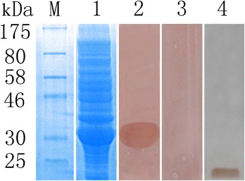
The TmP2 cDNA sequence consisted of an open reading frame of 366 bp encoding a putative protein with 121 amino acid residues. The protein was predicted to have a molecular weight of 12.62 kDa, a pI of 5.01, and weak hydropathicity. No signal peptides or transmembrane domains were predicted in this protein. The protein sequence of TmP2 was found to be highly homologous to those from T. solium (93 % identity), Echinococcus granulosus (81 %) and Hymenolepis microstoma (65 %), and it exhibited homology to the acidic ribosomal protein P2 from other parasites such as Spirometra erinaceieuropaei, Barentsia elongata and Caenorhabditis briggsae (51 %, 49 % and 47 % identity, respectively). Recombinant TmP2 was expressed as soluble protein with a molecular weight of approximately 32 kDa (Fig. 1). Due to an additional 20-kDa epitope tag fusion peptide, the molecular mass of TmP2 was ~12 kDa, similar to that predicted from its amino acid sequence. In Western blot analysis, a positive band of 32 kDa was observed when using goat anti-T. multiceps serum, suggesting a strong reactivity of this recombinant protein (Fig. 1). No signal was present when rTmP2 was incubated with native sera (Fig. 1). In addition, the total worm extract was blotted with anti-rTmP2 rabbit serum of approximately 12 kDa (Fig. 1).
Fig. 1.

SDS-PAGE and Western blotting analysis of recombinant TmP2 protein. M, Protein molecular weight markers; lane 1, the extracts of E. coli cells containing the pET-32a (+) expression vector with IPTG induction; lanes 2–3, rTmP2 protein probed with goat immune serum against T. multiceps (positive serum group; lane 2) or naïve goat serum (negative control, lane 3); lane 4, the total worm extract probed with goat immune serum against T. multiceps
Immunolocalization of P2 protein in T. multiceps
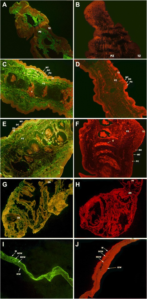
Protein P2 was highly localized to the parenchymatous zone (PZ) of both the adult parasite and the coenurus; furthermore, it was widely distributed in cystic wall of coenurus (Fig. 2). No fluorescence staining was detected with negative sera.
Fig. 2.

Immunolocalization of P2 protein in T. multiceps. The green fluorescence tint shows the location of TmP2 protein. a, antisera in cephalomeres of adult tapeworm; b, negative sera in cephalomeres of adult tapeworm; c, antisera in immature segments of adult tapeworm; d, negative sera in immature segments of adult tapeworm; e, antisera in mature segments of adult tapeworm; f, negative sera in mature segments of adult tapeworm. g, antisera in cephalomeres of coenurus; h, negative sera in cephalomeres of coenurus; i, antisera in cystic wall of coenurus; j, negative sera in cystic wall of coenurus. The magnification of all images is × 200. Arrows indicate the areas of the parasite: MT, microthrix; DC, distal cytoplasm; PC, perinuclear cytoplasm; TZ, tegument zone; PZ, parenchymatous zone; E, eggs; ZS, zone of scole; ZN, zone of neck; M, microtrichia; OCW, the outer layer of cystic wall; MCW, the middle layer of cystic wall; ICW, the inside layer of cystic wall
Establishment of indirect ELISA
The suitability of the recombinant TmP2 protein as a diagnostic antigen was tested based on indirect ELISA. Checkerboard titration tests indicated that the conditions that gave a highest P/N value (3.64) were when the coating antigen was 0.24 μg/well and the serum dilution was 1:10 (Table 1). In the optimized test conditions, a total of 24 negative serum samples were analyzed to obtain the cut-off value for the indirect ELISA, and OD450 value was 0.312 with a SD of 0.0622 (data not shown). All experiments were performed in triplicate. Thus, the cut-off value was 0.499 (mean + 3SD). Serum samples with an absorbance ≥0.499 were scored as coenurus antibody positive, otherwise they were determined to be coenurus antibody negative.
Table 1.
Determination of the coating concentration of protein and serum dilution in optimization of the indirect ELISA method
| Antisera at different dilutions | OD450 values of antigen at different coating concentrations | |||||
|---|---|---|---|---|---|---|
| 0.06 μg | 0.12 μg | 0.24 μg | 0.48 μg | 0.96 μg | 1.92 μg | |
| 1:5 (P) | 1.181 | 1.137 | 1.065 | 1.078 | 1.182 | 1.178 |
| 1:5 (N) | 0.427 | 0.361 | 0.319 | 0.307 | 0.478 | 0.487 |
| 1:10 (P) | 0.932 | 0.908 | 0.896 | 0.789 | 0.858 | 0.873 |
| 1:10 (N) | 0.357 | 0.288 | 0.246 | 0.238 | 0.357 | 0.371 |
| 1:20 (P) | 0.664 | 0.621 | 0.542 | 0.501 | 0.645 | 0.626 |
| 1:20 (N) | 0.238 | 0.244 | 0.191 | 0.180 | 0.275 | 0.245 |
| 1:40 (P) | 0.461 | 0.447 | 0.347 | 0.397 | 0.473 | 0.445 |
| 1:40 (N) | 0.198 | 0.165 | 0.131 | 0.148 | 0.223 | 0.227 |
| 1:80 (P) | 0.348 | 0.339 | 0.298 | 0.305 | 0.392 | 0.381 |
| 1:80 (N) | 0.157 | 0.143 | 0.115 | 0.122 | 0.134 | 0.139 |
Note: N, positive serum; P, negative serum; The values in bold represent optimum conditions of this indirect ELISA method
Sensitivity and specificity analysis of indirect ELISA
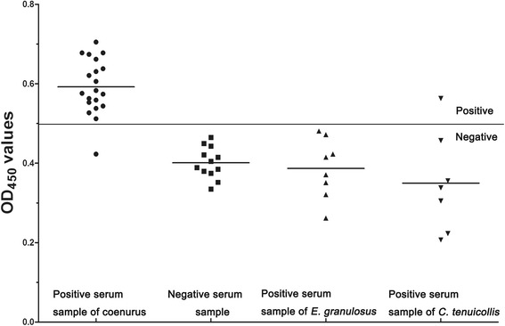
Specific IgG antibodies were determined in serum samples from goats infected with coenurus and C. tenuicollis, and from sheep infected with E. granulosus. (Fig. 3). Based on the cut-off of 0.499, a total of 20 serum samples from goats infected with coenurus were detected as positive, corresponding to a sensitivity of 95.0 % (19/20). There was cross-reactivity with serum from one C. tenuicollis-positive goat (n = 7) and no reactions with sera from E. granulosus-positive sheep (n = 8) and healthy goats (n = 12). Thus, the specificity of the ELISA using recombinant TmP2 antigen to detect anti-P2 antibody was 96.3 % (26/27).
Fig. 3.

The sensitivity, specificity and cross-reactivity of indirect ELISA. The bold horizontal line indicates the cut-off value (0.499)
Anti-TmP2 antibody in goats artificially infected with T. multiceps
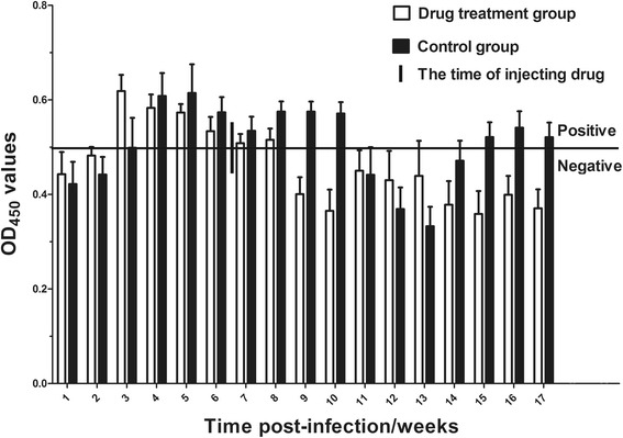
The regularities of serological antibodies are shown in Fig. 4 after the artificial infection of goats with T. multiceps. The following trends were observed: 3 weeks p.i., the drug treatment group and the control group were both positive for serum antibody to TmP2 (OD value >0.499, the cut-off value). At around 9 weeks p.i. (about 2 weeks post-injection of praziquantel), the antibody value for the drug-treated group dropped below the cut-off value; the antibody values remained below this critical value until the end of the experiment. In the control group, the antibody value dropped below the cut-off value 11 weeks p.i., but the value rose again and was detection positive from 15 weeks p.i. until the end of the experiment (17 weeks p.i.).
Fig. 4.

The regularities of serological antibody against TmP2 after the artificial infection of goats by T. multiceps. The bold horizontal line indicates the cut-off value (0.499)
Discussion
In recent years, studies concerning acidic ribosomal P2 proteins of parasites have mainly focused on Trypanosoma cruzi [37–39], Cryptosporidium parvum [29, 40], Toxoplasma gondii and Plasmodium falciparum [41, 42]. These studies confirmed that P2 protein could induce hosts to produce a strong humoral immune response, and the protein appears to constitute a potential target for host cell invasion inhibition in both T. gondii and P. falciparum infections [29, 41, 42]. Knowledge of P2 protein is very limited for the family Taeniidae, although there are few preliminary studies on T. solium [43–45] which confirmed that P2 is a main pathogenic factor of human cysticercosis, and demonstrated that a P2 fusion protein expressed in E. coli could be used as a diagnostic antigen for human neurocysticercosis [43]. In addition, Luo et al. (2003) [44] and Su et al. (2003) [45] showed that the recombinant P2 proteins of T. solium expressed in E. coli and Pichia pastoris were good immunogens. In the present study, we have cloned and expressed TmP2 from coenurus for the first time.
Immunofluorescence staining showed that, in P. falciparum, ribosomal protein P2 (PfP2) was present at the infected erythrocyte surface at the onset of cell division, and on the merozoite surface during the P. falciparum host infection process [41]. In the process of T. gondii host cell infection, P2 protein was confirmed to be localized at the surface and in the cytoplasm of the parasite [41]. In the present study, we observed that TmP2 was highly localized to the parenchymatous zone of both the adult parasite and the coenurus; besides, it was widely distributed in cystic wall of coenurus, commensurate with its ribosomal role. Whether P2 is present in the body of T. multiceps oncospheres during the oncosphere host-infection process, i.e. whether the P2 protein gathers at the body surface as in T. gondii and P. falciparum, requires further study.
Due to the fact that T. multiceps infected goats will not show obvious clinical symptoms, it is difficult to detect in the early infective stage. This stage involves the adherence and migration of oncospheres in the major blood vessels of the intestine, and then, frequently, oncosphere transport to the nervous system including the brain and spinal cord via the circulatory system [46]. Detection of an antibody against coenurus could be useful for early detection and treatment of the infection. ELISA, DIGFA and Dot-ELISA diagnostic methods have been established for coenurosis [14–16]. However, due to the cross-reaction of natural antigens, previous studies have failed to establish a reliable diagnostic method. Although indirect ELISA based on a recombinant antigen against coenurus has been developed for diagnosis, it has deficiencies including low sensitivity [47]. In our study, indirect ELISA of recombinant TmP2 was successfully established and optimized to detect coenurus in goats. The method had high sensitivity (95 %) and specificity (96.3 %) for 47 tested serum samples when compared with the results of necropsy. Moreover, there’s no cross reaction when using P2 protein for the detection of specificity of E. granulosus-positive sera. However, the indirect ELISA method against another protein (TmGST) of T. multiceps established based on the same sera showed one cross reaction (1/8) (unpublished). Serodiagnosis through indirect ELISA was successfully used in experimental coenurus infection in sheep [48], however, seropositivity was observed only from the 35th day p.i. In our experiment, the antibodies could be detected by indirect ELISA in the early stage of infection (3 weeks p.i), up to 17 weeks p.i.. The anti-TmP2 serum of goats fell below the cut-off value about 2 weeks post-injection of praziquantel in the drug treatment group, and the antibody values remained below the critical value until the end of the experiment. Therefore, we conclude that indirect ELISA can be applied to the evaluation of coenurosis after the drug treatment. We did not detect the anti-TmP2 antibody between 11 and 14 weeks p.i. in the control group; however, this phenomenon was not observed when we tested antibodies against another protein (TmGST) of T. multiceps by indirect ELISA (data not shown). So, one can speculate that, in this period, due to decreased P2 expression, we cannot positively detect the anti-TmP2 antibody. Although this is a potential weakness of the method, it can also be applied in clinic, because infected goats have shown obvious clinical symptoms between 11 and 14 weeks p.i.
Conclusions
Recombinant TmP2 is a suitable diagnostic antigen for coenurus infection. TmP2-based indirect ELISA for detection of coenurus in hosts is sensitive and specific, and detects the parasite from only 3 weeks post-infection. The method will be useful for the diagnosis of coenurosis and for validating the effectiveness of drug treatment of infections.
Acknowledgments
We are grateful to Dr Sanjie Cao, Tao Wang, Manli He and Yue Xie for their help and suggestions. This project was supported by a grant from the Program for Changjiang Scholars and Innovative Research Team in University (PCSIRT) (Grant no. IRT0848).
Footnotes
Competing interests
The authors declare that they have no competing interests.
Authors’ contributions
Xing Huang participated in the design of study, wrote the manuscript and performed the statistical analysis; Yingdong Yang, Yu Wang, Xiaobin Gu, Weimin Lai and Xuerong Peng contributed to animal care and experiments; Lin Chen contributed to study design and analyzed the data; Guangyou Yang conceived of the study, participated in its design and coordination; All authors read and approved the final manuscript.
Contributor Information
Xing Huang, Email: huangxing198308@163.com.
Lin Chen, Email: 77588606@qq.com.
Yingdong Yang, Email: yhhfg@163.com.
Xiaobin Gu, Email: 448070309@qq.com.
Yu Wang, Email: btyzwangyu@163.com.
Weimin Lai, Email: 451908892@qq.com.
Xuerong Peng, Email: 1295543513@qq.com.
Guangyou Yang, Email: guangyou1963@aliyun.com.
References
- 1.Ibechukwu BI, Onwukeme KE. Intraocular coenurosis: a case report. Br J Ophthalmol. 1991;75:430–1. doi: 10.1136/bjo.75.7.430. [DOI] [PMC free article] [PubMed] [Google Scholar]
- 2.El-On J, Shelef I, Cagnano E, Benifla M. Taenia multiceps: a rare human cestode infection in Israel. Vet Ital. 2008;44:621–31. [PubMed] [Google Scholar]
- 3.Christodoulopoulos G. Two rare clinical manifestations of coenurosis in sheep. Vet Parasitol. 2007;143:368–70. doi: 10.1016/j.vetpar.2006.09.010. [DOI] [PubMed] [Google Scholar]
- 4.Edwards GT, Herbert IV. Observations on the course of Taenia multiceps infections in sheep: clinical signs and post-mortem findings. Br Vet J. 1982;138:489–500. doi: 10.1016/s0007-1935(17)30934-x. [DOI] [PubMed] [Google Scholar]
- 5.Gauci C, Vural G, Oncel T, Varcasia A, Damian V, Kyngdon CT, et al. Vaccination with recombinant oncosphere antigens reduces the susceptibility of sheep to infection with Taenia multiceps. Int J Parasitol. 2008;38:1041–50. doi: 10.1016/j.ijpara.2007.11.006. [DOI] [PMC free article] [PubMed] [Google Scholar]
- 6.Hancock K, Pattabhi S, Greene RM, Yushak ML, Williams F, Khan A, et al. Characterization and cloning of GP50, a Taenia solium antigen diagnostic for cysticercosis. Mol Biochem Parasitol. 2004;133:115–24. doi: 10.1016/j.molbiopara.2003.10.001. [DOI] [PubMed] [Google Scholar]
- 7.Da Silva MR, Maia AA, Espíndola NM, Machado Ldos R, Vaz AJ, Henrique-Silva F. Recombinant expression of Taenia solium TS14 antigen and its utilization for immunodiagnosis of neurocysticercosis. Acta Trop. 2006;100:192–8. doi: 10.1016/j.actatropica.2006.10.009. [DOI] [PubMed] [Google Scholar]
- 8.Hancock K, Pattabhi S, Whitfield FW, Yushak ML, Lane WS, Garcia HH, et al. Characterization and cloning of T24, a Taenia solium antigen diagnostic for Cysticercosis. Mol Biochem Parasitol. 2006;147:109–17. doi: 10.1016/j.molbiopara.2006.02.004. [DOI] [PubMed] [Google Scholar]
- 9.Ferrer E, Bonay P, Foster-Cuevas M, González LM, Dávila I, Cortéz MM, et al. Molecular cloning and characterisation of Ts8B1, Ts8B2 and Ts8B3, three new members of the Taenia solium metacestode 8 kDa diagnostic antigen family. Mol Biochem Parasitol. 2007;152:90–100. doi: 10.1016/j.molbiopara.2006.12.003. [DOI] [PubMed] [Google Scholar]
- 10.Ferrer E, Martínez-Escribano JA, Barderas ME, González LM, Cortéz MM, Dávila I, et al. Peptide epitopes of the Taenia solium antigen Ts8B2 are immunodominant in human and porcine cysticercosis. Mol Biochem Parasitol. 2009;168:168–71. doi: 10.1016/j.molbiopara.2009.08.003. [DOI] [PubMed] [Google Scholar]
- 11.Lee EG, Lee MY, Chung JY, Je EY, Bae YA, Na BK, et al. Feasibility of baculovirus-expressed recombinant 10-kDa antigen in the serodiagnosis of Taenia solium neurocysticercosis. Trans R Soc Trop Med Hyg. 2005;99:919–26. doi: 10.1016/j.trstmh.2005.02.010. [DOI] [PubMed] [Google Scholar]
- 12.Virginio VG, Hernández A, Rott MB, Monteiro KM, Zandonai AF, Nieto A, et al. A set of recombinant antigens from Echinococcus granulosus with potential for use in the immunodiagnosis of human cystic hydatid disease. Clin Exp Immunol. 2003;132:309–15. doi: 10.1046/j.1365-2249.2003.02123.x. [DOI] [PMC free article] [PubMed] [Google Scholar]
- 13.Li J, Zhang WB, McManus DP. Recombinant antigens for immunodiagnosis of cystic echinococcosis. J Infect Dis. 2004;6:67–77. doi: 10.1251/bpo74. [DOI] [PMC free article] [PubMed] [Google Scholar]
- 14.Zhang XC. Preliminary study on the preparation of ELISA antigen for coenurosis in sheep. Chin J Vet Sci. 1986;6:404–11. [Google Scholar]
- 15.Wang CR, Yu WC, Qiu JH. The application of dot-immunogold filtration assay for the detection of coenurosis sheep. Chin J Vet Parasitol. 2001;9:12–4. [Google Scholar]
- 16.Yao XH, Yuan SX, Zhou W, Feng YJ, Yang JS. Study on the Application of Dot-ELISA for Diagnosing Coenurus. Jilin Anim Husb Vet Med. 2001;12:3–5. [Google Scholar]
- 17.Santos C, Ballesta JP. Ribosomal protein P0, contrary to phosphoproteins P1 and P2, is required for ribosome activity and Saccharomyces cerevisiae viability. J Biol Chem. 1994;269:15689–96. [PubMed] [Google Scholar]
- 18.Santos C, Ballesta JP. The highly conserved protein P0 carboxyl end is essential for ribosome activity only in the absence of proteins P1 and P2. J Biol Chem. 1995;270:20608–14. doi: 10.1074/jbc.270.35.20608. [DOI] [PubMed] [Google Scholar]
- 19.Remacha M, Jimenez-Diaz A, Bermejo B, Rodriguez-Gabriel MA, Guarinos E, Ballesta JP. Ribosomal acidic phosphoproteins P1 and P2 are not required for cell viability but regulate the pattern of protein expression in Saccharomyces cerevisiae. J Mol Cell Biol. 1995;15:4754–62. doi: 10.1128/MCB.15.9.4754. [DOI] [PMC free article] [PubMed] [Google Scholar]
- 20.Aguilar R, Montoya L, Sánchez de Jiménez E. Synthesis and phosphorylation of maize acidic ribosomal proteins. Plant Physiol. 1998;116:379–85. doi: 10.1104/pp.116.1.379. [DOI] [Google Scholar]
- 21.Bruner BF, Wynn DM, Reichlin M, Harley JB, James JA. Humoral antigenic targets of the ribosomal P0 lupus autoantigen are not limited to the carboxyl region. Ann NY Acad Sci. 2005;1051:390–403. doi: 10.1196/annals.1361.081. [DOI] [PubMed] [Google Scholar]
- 22.Bargis-Surgey P, Lavergne JP, Gonzalo P, Vard C, Filhol-Cochet O, Reboud JP. Interaction of elongation factor eEF2 with ribosomal P proteins. Eur J Biochem. 1999;262:606–11. doi: 10.1046/j.1432-1327.1999.00434.x. [DOI] [PubMed] [Google Scholar]
- 23.Szick K, Springer M, Bailey-Ser res J. Evolutionary analyses of the 12-kDa acidic ribosomal P-proteins reveal a distinct protein of higher plant ribosomes. Proc Natl Acad Sci U S A. 1998;95:2378–83. doi: 10.1073/pnas.95.5.2378. [DOI] [PMC free article] [PubMed] [Google Scholar]
- 24.Sun KH, Tang SJ, Lin ML, Wang YS, Sun GH, Liu WT. Monoclonal antibodies against human ribosomal P proteins penetrate into living cells and cause apoptosis of Jurkat T cells in culture. J Rheumatol. 2001;40:750–6. doi: 10.1093/rheumatology/40.7.750. [DOI] [PubMed] [Google Scholar]
- 25.Zampieri S, Degen W, Ghiradello A. Dephosphorylation of autoantigenic ribosomal P proteins during Fas-L induced apoptosis: a possible trigger for the development of the autoimmune response in patients with systemic lupus erythematosus. Ann Rheum Dis. 2001;60:72–6. doi: 10.1136/ard.60.1.72. [DOI] [PMC free article] [PubMed] [Google Scholar]
- 26.Barnard GF, Staniunas RJ, Bao S, Mafune K, Steele GD, Jr, Gollan JL, et al. Increased expression of human ribosomal phosphoprotein P0 messenger RNA in hepatocellular carcinoma and colon carcinoma. Cancer Res. 1992;52:3067–72. [PubMed] [Google Scholar]
- 27.Koren E, Reichlin MW, Koscec M, Fugate RD, Reichlin M. Autoantibodies to the ribosomal P proteins react with a plasma membrane-related target on human cells. J Clin Invest. 1992;89:1236–41. doi: 10.1172/JCI115707. [DOI] [PMC free article] [PubMed] [Google Scholar]
- 28.Honoré B, Vorum H, Pedersen AE, Buus S, Claësson MH. Changes in protein expression in p53 deleted spontaneous thymic lymphomas. Exp Cell Res. 2004;295:91–101. doi: 10.1016/j.yexcr.2003.11.029. [DOI] [PubMed] [Google Scholar]
- 29.Benitez A, Priest JW, Ehigiator HN, McNair N, Mead JR. Evaluation of DNA encoding acidic ribosomal protein P2 of Cryptosporidium parvum as a potential vaccine candidate for cryptosporidiosis. Vaccine. 2011;29:9239–45. doi: 10.1016/j.vaccine.2011.09.094. [DOI] [PMC free article] [PubMed] [Google Scholar]
- 30.Iborra S, Carrión J, Anderson C, Alonso C, Sacks D, Soto M. Vaccination with the Leishmania infantum acidic ribosomal P0 protein plus CpG oligodeoxynucleotides induces protection against cutaneous leishmaniasis in C57BL/6 mice but does not prevent progressive disease in BALB/c mice. Infect Immun. 2005;73:5842–52. doi: 10.1128/IAI.73.9.5842-5852.2005. [DOI] [PMC free article] [PubMed] [Google Scholar]
- 31.Tchorzewski M. The acidic ribosomal P proteins. Int J Biochem Cell Biol. 2002;34:911–5. doi: 10.1016/S1357-2725(02)00012-2. [DOI] [PubMed] [Google Scholar]
- 32.Martinez-Azorin F, Remacha M, Ballesta JP. Functional characterization of ribosomal P1/P2 proteins in human cells. Biochem J. 2008;413:527–34. doi: 10.1042/BJ20080049. [DOI] [PubMed] [Google Scholar]
- 33.Chen WJ, Niu DS, Zhang XY, Chen ML, Cui H, Wei WJ, et al. Recombinant 56-kilodalton major outer membrane protein antigen of Orientia tsutsugamushi Shanxi and its antigenicity. Infect Immun. 2003;71:4772–9. doi: 10.1128/IAI.71.8.4772-4779.2003. [DOI] [PMC free article] [PubMed] [Google Scholar]
- 34.Liddell S, Knox DP. Extracellular and cytoplasmic Cu/Zn superoxide dismutases from Haemonchus contortus. Parasitology. 1998;116:383–94. doi: 10.1017/S0031182098002418. [DOI] [PubMed] [Google Scholar]
- 35.Crowther JR, Walker JM. The ELISA guidebook. 2rd ed. Humana Press; 2009.
- 36.Shang SB, Li YF, Guo JQ, Wang ZT, Chen QX, Shen HG, et al. Development and validation of a recombinant capsid protein-based ELISA for detection of antibody to porcine circovirus type 2. Res Vet Sci. 2008;84:150–7. doi: 10.1016/j.rvsc.2007.02.007. [DOI] [PubMed] [Google Scholar]
- 37.Smulski C, Labovsky V, Levy G, Hontebeyrie M, Hoebeke J, Levin MJ. Structural basis of the cross-reaction between an antibody to the Trypanosoma cruzi ribosomal P2 beta protein and the human beta1 adrenergic receptor. Faseb J. 2006;20:1396–406. doi: 10.1096/fj.05-5699com. [DOI] [PubMed] [Google Scholar]
- 38.Pizarro JC, Boulot G, Bentley GA, Gómez KA, Hoebeke J, Hontebeyrie M, et al. Crystal structure of the complex mAb 17.2 and the C-terminal region of Trypanosoma cruzi P2β protein: implications in cross-reactivity. PLoS Negl Trop Dis. 2011;5:1–10. doi: 10.1371/journal.pntd.0001375. [DOI] [PMC free article] [PubMed] [Google Scholar]
- 39.Grippo V, Niborski LL, Gomez KA, Levin MJ. Human recombinant antibodies against Trypanosoma cruzi ribosomal P2β protein. Parasitology. 2011;138:736–47. doi: 10.1017/S0031182011000175. [DOI] [PubMed] [Google Scholar]
- 40.Priest JW, Kwon JP, Montgomery JM, Bern C, Moss DM, Freeman AR, et al. Cloning and characterization of the acidic ribosomal protein P2 of Cryptosporidium parvum, a new 17-kilodalton antigen. Clin Vaccine Immunol. 2010;17:954–65. doi: 10.1128/CVI.00073-10. [DOI] [PMC free article] [PubMed] [Google Scholar]
- 41.Das S, Basu H, Korde R, Tewari R, Sharma S. Arrest of nuclear division in Plasmodium through blockage of erythrocyte surface exposed ribosomal protein P2. PLoS Pathog. 2012;8:1–19. doi: 10.1371/annotation/913cb443-4033-4841-8666-1d348949a010. [DOI] [PMC free article] [PubMed] [Google Scholar]
- 42.Sudarsan R, Chopra RK, Khan MA, Sharma S. Ribosomal protein P2 localizes to the parasite zoite-surface and is a target for invasion inhibitory antibodies in Toxoplasma gondii and Plasmodium falciparum. Parasitol Int. 2014;64:43–9. doi: 10.1016/j.parint.2014.08.006. [DOI] [PubMed] [Google Scholar]
- 43.Kalinna BH, McManus DP. Cloning and characterization of a ribosomal P protein from Taenia solium, the aetiological agent of human cysticercosis. Biochem Biophys Res Commun. 1996;219:231–7. doi: 10.1006/bbrc.1996.0210. [DOI] [PubMed] [Google Scholar]
- 44.Luo XN, Cai XP, Cheng HT, Liu XT. Cloning and identification of phosphoprotein P2 of Cysticercus cellulosae and expression in E. coli. Chin J Zoonoses. 2003;19:80–4. [PubMed] [Google Scholar]
- 45.Su CX, Cai XP, Han XQ, Luo XL, Zheng YD, Dou YX. Expression of phosphoprotein P2 of Cysticercus cellulosae in Pichia pastoris and its application. Sheng Wu Gong Cheng Xue Bao. 2003;19:424–7. [PubMed] [Google Scholar]
- 46.Sharma DK, Chauhanb PPS. Coenurosis status in Afro-Asian region: A review. Small Rumin Res. 2006;64:197–202. doi: 10.1016/j.smallrumres.2005.05.021. [DOI] [Google Scholar]
- 47.An XX, Yang GY, Wang YW, Mu J, Yang AG, Gu XB, et al. Prokaryotic Expression of Tm7 Gene of T. multiceps and Establishment of Indirect ELISA Using the Expressed Protein. Acta Veterinariaet Zootechnica Sinica. 2011;42:1302–8. [Google Scholar]
- 48.Doganay A, Biyikoglu G, Oge H. Serodiagnosis of coenurosis by ELISA in experimentally infected lambs. Acta Parasitol Turc. 1999;23:185–9. [Google Scholar]


