

The patient
A 26-year-old man presented with a one-week history of lesions on the glans penis. The patient also stated that he had recurrent episodes of genital warts, which had been electrocauterized on previous occasions. He denied trauma, medication and history of another skin disorder. Physical examination revealed remarkably thick and hard, hyperkeratotic, oyster-shaped scales adherent to multiple erythematous lesions on the glans penis. Hyperpigmented, clustered verrucous papules, which coalesced to form plaques, were also detected on the shaft of his penis (Figure 1).
Figure 1.
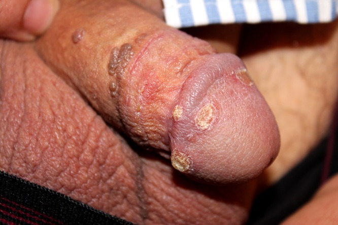
Thick, hyperkeratotic plaques on an erythematous base on the glans in association with warty, hyperpigmented papules on the shaft, of the penis. [Copyright: ©2015 Sezer et al.]
A shave biopsy from a hyperkeratotic lesion on the glans penis was performed; photomicrographs are presented in Figure 2A, B.
Figure 2.

(A) Marked papillomatosis, hyperkeratosis and congested blood vessels are observed at low magnification (hematoxylin and eosin stain; original magnification, X100). (B) Neutrophilic collections in the epidermis and stratum corneum (hematoxylin and eosin stain; original magnification, X400). [Copyright: ©2015 Sezer et al.]
What is your diagnosis?
Answer and explanation
Psoriasis verrucosa of the glans penis, in association with condyloma acuminatum
On histopathological examination, marked hyperkeratosis, parakeratosis and neutrophilic clusters in the stratum corneum were noted. Papillomatosis and acanthosis of the epidermis with neutrophilic collections in the upper stratum Malpighii along with vascular dilatation and perivascular lymphocytic infiltration of the dermis were also detected. Treponemal tests, namely VDRL and TPHA as well as immunohistochemical staining for the human papillomavirus (HPV), were negative.
Psoriasis verrucosa (PV) is a rare form of psoriasis, with only a few cases reported in the literature. Lesions are characterized by hard and thick hyperkeratotic plaques on the top of an erythematous base (as in our case). The lesions are located mainly on the trunk and extremities and, until now, penile involvement has not been reported. There is a male predominance (5:1), including our patient, and a history of long-term psoriasis (5–25 years) in all patients, excluding our case, who had rapid onset without previous psoriatic lesions [1–5]. Histopathological examination revealed combined features of psoriasis and verrucae, namely, marked papillomatosis with acanthosis, massive hyperkeratosis/parakeratosis with Munro’s microabscesses, Kogoj’s spongiform pustules in the stratum Malpighii, and dilated blood vessels in the upper dermis with lymphocytic inflammation [2,3]. Although condyloma acuminatum lesions, characterized by hyperpigmented, verrucous papules, were identified elsewhere on the penis in our patient, the lesion on the glans lacked koilocytosis, clumped keratohyalin granules, and negative immunohistochemistry for HPV, as well as neutrophilic microabscesses in the stratum corneum and epidermis, therefore arguing against a diagnosis of genital wart in this instance.
Associated features of PV such as obesity, cardiac dysfunction, lymphatic congestion and psychosis were absent in our case [1–4]. Some authors have suggested that local lymphatic disturbances and phlebitis may cause increased venous and lymphatic pressure, leading to leakage of plasma and proteins from blood, resulting in collagen fibrosis and epidermal hyperplasia [3]. We hypothesize that previous electrocauterization procedures for condyloma acuminatum may have resulted in lymphatic disturbances and activate PV via Koebnerization.
The treatment approaches for PV include oral retinoids, adalimumab, topical calcipotriol, corticosteroids and 5% crude coal tar [1–5]. We achieved marked regression of the lesions with a combination regimen of topical 5% salicylic acid and corticosteroid ointment including betamethasone valerate. Finally we suggest that a diagnosis of PV should be kept in mind in cases with oyster-like, hard, hyperkeratotic plaques on an erythematous base and histopathological findings including marked papillomatosis and neutrophilic microabscesses in the epidermis and stratum corneum.
Footnotes
Funding: None.
Competing interests: The authors have no conflicts of interest to disclose.
All authors have contributed significantly to this publication.
References
- 1.Wakamatsu K, Naniwa K, Hagiya K, Ichimiya M, Muto M. Psoriasis verrucosa. J Dermatol. 2010;37(12):1060–2. doi: 10.1111/j.1346-8138.2010.00944.x. [DOI] [PubMed] [Google Scholar]
- 2.Okuyama R, Tagami H. Psoriasis verrucosa in an obese Japanese man: a prompt clinical response observed with oral etretinate. J Eur Acad Dermatol Venereol. 2006;20(10):1359–61. doi: 10.1111/j.1468-3083.2006.01716.x. [DOI] [PubMed] [Google Scholar]
- 3.Nakamura S, Mihara M, Hagari Y, Shimao S. Psoriasis verrucosa showing peculiar histologic features. J Dermatol. 1994;21(2):102–5. doi: 10.1111/j.1346-8138.1994.tb01423.x. [DOI] [PubMed] [Google Scholar]
- 4.Maejima H, Katayama C, Watarai A, Nishiyama H, Katsuoka K. A case of psoriasis verrucosa successfully treated with adalimumab. J Drugs Dermatol. 2012;11:74–5. [PubMed] [Google Scholar]
- 5.Erkek E, Bozdogan O. Annular verrucous psoriasis with exaggerated papillomatosis. Am J Dermatopathol. 2001;23(2):133–5. doi: 10.1097/00000372-200104000-00008. [DOI] [PubMed] [Google Scholar]