Abstract
Feline morbillivirus (FmoPV) is an emerging virus in cats, which is associated with tubulointerstitial nephritis. To study the in vitro host range of FmoPV, we inoculated FmoPV strain SS1 to 32 cell lines originated from 13 species and cultured for 2 weeks, followed by RNA extraction and reverse-transcription-polymerase chain reaction for FmoPV detection. As a result, only cell lines derived from cats and African green monkeys were susceptible to FmoPV. FmoPV infects diverse feline cell lines: epithelial, fibroblastic, lymphoid and glial cells. These results indicate that the receptor (s) for FmoPV are ubiquitously expressed in cats. No infectivity of FmoPV was observed in human cell lines, which suggests least threatening of cross-species transmission of FmoPV from cats to humans.
Keywords: cat, cell tropism, cross-species transmission, feline morbillivirus, host range
Feline morbillivirus (FmoPV) is a negative-strand RNA virus in a morbillivirus family. Most morbilliviruses infect dendritic cells through the respiratory route and cause lymphopenia and immune suppression [1, 15, 18]. Subsequently, the virus is disseminated to peripheral tissues, resulting in large-scale infection of epithelial cells. FmoPV was initially reported in 2012 in Hong Kong, and this virus is considered to be associated with renal failure in cats, in particular, tubulointerstitial nephritis [19]. FmoPV RNA was detected in over 10% of stray cats in Hong Kong and mainland China. Furuya et al. also reported the existence of FmoPV infection in Japan [6]. By analyzing paraffin-embedded kidney tissues of cats with nephritis, 40% of domestic cats in Japan were positive for FmoPV. This pathological relationship is a big concern for veterinarians and cat owners, as tubulointerstitial nephritis in chronic renal disease is the most common renal failure observed in aged cats [4]. Recently, we demonstrated that although FmoPVs display genetic diversity in general, particular isolates from Japan and Hong Kong showed nearly identical nucleotide sequence [12]. This fact may suggest that there are natural vectors and/or reservoirs. Phylogenetic and recombination analyses indicated that the recombination has occurred within the F and H genes between Japan and Hong Kong isolates [11]. Although it was reported that Vero cell originated from African green monkey (AGM) is susceptible to FmoPV [19], it has never been examined whether FmoPV infects other species. In this investigation, we examined in vitro host range of FmoPV to assess the possibility of cross-species transmission.
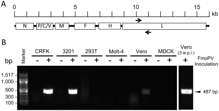
To prepare stock virus of FmoPV, we inoculated FmoPV strain SS1 [12] into Crandell-Rees feline kidney (CRFK) cells (ATCC, CCL-94). Two weeks post inoculation (w.p.i.), culture supernatants were harvested, filtered through a 450-nm membrane filter (PALL, Ann Arbor, MI, U.S.A.) and stored at −80°C as stock virus. The virus titer was determined as described before [7]. To study the in vitro host range of FmoPV, infectivity assay was performed using 32 cell lines originated from 13 species, i.e., human embryonic kidney (HEK) 293T cells (ATCC, CRL-11268), TE671 cells (human rhabdomyosarcoma) (ATCC, CCL-8805), HT1080 cells (human fibrosarcoma) (ATCC, CCL-12012), HeLa cells (human epithelioid carcinoma) (ATCC, CCL-2), MT-4 cells (human leukemia) (Public Health England, 08081402), Molt-4 cells (human leukemia) (ATCC, CRL-1582), Vero cells (AGM kidney cells) (ATCC, CCL-81), Cos-7 cells (AGM kidney cells) (ATCC, CRL-1651), CRFK cells (feline kidney cells) (ATCC, CCL-94), FEA cells (feline embryonic fibroblast), G355-5 cells (feline astrocyte) (ATCC, CRL-2033), AH927 cells (feline fibroblast), FER cells (feline embryonic fibroblast) (a kind gift from Prof. Jarrett), QN10S cells (feline fibroblast), fcwf-4 cells (feline whole fetus fibroblast) (ATCC, CRL-2787), 3201 cells (feline lymphoma) (ATCC, CRL-10909), MCC cells (feline lymphoma) [2], FL-4 cells (feline T cell) (ATCC, CRL-10772), FL-74 cells (feline lymphoma) (ATCC, CRL-8012), MDCK cells (canine kidney cells) (ATCC, CCL-34), Cf2Th cells (canine thymus fibroblast) (ATCC, CRL-1430), MDTF cells (Mus dunni tail fibroblast) (ATCC, CRL-2017), NIH3T3 cells (mouse fibroblast) (ATCC, CRL-1658), HSN cells (rat sarcoma) [3], C6 cells (rat glioma) (ATCC, CCL-107), SIRC cells (rabbit cornea) (ATCC, CCL-60), MPf cells (ferret brain fibroblast) (ATCC, CRL-1656), Mv1Lu cells (mink lung fibroblast) (ATCC, CCL-64), QT6 cells (quail fibrosarcoma) (ATCC, CRL-1708), MDBK cells (bovine kidney cells) (ATCC, CCL-22), FHK-Tcl3.1 cells (horse kidney cells) [8] and PK15 cells (porcine kidney cells) (ATCC, CCL-33) (Table 1). The cells were maintained in T25 flasks, trypsinized at 70 to 90% confluence and seeded T25 flasks with FmoPV strain SS1 at a multiplicity of infection of 1. The cultures were incubated at 37°C in a humidified atmosphere with 5% CO2 in air and observed daily for cytopathic effect under a light microscope. After serial passages for 2 weeks, we extracted RNA from the cells using QIAamp Viral RNA kit (Qiagen, Valencia, CA, U.S.A.) following the manufacturer’s instruction. Extracted RNA was reverse-transcribed using SuperScript III (Invitrogen, Carlsbad, CA, U.S.A.) with random primers, followed by PCR amplification targeting a 487 base pair (bp) fragment of the L gene by using specific primers (5′-GGAACATGGCCTCCTGTAGA-3′ and 5′-CTCCATTGGCAATCAGGTTT-3′) [6] (Fig. 1A). The PCR mixture (10 µl) contained cDNA, PCR buffer (10 nmol/l Tris-HCl, pH 8.3, 50 mmol/l KCl, 3 mmol/l MgCl2 and 0.01% gelatine), 200 µM of each dNTP and 1.0 units of ExTaq polymerase (TaKaRa, Otsu, Japan). PCR condition amplifying the DNA was as follows: 35 cycles of 94°C for 30 sec, 48°C for 30 sec and 72°C for 30 sec.
Table 1. Infectivity of feline morbillivirus in various cell lines.
| Cell line | Host | Origin | RT-PCR (Infectivity)* |
|---|---|---|---|
| 293T | Human | Embryonic kidney | - |
| TE671 | Human | Rhabdomyosarcoma | - |
| HT1080 | Human | Fibrosarcoma | - |
| Hela | Human | Adenocarcinoma | - |
| MT-4 | Human | Leukemia | - |
| Molt-4 | Human | Leukemia | - |
| Vero | AGM** | Kidney | + |
| Cos-7 | AGM** | Kidney | + |
| CRFK | Cat | Kidney | ++ |
| FEA | Cat | Embryonic fibroblast | ++ |
| G355-5 | Cat | Astrocyte | ++ |
| AH927 | Cat | Fibroblast | ++ |
| FER | Cat | Embryonic fibroblast | ++ |
| QN10s | Cat | Fibroblast | ++ |
| fcwf-4 | Cat | Fetus | ++ |
| 3201 | Cat | Lymphoma | ++ |
| MCC | Cat | Lymphoma | ++ |
| FL-4 | Cat | T cell | ++ |
| FL-74 | Cat | Lymphoma | ++ |
| MDCK | Dog | Kidney | - |
| Cf2Th | Dog | Thymus | - |
| MDTF | Mouse | Fibroblast | - |
| NIH3T3 | Mouse | Fibroblast | - |
| HSN | Rat | Sarcoma | - |
| C6 | Rat | Glioma | - |
| SIRC | Rabbit | Cornea | - |
| MPf | Ferret | Brain | - |
| Mv1Lu | Mink | Lung | - |
| QT6 | Quail | Fibrosarcoma | - |
| MDBK | Cattle | Kidney | - |
| FHK-Tcl 3.1 | Horse | Kidney | - |
| PK15 | Pig | Kidney | - |
* + and ++ indicate weak and strong bands, respectively. ** African green monkey.
Fig. 1.
The PCR amplification targeting a 487 bp fragment of the L gene (A) and gel electrophoresis analysis of representative samples (B). Only cell lines originated from AGMs and cats became positive for FmoPV at 2 w.p.i.. Three w.p.i., the band of FmoPV-infected Vero cells became stronger. Both kidney and lymphocyte cell lines derived humans were insensitive to FmoPV.
As a result, by RT-PCR amplifying the 487-bp fragment in the L gene of FmoPV, we revealed that only cell lines derived from cat and AGM were susceptible to FmoPV (Table 1). The difference of intensity of PCR bands is presumably due to replication efficacy in each cell line. Representative RT-PCR results are shown in Fig. 1B. Intensity of the PCR bands of FmoPV-infected Vero and COS-7 cells became stronger after three w.p.i. (Fig. 1 for Vero cells; data not shown for COS-7 cells), however, FmoPV was not detected from FmoPV-inoculated human cell lines and negative controls. CPE of FmoPV-infected Vero cells were also observed (data not shown). To further examine the possibility of viral adaptation in AGM cells to become infectious to human cells, we collected supernatants from FmoPV-infected Vero and Cos-7 cells and then inoculated them into human cell lines listed in Table 1. Again, we could not obtain any indication of FmoPV infection in human cell lines by RT-PCR.
Measles virus and canine distemper virus (CDV) use two cellular receptors: CD150 expressed on subsets of immune cells [17] and nectin-4 expressed in epithelial cells [9, 10]. Measles virus infects immune cells expressing CD150, and the infected immune cells travel to the draining lymph nodes. After amplifying the virus, cell-associated viremia spread the infection to other organs including kidney, gastrointestinal tract and liver. Late in pathogenesis, the virus infects airway epithelial cells expressing nectin-4 via the basolateral surface, followed by replication and shed apically from the infected cell into the airway lumen [10]. In this study, we found that FmoPV infected variety of cell types including epithelial, fibroblastic, lymphoid and glial cells. This indicates the FmoPV receptor (s) are expressed in a variety of cat tissues. The association between FmoPV and interstitial nephritis was reported, but relationships with other diseases are still unclear. CDV is known to cause lethal systemic diseases including central nervous system (CNS) disease in large felids [16]. FmoPV may have a potential to cause other diseases, such as CNS disease, since G355-5 cells originated from feline astrocytes are susceptible to FmoPV (Table 1).
The different morbilliviruses most likely evolved from a common ancestral virus that has adapted to their respective mammalian hosts, indicating that morbilliviruses have an intrinsic capacity to adapt to new host species [5]. Having cats might be a big zoonotic concern, because they are one of the most popular companion animals, and human may expose to FmoPV through physical contacts with cats. Our results indicate that FmoPV possibly infects AGMs but not humans. There was no adaptation to human cell lines through replication in AGM cell lines. Therefore, here, we provisionally conclude that there are least threatening of cross-species transmission of FmoPV from cats to humans. However, CDV is known to cause lethal diseases not only in carnivores but also in other species. CDV infects human cells only by one amino acid substitution [13, 14]. Thus, we cannot exclude the possibility that FmoPV would become a threat for other animals in the future. Although a current study showed that cell lines except for ones from cats and AGMs are insensitive to FmoPV in vitro, further investigation and monitoring might be needed.
Acknowledgments
We would like to express our gratitude to Makoto Ogawa for his kind help. We are grateful to Prof. Os Jarrett (University of Glasgow, Glasgow, U.K.) for providing AH927, FER, QN10S and MCC cells. We also thank Prof. Ken Maeda (Yamaguchi University, Yamaguchi, Japan) for providing FHK-Tcl3.1 cells. Shoichi Sakaguchi was supported by a fellowship of the Japan Society for the Promotion of Science.
REFERENCES
- 1.Beineke A., Puff C., Seehusen F., Baumgärtner W.2009. Pathogenesis and immunopathology of systemic and nervous canine distemper. Vet. Immunol. Immunopathol. 127: 1–18. doi: 10.1016/j.vetimm.2008.09.023 [DOI] [PubMed] [Google Scholar]
- 2.Cheney C. M., Rojko J. L., Kociba G. J., Wellman M. L., Di Bartola S. P., Rezanka L. J., Forman L., Mathes L. E.1990. A feline large granular lymphoma and its derived cell line. In Vitro Cell. Dev. Biol. 26: 455–463. doi: 10.1007/BF02624087 [DOI] [PubMed] [Google Scholar]
- 3.Currie G. A., Gage J. O.1973. Influence of tumour growth on the evolution of cytotoxic lymphoid cells in rats bearing a spontaneously metastasizing syngeneic fibrosarcoma. Br. J. Cancer 28: 136–146. doi: 10.1038/bjc.1973.131 [DOI] [PMC free article] [PubMed] [Google Scholar]
- 4.DiBartola S. P., Rutgers H. C., Zack P. M., Tarr M. J.1987. Clinicopathologic findings associated with chronic renal disease in cats: 74 cases (1973–1984). J. Am. Vet. Med. Assoc. 190: 1196–1202. [PubMed] [Google Scholar]
- 5.Di Guardo G., Marruchella G., Agrimi U., Kennedy S.2005. Morbillivirus infections in Aquatic Mammals: A Brief Overview. J. Vet. Med. A Physiol. Pathol. Clin. Med. 52: 88–93. doi: 10.1111/j.1439-0442.2005.00693.x [DOI] [PubMed] [Google Scholar]
- 6.Furuya T., Sassa Y., Omatsu T., Nagai M., Fukushima R., Shibutani M., Yamaguchi T., Uematsu Y., Shirota K., Mizutani T.2014. Existence of feline morbillivirus infection in Japanese cat populations. Arch. Virol. 159: 371–373. doi: 10.1007/s00705-013-1813-5 [DOI] [PubMed] [Google Scholar]
- 7.Koide R., Sakaguchi S., Miyazawa T.2015. Basic biological characterization of feline morbillivirus. J. Vet. Med. Sci. 77: 565–569. doi: 10.1292/jvms.14-0623 [DOI] [PMC free article] [PubMed] [Google Scholar]
- 8.Maeda K., Yasumoto S., Tsuruda A., Andoh K., Kai K., Otoi T., Matsumura T.2007. Establishment of a novel equine cell line for isolation and propagation of equine herpesviruses. J. Vet. Med. Sci. 69: 989–991. doi: 10.1292/jvms.69.989 [DOI] [PubMed] [Google Scholar]
- 9.Mühlebach M. D., Mateo M., Sinn P. L., Prüfer S., Uhlig K. M., Leonard V. H. J., Navaratnarajah C. K., Frenzke M., Wong X. X., Sawatsky B., Ramachandran S., McCray P. B., Cichutek K., von Messling V., Lopez M., Cattaneo R.2011. Adherens junction protein nectin-4 is the epithelial receptor for measles virus. Nature 480: 530–533. [DOI] [PMC free article] [PubMed] [Google Scholar]
- 10.Noyce R. S., Delpeut S., Richardson C. D.2013. Dog nectin-4 is an epithelial cell receptor for canine distemper virus that facilitates virus entry and syncytia formation. Virology 436: 210–220. doi: 10.1016/j.virol.2012.11.011 [DOI] [PubMed] [Google Scholar]
- 11.Park E.S., Suzuki M., Kimura M., Maruyama K., Mizutani H., Saito R., Kubota N., Furuya T., Mizutani T., Imaoka K., Morikawa S.2014. Identification of a natural recombination in the F and H genes of feline morbillivirus. Virology 468-470: 524–531. doi: 10.1016/j.virol.2014.09.003 [DOI] [PubMed] [Google Scholar]
- 12.Sakaguchi S., Nakagawa S., Yoshikawa R., Kuwahara C., Hagiwara H., Asai K.I., Kawakami K., Yamamoto Y., Ogawa M., Miyazawa T.2015. Genetic diversity of feline morbilliviruses isolated in Japan. J. Gen. Virol. 96: 681–687. doi: 10.1099/vir.0.071688-0 [DOI] [PubMed] [Google Scholar]
- 13.Sakai K., Nagata N., Ami Y., Seki F., Suzaki Y., Iwata-Yoshikawa N., Suzuki T., Fukushi S., Mizutani T., Yoshikawa T., Otsuki N., Kurane I., Komase K., Yamaguchi R., Hasegawa H., Saijo M., Takeda M., Morikawa S.2013. Lethal canine distemper virus outbreak in cynomolgus monkeys in Japan in 2008. J. Virol. 87: 1105–1114. doi: 10.1128/JVI.02419-12 [DOI] [PMC free article] [PubMed] [Google Scholar]
- 14.Sakai K., Yoshikawa T., Seki F., Fukushi S., Tahara M., Nagata N., Ami Y., Mizutani T., Kurane I., Yamaguchi R., Hasegawa H., Saijo M., Komase K., Morikawa S., Takeda M.2013. Canine distemper virus associated with a lethal outbreak in monkeys can readily adapt to use human receptors. J. Virol. 87: 7170–7175. doi: 10.1128/JVI.03479-12 [DOI] [PMC free article] [PubMed] [Google Scholar]
- 15.Sato H., Yoneda M., Honda T., Kai C.2012. Morbillivirus receptors and tropism: multiple pathways for infection. Front. Microbiol. 3: 75. doi: 10.3389/fmicb.2012.00075 [DOI] [PMC free article] [PubMed] [Google Scholar]
- 16.Seimon T. A., Miquelle D. G., Chang T. Y., Newton A. L., Korotkova I., Ivanchuk G., Lyubchenko E., Tupikov A., Slabe E., McAloose D.2013. Canine distemper virus: an emerging disease in wild endangered Amur tigers (Panthera tigris altaica). MBio 4: e00410–e00413. doi: 10.1128/mBio.00410-13 [DOI] [PMC free article] [PubMed] [Google Scholar]
- 17.Tatsuo H., Ono N., Yanagi Y.2001. Morbilliviruses use signaling lymphocyte activation molecules (CD150) as cellular receptors. J. Virol. 75: 5842–5850. doi: 10.1128/JVI.75.13.5842-5850.2001 [DOI] [PMC free article] [PubMed] [Google Scholar]
- 18.von Messling V., Milosevic D., Devaux P., Cattaneo R.2004. Canine distemper virus and measles virus fusion glycoprotein trimers: partial membrane-proximal ectodomain cleavage enhances function. J. Virol. 78: 7894–7903. doi: 10.1128/JVI.78.15.7894-7903.2004 [DOI] [PMC free article] [PubMed] [Google Scholar]
- 19.Woo P. C. Y., Lau S. K. P., Wong B. H. L., Fan R. Y. Y., Wong A. Y. P., Zhang A. J. X., Wu Y., Choi G. K. Y., Li K. S. M., Hui J., Wang M., Zheng B.J., Chan K. H., Yuen K.Y.2012. Feline morbillivirus, a previously undescribed paramyxovirus associated with tubulointerstitial nephritis in domestic cats. Proc. Natl. Acad. Sci. U.S.A. 109: 5435–5440. doi: 10.1073/pnas.1119972109 [DOI] [PMC free article] [PubMed] [Google Scholar]


