Vascularized lymph node transfer of lymph nodes from donor sites to affected sites can restore lymphatic flow and effectively treat lymphedema. A documented risk of vascularized lymph node transfer is the development of new lymphedema at or around the lymph node harvest donor site or limb. Studies have reported rare instances of donor-site lymphedema following lymph node flap harvest from axillary or groin donor sites.1–3
The supraclavicular area has been described previously as a donor site without risk of secondary lymphedema in the surrounding tissues, with some surgeons favoring this donor site because of the perceived lack of risk.4 We describe a patient who presented with lymphedema of the right arm following vascularized lymph node transfer from the right supraclavicular donor area to the left groin. The development of lymphedema in the right upper extremity following a supraclavicular node harvest challenges this previous notion that the supraclavicular area is without risk of donor-site lymphedema. Careful patient selection, surgical expertise, and methods such as reverse lymph node mapping may reduce this risk.5–8
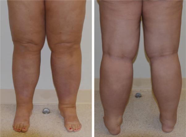
A 55-year-old woman presented to our office after she developed lymphedema of the right arm approximately 2 years after she had vascularized lymph node transfer performed by another surgeon. She had initially developed left leg lymphedema after an epidural procedure. In the following year, the patient also developed lymphedema in the right leg (Fig. 1). The vascularized lymph node transfer procedure from the right supraclavicular fossa to the left groin was then performed by the other surgeon to treat the swelling (Fig. 2). The patient's postoperative course was complicated by the accumulation of seroma containing milky fluid at the supraclavicular donor site, which resolved approximately 4 weeks after surgery with conservative treatment. Approximately 6 months after the vascularized lymph node transfer surgery, the patient developed lymphedema in her right arm. A volume excess of 1055 cc was present on follow-up examination (Fig. 3). Lymphoscintigraphic imaging before and after the vascularized lymph node transfer surgery revealed a significant decrease of tracer migration in the right arm and loss of visualization of tracer in the right axillary lymph nodes after the operation, consistent with lymphedema (Fig. 4).
Fig. 1.
Patient with bilateral lower extremity lymphedema.
Fig. 2.

Right supraclavicular lymph node transfer donor site.
Fig. 3.

Right upper extremity lymphedema following vascularized lymph node transfer from the right supraclavicular area.
Fig. 4.
Lymphoscintigraphic findings before (left) and after (right) supraclavicular lymph node harvest. Note loss of tracer uptake in the right axilla in the postoperative image.
Effective treatments for both congenital and secondary lymphedema have been documented extensively in the medical literature. Multiple studies have documented the effectiveness of conservative lymphedema therapy, vascularized lymph node transfer, lymphaticovenous anastomosis, and suction-assisted protein lipectomy for properly selected patients with lymphedema.5–14 Vascularized lymph node transfer involves transfer of lymph nodes and the surrounding soft tissue as a microsurgical free flap from a donor site to the affected area. This technique is most effective for the treatment of fluid-predominant lymphedema, and can reduce the need for compression garment use and lymphedema therapy. Furthermore, vascularized lymph node transfer can improve patient quality of life and dramatically reduce the risk of dangerous lymphedema cellulitis in affected individuals.5–14
This case challenges the previous notion that the supraclavicular donor site is free from postoperative lymphedema risk. Careful patient selection and anatomical dissection, surgeon experience with the vascularized lymph node transfer procedure, and the use of reverse lymphatic mapping may reduce such donor-site risk.
DOI: 10.1097/PRS.0000000000001253
Footnotes
DISCLOSURE
The authors have no financial interest to declare in relation to the content of this article.
Contributor Information
Ming Lee, Emory University School of Medicine
Evan McClure, Emory University School of Medicine, and Goizueta Business School, Emory University
Erik Reinertsen, Emory University School of Medicine, Wallace H. Coulter Department of Biomedical Engineering, at Emory University, and Georgia Institute of Technology, Atlanta, Ga.
Jay W. Granzow, Division of Plastic Surgery, University of California, Los Angeles, Harbor–UCLA Medical Center and UCLA David Geffen School of Medicine, Los Angeles, Calif.
REFERENCES
- 1.Viitanen TP, Mäki MT, Seppänen MP, Suominen EA, Saaristo AM. Donor-site lymphatic function after microvascular lymph node transfer. Plast Reconstr Surg. 2012;130:1246–1253. doi: 10.1097/PRS.0b013e31826d1682. [DOI] [PubMed] [Google Scholar]
- 2.Vignes S, Blanchard M, Yannoutsos A, Arrault M. Complications of autologous lymph-node transplantation for limb lymphoedema. Eur J Vasc Endovasc Surg. 2013;45:516–520. doi: 10.1016/j.ejvs.2012.11.026. [DOI] [PubMed] [Google Scholar]
- 3.Pons G, Masia J, Loschi P, Nardulli ML, Duch J. A case of donor-site lymphoedema after lymph node-superficial circumflex iliac artery perforator flap transfer. J Plast Reconstr Aesthet Surg. 2014;67:119–123. doi: 10.1016/j.bjps.2013.06.005. [DOI] [PubMed] [Google Scholar]
- 4.Althubaiti GA, Crosby MA, Chang DW. Vascularized supraclavicular lymph node transfer for lower extremity lymphedema treatment. Plast Reconstr Surg. 2013;131:133e–135e. doi: 10.1097/PRS.0b013e318272a1b4. [DOI] [PubMed] [Google Scholar]
- 5.Granzow JW, Soderberg JM, Kaji AH, Dauphine C. An effective system of surgical treatment of lymphedema. Ann Surg Oncol. 2014;21:1189–1194. doi: 10.1245/s10434-014-3515-y. [DOI] [PubMed] [Google Scholar]
- 6.Granzow JW, Soderberg JM, Kaji AH, Dauphine C. Review of current surgical treatments for lymphedema. Ann Surg Oncol. 2014;21:1195–1201. doi: 10.1245/s10434-014-3518-8. [DOI] [PubMed] [Google Scholar]
- 7.Granzow JW, Soderberg JM, Dauphine C. A novel two-stage surgical approach to treat chronic lymphedema. Breast J. 2014;20:420–422. doi: 10.1111/tbj.12282. [DOI] [PubMed] [Google Scholar]
- 8.Dayan JH, Dayan E, Smith ML. Reverse lymphatic mapping: A new technique for maximizing safety in vascularized lymph node transfer. Plast Reconstr Surg. 2015;135:277–285. doi: 10.1097/PRS.0000000000000822. [DOI] [PubMed] [Google Scholar]
- 9.Cheng MH, Huang JJ, Huang JJ, et al. A novel approach to the treatment of lower extremity lymphedema by transferring a vascularized submental lymph node flap to the ankle. Gynecol Oncol. 2012;126:93–98. doi: 10.1016/j.ygyno.2012.04.017. [DOI] [PubMed] [Google Scholar]
- 10.Basta MN, Gao LL, Wu LC. Operative treatment of peripheral lymphedema: A systematic meta-analysis of the efficacy and safety of lymphovenous microsurgery and tissue transplantation. Plast Reconstr Surg. 2014;133:905–913. doi: 10.1097/PRS.0000000000000010. [DOI] [PubMed] [Google Scholar]
- 11.Patel KM, Lin CY, Cheng MH. From theory to evidence: Long-term evaluation of the mechanism of action and flap integration of distal vascularized lymph node transfers. J Reconstr Microsurg. 2015;31:26–30. doi: 10.1055/s-0034-1381957. [DOI] [PubMed] [Google Scholar]
- 12.Becker C, Assouad J, Riquet M, Hidden G. Postmastectomy lymphedema: Long-term results following microsurgical lymph node transplantation. Ann Surg. 2006;243:313–315. doi: 10.1097/01.sla.0000201258.10304.16. [DOI] [PMC free article] [PubMed] [Google Scholar]
- 13.Lin CH, Ali R, Chen SC, et al. Vascularized groin lymph node transfer using the wrist as a recipient site for management of postmastectomy upper extremity lymphedema. Plast Reconstr Surg. 2009;123:1265–1275. doi: 10.1097/PRS.0b013e31819e6529. [DOI] [PubMed] [Google Scholar]
- 14.Patel KM, Cheng MH. A prospective evaluation of lymphedema-specific quality of life outcomes following vascularized lymph node transfer. Plast Reconstr Surg. 2014;133:1008. doi: 10.1245/s10434-014-4276-3. [DOI] [PubMed] [Google Scholar]


