Abstract
Varicella zoster virus (VZV) is a highly infectious human herpesvirus that is the causative agent for chicken pox and shingles. VZV encodes a functional thymidylate synthase (TS), which is the sole enzyme that produces dTMP from dUMP de novo. To study substrate binding, the complex structure of TSVZV with dUMP was determined to a resolution of 2.9 Å. In the absence of a folate co-substrate, dUMP binds in the conserved TS active site and is coordinated similarly as in the human encoded TS (TSHS) in an open conformation. The interactions between TSVZV with dUMP and a cofactor analog, raltitrexed, were also studied using differential scanning fluorimetry (DSF), suggesting that TSVZV binds dUMP and raltitrexed in a sequential binding mode like other TS. The DSF also revealed interactions between TSVZV and in vitro phosphorylated brivudine (BVDUP), a highly potent anti-herpesvirus drug against VZV infections. The binding of BVDUP to TSVZV was further confirmed by the complex structure of TSVZV and BVDUP solved at a resolution of 2.9 Å. BVDUP binds similarly as dUMP in the TSHS but it induces a closed conformation of the active site. The structure supports that the 5-bromovinyl substituent on BVDUP is likely to inhibit TSVZV by preventing the transfer of a methylene group from its cofactor and the subsequent formation of dTMP. The interactions between TSVZV and BVDUP are consistent with that TSVZV is indeed a target of brivudine in vivo. The work also provided the structural basis for rational design of more specific TSVZV inhibitors.
Introduction
Varicella zoster virus (VZV) is a highly contagious human herpesvirus that causes varicella, which is commonly known as chicken pox [1]. Like all other human herpesviruses, VZV infection is life-long and the virus establishes its latency in dorsal root ganglia [2, 3]. Reactivation of VZV from latency can cause herpes zoster or shingles, which occurs more frequently in older adults and immunocompromised individuals [1]. The herpes zoster lesions have been associated with acute and persisting pain that may last beyond a month after the initial outbreak [4].
Herpesviral DNA synthesis is pivotal for the production of new infectious virus particles in the host cells. This is governed by activities of DNA synthesis proteins, which have been the focus in the development of many anti-herpesvirus drugs [5, 6]. A large proportion of the clinically approved anti-herpesvirus drugs are nucleoside analogs that have inhibitory actions against the viral DNA polymerase upon their conversions to the nucleotide triphosphosphates [7, 8]. These include the acyclic nucleoside analogs aciclovir, valaciclovir, penciclovir, famiciclovir, ganciclovir and valgancyclovir, the deoxycytidine analog cidofovir, and the 5-substituted pyrimidine nucleoside analogs idoxuridine (IDU) and brivudine (BVDU) [7, 8]. The safety and selectivity of most of these nucleoside analogs are dependent on the first phosphorylation step by the viral thymidine kinase (TK) that is only encoded in a virally infected cell [9, 10].
Thymidylate synthase (TS) is a highly conserved protein found through all kingdoms of life including human, mouse, bacteria, protozoa and some viruses. It catalyzes the transfer of the methylene group from cofactor 5,10-methylenetetrahydrofolate (mTHF) to substrate deoxyuridine monophosphate (dUMP) [11]. In this process, deoxythymidine monophosphate (dTMP) is produced de novo and mTHF is converted to dihydrofolate (DHF). Subsequently, dTMP undergoes another two successive phosphorylation steps to produce deoxythymidine triphosphate (dTTP), which is added to the elongating DNA chain by the DNA polymerase [11].
Due to its essential role in the DNA replication, the human TS (TSHS) has been extensively studied and is a well-proven drug target for cancer therapy [12]. Inhibition of TSHS has been shown to lead to a deficiency in dTTP and DNA damage due to insertion of deoxyuridine triphosphate (dUTP) into DNA. This results in a cell death, so called thymineless death [13]. Different TSHS inhibitors have been derived from modifications of its natural substrate dUMP and cofactor mTHF. These include the nucleobase analog 5-fluorouridine and the folate analogs raltitrexed and pemetrexed, which are currently used in treatments against solid tumors [14–16]. Molecular features of TSHS and other TS have been elucidated in complex structures with different substrates and/or inhibitors [17–30]. These structures have contributed greatly to rational structure based drug designs that improved the specificity and efficacy of TSHS inhibitors [31].
For reasons not completely clear, only VZV and Kaposi’s sarcoma associated herpesvirus (KSHV) encode a TS (TSVZV and TSKSHV respectively) [32, 33]. Both TSVZV and TSKSHV share more than 50% sequence identity with TSHS but unlike the well-studied human counterpart, the homologous herpesviral TSs remain poorly characterized and no structural information is available on these proteins. TSVZV has also been shown to be non-essential for the viral replication in vitro [34]. In an attempt to gain insights into TSVZV and to differentiate TSVZV from TSHS, biophysical analysis and structural analyzes of TSVZV with its substrate dUMP and a TSHS inhibitor raltitrexed were carried out. The structures of TSVZV and TSVZV in complex with dUMP were solved to a resolution of 3.1 Å and 2.9 Å respectively. This constitutes the first crystal structure of an eukaryotic virus TS. The biophysical assays and the complex structure of TSVZV with dUMP demonstrated similar modes of substrate binding for TSVZV and TSHS. Structural comparisons of TSVZV and TSHS also revealed similar substrate binding details but with slight amino acids variations in the TS active site. Biophysical and crystallographic studies of TSVZV with an in vitro phosphorylated BVDU supports binding of phosphorylated BVDU to TSVZV. This study also provided further support for that TSVZV is a potential target enzyme for BVDU after metabolic activation in infected cells.
Results
Structure of apo TSVZV
The structure of the apo TSVZV (apo-TSVZV) was determined and refined to a final resolution of 3.1 Å, with four molecules in each asymmetric unit (Table 1 and S1 Fig). Apo-TSVZV was crystallized with a protein construct comprising of amino acids 8 to 295, which lacks the N-terminus seven amino acids and C-terminus six amino acids. This model has electron densities for amino acids 15–295, except for a missing loop containing amino acids 38–39 in one of the four molecules in the asymmetric unit. The four TSVZV monomers in the asymmetric unit are arranged as two homo-dimers. The monomers are virtually the same and the Cα atoms superimpose with a root mean square deviation (rmsd) of 0.78 Å.
Table 1. Data collection and refinement statistics on the apo and complex structures of TSVZV.
| Apo TSVZV (4XSE) | TSVZV+dUMP (4XSD) | TSVZV+BVDUP (4XSC) | |
|---|---|---|---|
| Data Collection | |||
| Beamline | AS MX1 | AS MX1 | AS MX1 |
| Wavelength (Å) | 0.9537 | 0.9537 | 0.9537 |
| Resolution Range (Å) | 29.7–3.1 (3.1–3.2) a | 30.0–2.9 (2.9–3.0)b | 30.0–2.9 (2.9–3.0) b |
| Space group | p32 | p32 | p32 |
| Unit cell | 153.4 Å 153.4 Å 89.2 Å | 150.2 Å 150.2 Å 89.2 Å | 149.5 Å 149.5 Å 89.0 Å |
| 90.0° 90.0° 120.0° | 90.0° 90.0° 120.0° | 90.0° 90.0° 120.0° | |
| Total reflections | 155476 | 1195011 | 94756 |
| Unique reflections | 42243 | 49612 | 46238 |
| I/I(σ) | 8.5 (2.4) a | 19.8 (3.9) b | 8.1 (1.9) b |
| Multiplicity | 3.7 (3.4) a | 5.5 (5.2) b | 1.5 (1.5) b |
| Completeness (%) | 99.2 (97.5) a | 100.0 (99.9) b | 96.2 (95.3) b |
| Rmerge c (%) | 12.7 (47.5) a | 6.9 (38.0) b | 9.7 (40.3) b |
| Refinement | |||
| Rfactor d (%) | 23.8 | 24.0 | 19.6 |
| Rfree e (%) | 26.8 | 28.0 | 23.5 |
| Protein residues | 1116 | 1081 | 1244 |
| Solvent | 0 | 9 | 30 |
| Ligands | 4 phosphate ions | 4 dUMP | 4 BVDUP |
| 4 1PE | |||
| RMSD bonds (Å) | 0.5 | 0.0045 | 0.002 |
| RMSD angles (°) | 1.134 | 0.882 | 0.585 |
| Ramachandran favored (%) | 95 | 96 | 94.4 |
| Ramachandran allowed (%) | 5 | 4 | 5.1 |
| Ramachandran outliers (%) | 0 | 0 | 0.5 |
| Rotamer outliers (%) | 4.4 | 6.4 | 0.8 |
a The values in the parentheses are for the highest resolution shell (3.1–3.2 Å).
b The values in the parentheses are for the highest resolution shell (2.9–3.0 Å).
c , where I i is the ith intensity measurement of reflection hkl, Ī(hkl) is the mean intensity measurement of the symmetry related or replicated reflections of the unique reflection hkl.
d , where F obs and F calc are the observed and calculated structure factors respectively.
e Rfree is equivalent to Rfactor but 5% of the measured reflections have been excluded from refinement and set aside for cross validation.
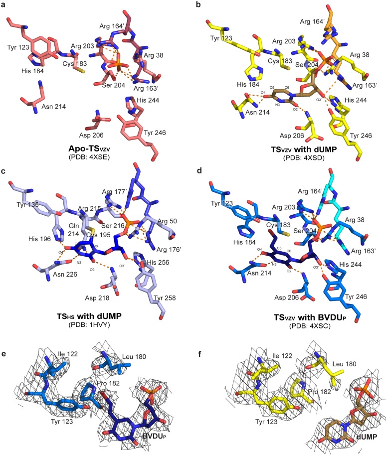
The structures of apo-TSVZV and the human encoded apo TS (TSHS) superimpose with a rmsd of 0.9 Å for 558 residues, with the highest similarity at the active sites [22]. A phosphate ion was found at one end of the active site in each of the TSVZV monomers and is held in place by Arg 203, Ser 204, as well as Arg 163’ and Arg 164’ that are located on the loop from the dimer partner (Fig 1a).
Fig 1. Stereo views of the TSVZV and TSHS active sites.
Corresponding stereo views of the ligands and amino acids lining the TS active sites in the structures of (a) apo-TSVZV with a phosphate ion, (b) TSVZV with dUMP, (c) TSHS with dUMP (PDB ID: 1HVY) [35] and (d) TSVZV with BVDUP. The polar interactions between the amino acids and the different ligands are illustrated by orange dotted lines. 2F0-FC electron densities of the binding (e) BVDUP and (f) dUMP with the surrounding hydrophobic amino acids are also shown (contoured at 1σ with carved = 2.0).
Complex structure of TSVZV with dUMP
TSVZV was crystallized in a second crystal form that was used for subsequent determination of the structure of TSVZV in complex with dUMP (TSVZV+dUMP). The TSVZV+dUMP structure was determined and refined to a resolution of 2.9 Å (Table 1). Each asymmetric unit contains four TSVZV subunits arranged into two homo-dimers. Each subunit is made up of amino acids 15 to 295. This is with exception to amino acids 98 to 117 in one monomer of the TSVZV dimer, and amino acids 38 to 39 and 136 to 139 in the other monomer of the TSVZV dimer. Electron densities in these regions are not continuous and could therefore not be modeled.
A molecule of dUMP binds in the TSVZV active site, where the phosphate group is coordinated by Arg 38, Arg 203, and Arg 163’ of the dimer partner (Fig 1b). O3’ of the pentose sugar group makes a hydrogen bond to the hydroxyl group of Tyr 246 and the pyrimidine ring forms hydrogen bonds with Asp 206 and Asn 214 (Fig 1b). O2 interacts with the main chain amide group of Asp 206 while O4 binds Nδ1 and Oδ1 of Asn 214.
Comparison of the apo-TSVZV and TSVZV+dUMP structures reveals no major conformational difference upon the binding of dUMP (Fig 1a and 1b). Cα backbones of TSVZV monomers in both structures overlay well with an average rmsd of 0.69 Å. Only small side chain rearrangements are seen for the amino acids lining the active site. The most noticeable changes are observed for amino acids interacting directly with dUMP (Fig 1a and 1b). In the presence of dUMP, the side chains of Asp 206, Asn 214 and Tyr 246 shifted for direct interactions with the pentose sugar and pyrimidine ring. The loop spanning amino acids 204 to 206 also inclined towards dUMP for direct interactions. The side chain configurations of Arg 163’ and Arg 164’ from the other monomer are also slightly displaced. Although Tyr 123 does not make direct interactions with dUMP, the tyrosine ring is rotated in the presence of dUMP.
Structural comparison of the coordination of dUMP in TSVZV and TSHS
Complex structures of different TS have been described to exist in either an open or a closed conformation [22, 35]. Both conformations are structurally conserved but the thiol group of the catalytic cysteine in the closed conformation is in an active complex and binds covalently with C6 of the dUMP pyrimidine ring [22, 36]. The catalytic cysteine corresponds to Cys 183 in TSVZV or Cys 195 in TSHS (Fig 1b and 1c). The structure of TSVZV+dUMP superimposes well with the open and closed conformations of TSHS with an average rmsd of 0.7 Å and 0.9 Å along the Cα backbone respectively.
The coordination of dUMP in the active site of TSVZV was compared with the closed TSHS, since the latter was reported to represent the conformation of a catalytically active TSHS [22]. The overall coordination of dUMP in the active sites of both TSVZV and TSHS is highly similar with slight differences. The dUMP phosphate group is coordinated by three conserved arginines (Arg 38, Arg 203, and Arg 164’) in TSVZV but four conserved arginines (Arg 50, Arg 215, Arg 176’ and Arg 177’) in TSHS (Fig 1b and 1c). O2 of the dUMP pyrimidine ring binds to the main chain amide of Asp 206 in TSVZV and the corresponding Asp 218 in TSHS. Both Asn 214 in TSVZV and the corresponding Asn 226 in TSHS bind to O4 of the dUMP pyrimidine ring.
Differences are also observed in the coordination of dUMP in TSVZV and TSHS (Fig 1b and 1c). While the catalytic cysteine is conserved in TSVZV, it does not appear to be covalently bound to dUMP as in some analog complexes from other TS (Fig 1b) [22, 36]. Distance of the catalytic cysteine thiol group to C6 of the dUMP binding to TSHS in the closed TSHS structure (PDB ID: 1HVY) was reported to be 2.1 Å [22]. The distance between the thiol group of the corresponding Cys 183 and C6 of dUMP in the TSVZV+dUMP structure varies across the four TSVZV subunits in the asymmetric unit, ranging from 3.2 Å to 3.7 Å and averages at 3.4 Å. This is more similar to the structure of TSHS in complex with only dUMP (PDB ID: 3HB8), where the distance between the catalytic thiol and C6 of the dUMP is also approximately 3.4 Å. This is consistent with the proposal that folate is required for formation of the covalent bond between dUMP and the catalytic cysteine [35]. In addition, O3’ of the dUMP pentose sugar group is hydrogen-bonded to His 246 in TSVZV and the corresponding His 258 in TSHS. However, O3’ is also hydrogen-bonded to Tyr 256 in TSHS but not the corresponding Tyr 244 in TSVZV. The distance between O3’ and NE2 on Tyr 244 in TSVZV is 3.5 Å, which is too far for Tyr 244 to be a hydrogen bond donor. An additional hydrogen bond also exists between N3 of the pyrimidine ring and Oδ1 of Asn 226 in TSHS but not between N3 and the corresponding Asn 214 in TSVZV. Another distinctive difference is located at His 184 (His 196 in TSHS). In TSHS, there exists a direct interaction between the imidazole ring of His 196 and O4 of the pyrimidine ring but in TSVZV, the corresponding His 184 is tilted and does not make any direct interactions with dUMP (Fig 1c and 1d).
Structure of TSVZV in complex with BVDUP
The structure of TSVZV in complex BVDUP was also determined by soaking BVDUP with crystals of the second crystal form. The complex structure was determined and refined to a resolution of 2.9 Å (Table 1). There are also four TSVZV monomers that are arranged into two homo-dimers in each asymmetric unit. Each monomer is made up of amino acids 15 to 295, except the disordered amino acids 98 to 117 in one monomer of the TSVZV dimer.
The active site of each monomer contains a molecule of BVDUP (Fig 1d). However, there is no electron density for the bromide atom in two of the four subunits in the asymmetric unit. The missing bromide atom in BVDUP is likely attributed to radiation damage during data collection [37, 38]. A molecule of PEG 400, that is likely to be contributed by the crystallization buffer, is also found in the active site. It binds to BVDUP and is located in the TSVZV active site where the folate binds.
The BVDUP phosphate group is coordinated by the conserved Arg 38, Arg 203 and Ser 204 from one monomer, and Arg 163’ and Arg 164’ from the other monomer (Fig 1d). The pentose sugar on BVDUP interacts with the surrounding residues through hydrogen bonds involving O3’. O3’ is hydrogen-bonded to both the hydroxyl group on Tyr 246 and Nε2 on the imidazole ring of His 244. BVDUP is also held in place through interactions involving the pyrimidine ring. O2 of the BVDUP pyrimidine ring is hydrogen-bonded to the main chain amide of Asp 206. O4 and N3 of the BVDUP pyrimidine ring are also hydrogen-bonded to Nδ1 and Oδ1 of Asn 214 respectively. The 5-bromovinyl substituent on BVDUP stretches towards and binds the hydrophobic patch on the active site that is lined by amino acids Tyr 100, Ile 122, Tyr 123, Leu 180 and Pro 182 (Fig 1e).
No major conformational difference is observed between the structures of TSVZV+BVDUP and TSVZV+dUMP. However, the position and coordination of BVDUP deviate somewhat from dUMP in the active site of TSVZV. As a result, the direct interactions of BVDUP and TSVZV is not conserved with that observed between dUMP and TSVZV (Fig 1b and 1d). The O3’ of the pentose sugar in BVDUP is hydrogen-bonded to His 244 and Tyr 246 but that in dUMP only interacts with Tyr 246. An additional interaction also exists between N3 of the BVDUP pyrimidine ring and Oδ1 of Asn 214. The distance between the thiol group atom on Cys 183 to C6 of the pyrimidine ring in dUMP and BVDUP decreased from 3.4 Å to 3.0 Å respectively. The 5-bromovinyl substituent in BVDUP also caused slight side chain perturbations at Tyr 100, Ile 122, Tyr 123, Leu 180 and Pro 182 (Fig 1e and 1f).
In contrast, the coordination of BVDUP in TSVZV+BVDUP is more similar with the coordination of dUMP in the closed-TSHS structure (PDB ID: 1HVY) [22] (Fig 1c and 1d). The coordination of the phosphate groups, pentose sugars and pyrimidine rings of both BVDUP and dUMP is highly conserved in both TSVZV and TSHS. This is with exception to His 184. In TSHS, O4 of the dUMP pyrimidine ring is hydrogen-bonded to Nε2 of His 196 but in TSVZV, the corresponding His 184 is not hydrogen-bonded to O4 of the BVDUP pyrimidine ring. The imidazole ring of His 184 in three of the four TSVZV subunits in the asymmetric unit is rotated approximately 50–60° about Cβ from the corresponding His 196 in TSHS and does not bind to O4 of the BVDUP pyrimidine ring directly. Even though the conformation of His 184 in one of the TSVZV subunits in the asymmetric unit is more similar to the corresponding His 196 in TSHS, the distance between His 184 and BVDUP is still too far for direct interaction.
Binding studies of TSVZV using differential scanning fluorimetry (DSF)
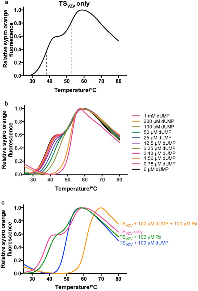
To study the binding of ligands to TSVZV, we explored a thermal stability shift assay based on differential scanning fluorimetry (DSF). In the absence of any ligand, DSF shows that TSVZV has a dual-transition melting curve that is made up of two sigmoidal curves (Fig 2a). The melting temperature (Tm) of the first and second melting transition of TSVZV in the absence of any ligand were estimated to be 38°C and 53°C respectively (Fig 2a). When TSVZV was titrated with different concentrations of dUMP, a shift of the first transition of the TSVZV melting curve was seen, while the second melting transition remained unchanged (Fig 2b). The first transition eventually disappeared with 100 μM of dUMP and further increase of the dUMP concentration could not increase the Tm beyond the second melting transition (Fig 2b).
Fig 2. DSF melting curves of TSVZV with dUMP.
(a) The DSF melting curve of apo-TSVZV has two melting transitions and two Tm can be estimated from the mid-slope of each sigmoidal transition. Dotted lines were added to the mid-slopes of each melting curve as references to the TSVZV Tm for each melting transition. (b) Increasing concentrations of dUMP only increased the Tm of the first melting transition. 100 μM of dUMP was sufficient to stabilize the first melting transition of TSVZV to produce a single melting transition. Further increase of the dUMP concentration could not increase the Tm beyond the second melting transition. (c) Raltitrexed further stabilized TSVZV in the presence of 100 μM of dUMP in the DSF. In the absence of dUMP, raltitrexed could not stabilize TSVZV in DSF.
The Tm of TSVZV is 50°C in the presence of 100 μM dUMP (Fig 2c). When 100 μM raltitrexed and 100 μM dUMP was added to TSVZV, the Tm of TSVZV increased from 50°C to 61°C (Fig 2c). In the absence of dUMP, the addition of 100 μM raltitrexed produced a dual-transition melting curve that is largely similar to the melting curve of TSVZV without any ligand (Fig 2c). No significant stabilization effect on the first and second melting transitions was observed with raltitrexed (Fig 2c). This is consistent with the notion where dUMP must be bound in order for the successive binding of raltitrexed in the active site of TSHS [18].
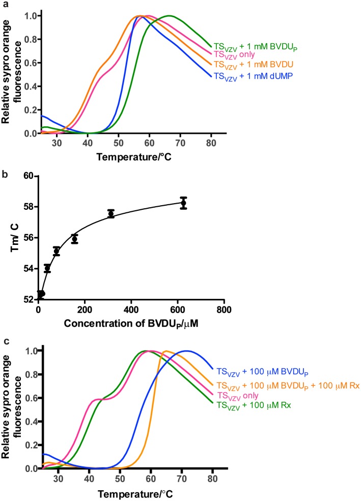
We were also interested in whether BVDU, a nucleoside based VZV drug with some similarities to the substrate dUMP, could also bind TSVZV. The binding of TSVZV to BVDU before (BVDU) and after in vitro phosphorylation (BVDUP) with TKHS was therefore studied with DSF. The addition of 100 μM BVDU to TSVZV produced a dual-transition melting curve that is similar to the ligand free TSVZV (Fig 3a). On the contrary, BVDUP produced a single melting transition with a Tm of approximately 57°C in DSF. This observed stabilization of TSVZV by BVDUP is approximately 7°C higher than the stabilization of TSVZV by 100 μM dUMP (Fig 3a). Varying concentrations of BVDUP produced a dose responsive increment in the Tm of TSVZV (Fig 3b). When 100 μM of raltitrexed was added to TSVZV and 100 μM of BVDUP, the Tm of TSVZV increased from 57°C to 64°C (Fig 3c).
Fig 3. DSF melting curves of TSVZV with BVDUP.
(a) BVDU could not stabilize TSVZV before phosphorylation but after an in vitro phosphorylation with TKHS, BVDUP increased the Tm of TSVZV by a larger extent than dUMP. (b) Varying concentrations of BVDUP increased the Tm of TSVZV in a dose responsive manner. (c) Further stabilization of TSVZV was observed in the presence of 100 μM raltitrexed with 100 μM of BVDUP.
Discussion
TS is a highly conserved enzyme in many organisms including human, mouse, bacteria, protozoa and some viruses. TS is essential for the de novo synthesis of dTMP and its expression is up-regulated in actively replicating cells. It is an attractive drug target and several TS inhibitors including 5-fluorouracil, raltitrexed and pemetrexed are extensively used for treatments of solid tumors [39, 40]. Structural information of TS from several different species in complex with its substrates and inhibitors is also available [17–30]. TS is seemingly only present in two human herpesviruses, VZV and KSHV [32, 33]. Both TSVZV and TSKSHV share high sequence similarities with TSHS, with at least 80% sequence identity at the TS active site. Enzymatic studies have showed that TSVZV and TSKSHV display similar catalytic kinetics as human TS in the presence of dUMP and mTHF [32]. However, both herpesviral TS remain poorly characterized till date.
In an attempt to further characterize TSVZV, the crystal structures of the apo-, dUMP and BVDUP bound TSVZV were determined. These constitute the first crystal structure of an eukaryotic viral TS. The overall structure is, as expected, well conserved with the human TS (S1 Fig) [35]. However, the TSVZV+dUMP and TSVZV+BVDUP structures have disordered regions that cannot be modeled in two of the four subunits of the crystallographic asymmetric unit. It is tempting to speculate that the disorder is correlated to the binding of the dUMP/BVDUP. However, these disordered regions are observed only in one of the monomer of the TSVZV dimer. Although the binding of dUMP and BVDUP appears to induce minor conformational changes to the amino acids lining the active site, the positioning of both dUMP/BVDUP and the amino acids lining the active site are largely conserved in all the monomers (Fig 1). An inspection of the crystal packing in the unit cell of the TSVZV+dUMP and TSVZV+BVDUP structures reveals that these disordered regions are located on the protein surfaces that lack crystal contact with a neighboring monomer. Thus, the observed structural disorder is most likely due to differences in crystal packing and crystal contacts of the two molecules and not due to the binding of dUMP/BVDUP to TSVZV.
The TSVZV+dUMP structure adopts an open conformation, as concluded from the lack of covalent bonds between the thiol group of the catalytic cysteine and C6 of the dUMP pyrimidine ring (Fig 1b). The presence of the cofactor and the C-terminal loop have been proposed to be required for the closed conformation [41]. However, the TSVZV protein construct that produced well diffracting crystals for structural determination of TSVZV lacks the last six amino acids at the C-terminal. These six amino acids correspond to the C-terminal loop that has been reported to act as a lid over the TS active site and assist in folate binding [22]. Thus, the reason for an open conformation in TSvzv+dUMP is likely partly due to the absence of the folate cofactor in the active site and the C-terminal loop in the TSVZV structures.
The interactions between dUMP/BVDUP and an anti-folate drug raltitrexed with TSVZV were characterized with DSF. While the DSF demonstrated that dUMP/BVDUP and raltitrexed are able to bind to TSVZV in vitro, it also showed that raltitrexed is not able to bind TSVZV in the absence of the nucleotides (Figs 2c and 3c). This is consistent with the well-characterized sequential binding mode in other TSs [18]. Thus, the lack of binding of raltitrexed to TSVZV in the DSF is likely due to the requirement for bound dUMP in the active site before raltitrexed can bind.
Nucleoside analogs have routinely been used for treatments of viral infections [7, 42]. Many of these drugs are administered as a pro-drug and requires an activation step through phosphorylation by the viral or human TK [43]. To study the possibility of TSVZV being a target protein of nucleoside analogs, which are often not commercially available in their phosphorylated form, in vitro phosphorylation by TKHS was performed prior to the binding studies. The assay was tested with deoxythymidine, which should be phosphorylated by TKHS to TMP in the presence of ATP. The in vitro phosphorylation was validated with DSF, as deoxythymidine is unable to bind to TSVZV while dTMP gave significant shifts (S2 Fig).
Several clinically approved anti-herpesviral nucleoside analogs are 5-substituted pyrimidine nucleosides, which are modifications of the TS binding dUMP. BVDU is a clinically approved 5-substituted pyrimidine nucleoside analog that displays the highest potency against HSV-1 and VZV [42, 44]. It has also been shown to be an inhibitor of TSHS [45]. Our DSF experiments demonstrated that BVDU is able to bind TSVZV after in vitro phosphorylation by TKHS (Fig 3a). This supports that the nucleoside analog has been successfully phosphorylated by TKHS in vitro. The successful phosphorylation of BVDUP was eventually confirmed by the presence of the phosphate group on BVDUP in the complex structure of TSVZV with BVDUP (Fig 1d). This may seem contradicting to previous studies that have reported a selective phosphorylation of BVDU by the herpesviral TK but TKHS has been shown to be catalytically active towards BVDU, albeit with low efficiency [10, 46]. The effectiveness of TKHS on BVDU may be attributed to the higher concentration of the kinase used in vitro and the efficiency of TKHS activity on BVDU is likely to diminish in vivo.
The binding of BVDUP to TSVZV was further confirmed with the complex structure, TSvzv+BVDUp. The coordination of BVDUP is highly similar to the coordination of dUMP in TSVZV (Fig 1b and 1d). Though BVDUP is not covalently bound to the catalytic thiol, a comparison of the structure of TSvzv+BVDUP with the closed TSHS shows that the coordination of BVDUP by TSVZV is more conserved with the coordination of dUMP in the closed-TSHS (Fig 1c and 1d). Even in the absence of the folate cofactor and the C-terminal loop, BVDUP appears to be held in a closed conformation by the additional hydrophobic interactions contributed by its 5-bromovinyl substituent (Fig 1e). The presence of a bound PEG 400 in the TSVZV active site, in the region where folate binds, may have also helped in stabilizing the closed conformation. However, rather than a catalytically active closed conformation, the BVDUP bound TSVZV structure is likely to represent a catalytically inhibited TSVZV. Together, these data suggest that phosphorylated BVDU inhibits TSVZV by binding competitively to the active site and yield further support for that TSVZV could be a target protein for BVDU in the cell after the activation by the herpesvirus TK.
BVDUP has been previously demonstrated to inhibit the catalytic activities of both TSVZV and TSHS in vitro [32, 47]. The high degree of structural conservation and the lack of selectivity of BVDUP between TSVZV and TSHS could potentially lead to TSHS mediated adverse effects. However, structural differences observed between TSVZV and TSHS may confer additional selectivity of BVDUP towards TSVZV. His 184 do not share the same conformation as the corresponding His 196 in TSHS (Fig 1c and 1d). As a result, the imidazole ring of His 184 does not interact with O4 of the dUMP/BVDUP pyrimidine ring. Even though the amino acids lining the active sites of TSVZV and TSHS are highly conserved, there is a single amino acid substitution at Tyr 100 in TSVZV that corresponds to Asn 112 in TSHS. This could potentially aid in the design of more specific inhibitors towards TSVZV. Regardless of the structural differences between TSVZV and TSHS, the potency of BVDU towards VZV infected cells is likely due to the preferential phosphorylation by herpesvirus TK [10, 46]. Nevertheless, further studies are required to evaluate whether TSVZV is indeed a physiological relevant target protein for the mono-phosphorylated BVDU.
In conclusion, substrate binding in TSVZV has been characterized with a thermal shift assay and the apo and complex structures with nucleotides have been determined. The different TSVZV structures share features with the evolutionary conserved TSHS but also show some small but significant differences. Binding between TSVZV and an activated herpesviral drug BVDU was demonstrated and a complex structure with an in vitro phosphorylated BVDU was determined. Together this work adds further support BVDU as a good inhibitor for TSVZV and provides the structural basis for structure driven design of more specific TSVZV inhibitors.
Materials and Methods
Cell culture and harvest
The starter culture supplemented with 50 μg/mL kanamycin (Sigma Aldrich) and 34 μg/mL chloroamphenicol (Sigma Aldrich) was inoculated with a streak from a TSVZV glycerol stock. The culture was incubated overnight at 37°C, 220 rpm. 2 mL of the starter culture was added into each flask containing 750 mL of fresh TB, 50 μg/mL kanamycin and 34 μg/mL chloroamphenicol. The cultures were incubated at 37°C, 180 rpm until OD600 was 0.8. Expression was induced overnight with 0.5 mM IPTG at 18°C and harvested by centrifugation (4500 g for 10 minutes at 15°C) the next day. The cell pellet was flash frozen with liquid nitrogen, pounded and stored at -20°C.
Protein purification
TSVZV was purified with a two-step standard purification protocol, consisting of an immobilized metal affinity chromatography step and a size exclusion chromatography step using the ÄKTAxpress protein purification systems at 4°C. Ice-cold lysis buffer (100 mM Na-HEPES pH 8.0 (Sigma Aldrich), 500 mM NaCl (Sigma Aldrich), 10 mM imidazole pH 8.0 (Sigma Aldrich), 10% glycerol (Sigma Aldrich) and 0.5 mM TCEP (Sigma Aldrich)) supplemented with 0.1 mg/mL lysozyme (Sigma Aldrich), 1 μL/mL protease inhibitor cocktail (Nacalai) and 125 U/mL benzonase (Merck Millipore) was added to the frozen TSVZV cell pellet. The cell lysate was sonicated before clarifying by centrifugation at 47000 g for 25 minutes at 4°C and filtration of the supernatant with an 1.2 μm syringe filter. The filtrate was loaded onto a pre-equilibrated (10 mM Na-HEPES pH 7.5, 500 mM NaCl, 10 mM imidazole pH 7.5, 10% glycerol and 0.5 mM TCEP) 5 mL HiTrap IMAC HP column (GE Healthcare) and the column was washed with 25 mL of purification wash buffer (10 mM Na-HEPES pH 7.5, 500 mM NaCl, 10 mM imidazole pH 7.5, 10% glycerol). The protein was eluted with a step elution of 156 mM and 335 mM imidazole. The samples eluted with 335 mM imidazole were pooled and concentrated to 5 mL before loading onto a pre-equilibrated (20 mM Na-HEPES pH 7.5, 300 mM NaCl, 10% glycerol and 0.5 mM TCEP) HiLoad 16/600 Superdex 200 gel filtration column (GE Healthcare). The fractions were analyzed on a 4–12% NuPAGE Bis-Tris gel (Life Technologies) and the purest fractions were pooled and concentrated to 12 mg/mL with a 10 kD MWCO protein concentrator (GE Healthcare). The concentrated TSVZV samples were flash frozen in liquid nitrogen and stored at -80°C.
In vitro phosphorylation of nucleosides using TKHS
Deoxythymidine was phosphorylated by incubating 1 mM deoxythymidine (Sigma-Aldrich) with 1 mM ATP (Sigma-Aldrich) and 40 μg of recombinant purified TKHS (gift from Chen Dan, Nanyang Technological University, Singapore) overnight at 37°C in a reaction buffer (20 mM HEPES pH 7.5, 300 mM NaCl, 25 mM MgCl2). The reaction was stopped by heating the samples at 95°C for 15 minutes. The denatured TKHS was removed by centrifugation at 13000 rpm for 5 minutes. The supernatant was removed and used for the crystallization and differential scanning fluorimetry (DSF) experiments with TSVZV. The phosphorylated BVDU (BVDUP) was prepared by incubating BVDU (Santa Cruz Biotechnology) with TKHS as with deoxythymidine.
Crystallization and data collection
The apo-TSVZV was crystallized in two crystal forms. The first crystal form was obtained by mixing 200 nL of the crystallization solution (12% Ethylene glycol, 0.1 M HEPES pH 7.5) with 100 nL of the protein solution (12 mg/mL) in a 96-wells sitting drop Intelli-plate (Art Robbins) at 20°C. The second crystal form was obtained by adding 200 nL of the crystallization solution (0.1 M Tris-HCl pH 8.0 and 40% PEG 300) to 100 nL of the protein solution (12 mg/mL) in a 96-wells sitting drop Intelli-plate at 20°C. Crystals of the second crystal form were soaked with 1 mM dUMP for three minutes and 1 mM BVDUP for six minutes for the structures of the complex of TSVZV with dUMP (TSVZV+dUMP) and TSVZV with BVDUP (TSVZV+BVDUP) respectively. The crystals were then flash frozen with liquid nitrogen. Diffraction data were collected at the MX2 Micro Crystallography beam line at the Australian Synchrotron, Australia using the Blu-Ice data collection software [48]. All diffraction datasets were integrated and scaled with HKL2000 [49].
Structure determination
The apo structure of TSVZV was determined by molecular replacement with Phaser, using a molecule of TSHS (PDB ID: 1HZW) as the search model [35]. Inspection of the electron density maps revealed spherical positive electron densities at one end of the TSVZV active site and a phosphate ion was manually modeled into the positive electron densities using coot [50]. The structure of TSVZV with phosphate was further refined with phenix.refine from the Phenix suite [51, 52].
The structures of TSVZV in complex with dUMP (TSVZV+dUMP) and BVDUP (TSVZV+BVDUP) were phased with molecular replacement using Phaser from the CCP4 suite [53, 54]. A TSVZV monomer from the apo-TSVZV structure was used as the molecular replacement search model. Inspection of the electron density maps of TSVZV+dUMP and TSVZV+BVDUP revealed clear electron densities for dUMP and BVDUP at their respective TSVZV active sites. dUMP and BVDUP were manually modeled into the respective positive electron densities using coot [50]. Iterations of automated and manual refinements of the structure were performed with phenix.refine and coot [50, 52]. The final structure refinement of the TSVZV structures were validated with MolProbity [55]. All figures of the TSVZV structures were displayed with pymol [56]. The apo TSVZV, TSVZV+dUMP and TSVZV+BVDUP structures were deposited on the PDB as 4XSE, 4XSD and 4XSC.
Differential scanning fluorimetry
TSVZV was diluted to 0.2 mg/mL with the DSF buffer (20 mM HEPES pH 7.5 and 300 mM NaCl) supplemented with 5x SYPROrange (Life Technologies) and was added to the 96-wells PCR plate. Triplicates were performed. Samples were heated from 25°C to 80°C, in increments of 1°C. The average absolute fluorescence intensity of the triplicates was plotted against the temperature (°C) on Prism 6 (GraphPad, Prism). The curves were then normalized with Prism 6 to obtain the DSF melting curves of TSVZV. The melting temperatures (Tm) of TSVZV were estimated from the mid-point of each sigmoidal curve.
The effect of different ligands on TSVZV was also performed with DSF as above, with slight modifications. In addition to diluting TSVZV in the DSF buffer, TSVZV was mixed with 1 mM dUMP (Sigma-Aldrich), 1 mM dTMP (Sigma-Aldrich), 1 mM deoxythymidine (Sigma-Aldrich) before and after phosphorylation or 1 mM BVDU (Santa Cruz) before and after phosphorylation. In the binding studies of raltitrexed with TSVZV, the DSF experiment was repeated with 100 μM of raltitrexed (Selleck Chemicals) in the presence and absence of 100 μM dUMP or BVDUP. The dose responsive DSF of TSVZV with dUMP was obtained by mixing TSVZV with dUMP at varying concentrations of 1 mM, 200 μM, 100 μM, 50 μM, 25 μM, 12.5 μM, 6.25 μM, 3.125 μM, 1.56 μM, 0.78 μM and 0 μM. Likewise, the dose responsive DSF of TSVZV with the phosphorylated BVDU was also performed by mixing TSVZV with 625 μM, 313 μM, 156 μM, 78 μM and 39 μM of BVDUP. The normalized melting curves and the Tm of TSVZV in the presence of different ligands were obtained as described above.
Supporting Information
Two orientations of the apo-TSVZV dimer are illustrated in cartoon. A phosphate ion has been found in each TSVZV active site and both phosphate ions are displayed as sticks.
(TIF)
Deoxythymidine did not stabilize TSVZV before phosphorylation but after an in vitro phosphorylation with TKHS, it stabilizes TSVZV to a similar extent as dTMP.
(TIF)
Acknowledgments
We would like to thank Jurgen Haas (University of Edinburgh, UK) for providing the full-length clone of TS from VZV, the Protein Production Platform (Nanyang Technological University, Singapore) for the initial cloning and small-scale expression screening, and Chen Dan (Nanyang Technological University, Singapore) for the providing the recombinant purified TKHS. Portions of this research were undertaken on the MX1 and MX2 beam lines at the Australian Synchrotron, Victoria, Australia.
Data Availability
All relevant data are within the paper and its Supporting Information files.
Funding Statement
The authors have no support or funding to report.
References
- 1. Gilden DH, Kleinschmidt-DeMasters B, LaGuardia JJ, Mahalingam R, Cohrs RJ. Neurologic complications of the reactivation of varicella–zoster virus. New England Journal of Medicine. 2000;342(9):635–45. [DOI] [PubMed] [Google Scholar]
- 2. Gilden DH, Vafai A, Shtram Y, Becker Y, Devlin M, Wellish M. Varicella-zoster virus DNA in human sensory ganglia. 1983. [DOI] [PubMed] [Google Scholar]
- 3. Lungu O, Annunziato PW, Gershon A, Staugaitis SM, Josefson D, LaRussa P, et al. Reactivated and latent varicella-zoster virus in human dorsal root ganglia. Proceedings of the National Academy of Sciences. 1995;92(24):10980–4. [DOI] [PMC free article] [PubMed] [Google Scholar]
- 4. Stankus SJ, Dlugopolski M, Packer D. Management of herpes zoster (shingles) and postherpetic neuralgia. American family physician. 2000;61(8):2437–44, 47–8. [PubMed] [Google Scholar]
- 5. Weller SK, Coen DM. Herpes simplex viruses: mechanisms of DNA replication. Cold Spring Harbor perspectives in biology. 2012;4(9):a013011 10.1101/cshperspect.a013011 [DOI] [PMC free article] [PubMed] [Google Scholar]
- 6. Mocarski ESJ. Comparative analysis of herpesvirus-common proteins In: Arvin A, Campadelli-Fiume G, Mocarski E, Moore PS, Roizman B, Whitley R, et al. , editors. Human Herpesviruses: Biology, Therapy, and Immunoprophylaxis. Cambridge: Cambridge University Press; 2007. [PubMed] [Google Scholar]
- 7. De Clercq E. Antiviral drugs in current clinical use. Journal of Clinical Virology. 2004;30(2):115–33. [DOI] [PubMed] [Google Scholar]
- 8. De Clercq E. Antivirals and antiviral strategies. Nat Rev Micro. 2004;2(9):704–20. http://www.nature.com/nrmicro/journal/v2/n9/suppinfo/nrmicro975_S1.html. [DOI] [PMC free article] [PubMed] [Google Scholar]
- 9. Fyfe J, Keller P, Furman P, Miller R, Elion G. Thymidine kinase from herpes simplex virus phosphorylates the new antiviral compound, 9-(2-hydroxyethoxymethyl) guanine. Journal of Biological Chemistry. 1978;253(24):8721–7. [PubMed] [Google Scholar]
- 10. Cheng Y, Dutschman G, Fox J, Watanabe K, Machida H. Differential activity of potential antiviral nucleoside analogs on herpes simplex virus-induced and human cellular thymidine kinases. Antimicrobial agents and chemotherapy. 1981;20(3):420–3. [DOI] [PMC free article] [PubMed] [Google Scholar]
- 11. Evans DR, Guy HI. Mammalian Pyrimidine Biosynthesis: Fresh Insights into an Ancient Pathway. Journal of Biological Chemistry. 2004;279(32):33035–8. 10.1074/jbc.R400007200 [DOI] [PubMed] [Google Scholar]
- 12. Rose MG, Farrell MP, Schmitz JC. Thymidylate synthase: a critical target for cancer chemotherapy. Clinical colorectal cancer. 2002;1(4):220–9. [DOI] [PubMed] [Google Scholar]
- 13. Ostrer L, Hamann BL, Khodursky A. Perturbed states of the bacterial chromosome: a thymineless death case study. Frontiers in microbiology. 2015;6 10.3389/fmicb.2015.00363 [DOI] [PMC free article] [PubMed] [Google Scholar]
- 14. Jackman A, Farrugia D, Gibson W, Kimbell R, Harrap K, Stephens T, et al. ZD1694 (Tomudex): a new thymidylate synthase inhibitor with activity in colorectal cancer. European journal of cancer. 1995;31(7):1277–82. [DOI] [PubMed] [Google Scholar]
- 15. Adjei AA. Pemetrexed (ALIMTA), a novel multitargeted antineoplastic agent. Clinical Cancer Research. 2004;10(12):4276s–80s. [DOI] [PubMed] [Google Scholar]
- 16. Klaassen D, MacIntyre J, Catton G, Engstrom P, Moertel C. Treatment of locally unresectable cancer of the stomach and pancreas: a randomized comparison of 5-fluorouracil alone with radiation plus concurrent and maintenance 5-fluorouracil—an Eastern Cooperative Oncology Group study. Journal of Clinical Oncology. 1985;3(3):373–8. [DOI] [PubMed] [Google Scholar]
- 17. Hardy LW, Finer-Moore JS, Montfort WR, Jones MO, Santi DV, Stroud RM. Atomic structure of thymidylate synthase: target for rational drug design. Science. 1987;235(4787):448–55. [DOI] [PubMed] [Google Scholar]
- 18. Finer-Moore JS, Montfort WR, Stroud RM. Pairwise specificity and sequential binding in enzyme catalysis: thymidylate synthase. Biochemistry. 1990;29(30):6977–86. [DOI] [PubMed] [Google Scholar]
- 19. Matthews D, Appelt K, Oatley S, Xuong NH. Crystal structure of Escherichia coli thymidylate synthase containing bound 5-fluoro-2′-deoxyuridylate and 10-propargyl-5, 8-dideazafolate. J Mol Biol. 1990;214(4):923–36. [DOI] [PubMed] [Google Scholar]
- 20. Finer-Moore J, Fauman EB, Foster PG, Perry KM, Santi DV, Stroud RM. Refined Structures of Substrate-bound and Phosphate-bound Thymidylate Synthase from Lactobacillus casei. J Mol Biol. 1993;232(4):1101–16. [DOI] [PubMed] [Google Scholar]
- 21. Schiffer CA, Clifton IJ, Davisson VJ, Santi DV, Stroud RM. Crystal structure of human thymidylate synthase: a structural mechanism for guiding substrates into the active site. Biochemistry. 1995;34(50):16279–87. [DOI] [PubMed] [Google Scholar]
- 22. Phan J, Koli S, Minor W, Dunlap RB, Berger SH, Lebioda L. Human thymidylate synthase is in the closed conformation when complexed with dUMP and raltitrexed, an antifolate drug. Biochemistry. 2001;40(7):1897–902. [DOI] [PubMed] [Google Scholar]
- 23. Phan J, Steadman DJ, Koli S, Ding WC, Minor W, Dunlap RB, et al. Structure of human thymidylate synthase suggests advantages of chemotherapy with noncompetitive inhibitors. Journal of Biological Chemistry. 2001;276(17):14170–7. [DOI] [PubMed] [Google Scholar]
- 24. Mathews II, Deacon AM, Canaves JM, McMullan D, Lesley SA, Agarwalla S, et al. Functional analysis of substrate and cofactor complex structures of a thymidylate synthase-complementing protein. Structure. 2003;11(6):677–90. [DOI] [PubMed] [Google Scholar]
- 25. Basta T, Boum Y, Briffotaux J, Becker HF, Lamarre-Jouenne I, Lambry J-C, et al. Mechanistic and structural basis for inhibition of thymidylate synthase ThyX. Open biology. 2012;2(10):120120 10.1098/rsob.120120 [DOI] [PMC free article] [PubMed] [Google Scholar]
- 26. Yuthavong Y, Tarnchompoo B, Vilaivan T, Chitnumsub P, Kamchonwongpaisan S, Charman SA, et al. Malarial dihydrofolate reductase as a paradigm for drug development against a resistance-compromised target. Proceedings of the National Academy of Sciences. 2012;109(42):16823–8. [DOI] [PMC free article] [PubMed] [Google Scholar]
- 27. Baugh L, Gallagher LA, Patrapuvich R, Clifton MC, Gardberg AS, Edwards TE, et al. Combining functional and structural genomics to sample the essential Burkholderia structome. PloS one. 2013;8(1):e53851 10.1371/journal.pone.0053851 [DOI] [PMC free article] [PubMed] [Google Scholar]
- 28. Wilk P, Jarmuła A, Ruman T, Banaszak K, Rypniewski W, Cieśla J, et al. Crystal structure of phosphoramide-phosphorylated thymidylate synthase reveals pSer127, reflecting probably pHis to pSer phosphotransfer. Bioorganic chemistry. 2014;52:44–9. 10.1016/j.bioorg.2013.11.003 [DOI] [PubMed] [Google Scholar]
- 29. Dowierciał A, Wilk P, Rypniewski W, Rode W, Jarmuła A. Crystal Structure of Mouse Thymidylate Synthase in Tertiary Complex with dUMP and Raltitrexed Reveals N-Terminus Architecture and Two Different Active Site Conformations. BioMed research international. 2014;2014. [DOI] [PMC free article] [PubMed] [Google Scholar]
- 30. Brunn ND, Dibrov SM, Kao MB, Ghassemian M, Hermann T. Analysis of mRNA recognition by human thymidylate synthase. Bioscience reports. 2014;34(6):905–13. [DOI] [PMC free article] [PubMed] [Google Scholar]
- 31. Costi MP, Tondi D, Rinaldi M, Barlocco D, Pecorari P, Soragni F, et al. Structure-based studies on species-specific inhibition of thymidylate synthase. Biochimica et Biophysica Acta (BBA)-Molecular Basis of Disease. 2002;1587(2):206–14. [DOI] [PubMed] [Google Scholar]
- 32. Gáspár G, De Clercq E, Neyts J. Human herpesvirus 8 gene encodes a functional thymidylate synthase. Journal of virology. 2002;76(20):10530–2. [DOI] [PMC free article] [PubMed] [Google Scholar]
- 33. Thompson R, Honess R, Taylor L, Morran J, Davison A. Varicella-zoster virus specifies a thymidylate synthetase. J Gen Virol. 1987;68:1449–55. [DOI] [PubMed] [Google Scholar]
- 34. Cohen JI, Seidel KE. Generation of varicella-zoster virus (VZV) and viral mutants from cosmid DNAs: VZV thymidylate synthetase is not essential for replication in vitro. Proceedings of the National Academy of Sciences. 1993;90(15):7376–80. [DOI] [PMC free article] [PubMed] [Google Scholar]
- 35. Almog R, Waddling CA, Maley F, Maley GF, Van Roey P. Crystal structure of a deletion mutant of human thymidylate synthase Δ (7–29) and its ternary complex with Tomudex and dUMP. Protein Science. 2001;10(5):988–96. 10.1110/ps.47601 [DOI] [PMC free article] [PubMed] [Google Scholar]
- 36. Sayre PH, Finer-Moore JS, Fritz TA, Biermann D, Gates SB, MacKellar WC, et al. Multi-targeted antifolates aimed at avoiding drug resistance form covalent closed inhibitory complexes with human and< i> Escherichia coli thymidylate synthases. J Mol Biol. 2001;313(4):813–29. [DOI] [PubMed] [Google Scholar]
- 37. McGeehan JE, Carpentier P, Royant A, Bourgeois D, Ravelli RB. X-ray radiation-induced damage in DNA monitored by online Raman. Journal of synchrotron radiation. 2007;14(1):99–108. [DOI] [PubMed] [Google Scholar]
- 38. Beck T, Gruene T, Sheldrick GM. The magic triangle goes MAD: experimental phasing with a bromine derivative. Acta Crystallographica Section D: Biological Crystallography. 2010;66(4):374–80. [DOI] [PMC free article] [PubMed] [Google Scholar]
- 39. Noordhuis P, Holwerda U, Van der Wilt C, Van Groeningen C, Smid K, Meijer S, et al. 5-Fluorouracil incorporation into RNA and DNA in relation to thymidylate synthase inhibition of human colorectal cancers. Annals of oncology. 2004;15(7):1025–32. [DOI] [PubMed] [Google Scholar]
- 40. Wishart DS, Knox C, Guo AC, Cheng D, Shrivastava S, Tzur D, et al. DrugBank: a knowledgebase for drugs, drug actions and drug targets. Nucleic Acids Res. 2008;36(suppl 1):D901–D6. [DOI] [PMC free article] [PubMed] [Google Scholar]
- 41. Perry KM, Carreras CW, Chang LC, Santi DV, Stroud RM. Structures of thymidylate synthase with a C-terminal deletion: role of the C-terminus in alignment of 2'-deoxyuridine 5'-monophosphate and 5, 10-methylenetetrahydrofolate. Biochemistry. 1993;32(28):7116–25. [DOI] [PubMed] [Google Scholar]
- 42. De Clercq E, Holý A. Acyclic nucleoside phosphonates: a key class of antiviral drugs. Nature Reviews Drug Discovery. 2005;4(11):928–40. [DOI] [PubMed] [Google Scholar]
- 43. Stein DS, Moore KH. Phosphorylation of nucleoside analog antiretrovirals: a review for clinicians. Pharmacotherapy: The Journal of Human Pharmacology and Drug Therapy. 2001;21(1):11–34. [DOI] [PubMed] [Google Scholar]
- 44. De Clercq E, Descamps J, De Somer P, Barr P, Jones A, Walker R. (E)-5-(2-Bromovinyl)-2'-deoxyuridine: a potent and selective anti-herpes agent. Proceedings of the National Academy of Sciences. 1979;76(6):2947–51. [DOI] [PMC free article] [PubMed] [Google Scholar]
- 45. Balzarini J, De Clercq E, Verbruggen A, Ayusawa D, Shimizu K, Seno T. Thymidylate synthase is the principal target enzyme for the cytostatic activity of (E)-5-(2-bromovinyl)-2'-deoxyuridine against murine mammary carcinoma (FM3A) cells transformed with the herpes simplex virus type 1 or type 2 thymidine kinase gene. Molecular pharmacology. 1987;32(3):410–6. [PubMed] [Google Scholar]
- 46. Cheng Y-C, Dutschman G, De Clercq E, Jones A, Rahim S, Verhelst G, et al. Differential affinities of 5-(2-halogenovinyl)-2'-deoxyuridines for deoxythymidine kinases of various origins. Molecular pharmacology. 1981;20(1):230–3. [PubMed] [Google Scholar]
- 47. Yokota Y, Konno K, Shigeta S. Inhibition of thymidylate synthetase activity induced in varicella-zoster virus infected cells by (E)-5-(2-bromovinyl)-2'-deoxyuridine. Antiviral chemistry & chemotherapy. 1994;5(3):191–4. [Google Scholar]
- 48. McPhillips TM, McPhillips S E, Chiu H J, Cohen A E, Deacon A M, Ellis P J, et al. Blu-Ice and the Distributed Control System: software for data acquisition and instrument control at macromolecular crystallography beamlines. Journal of Synchrotron Radiation. 2002;9:401–6. [DOI] [PubMed] [Google Scholar]
- 49. Otwinowski Z, Minor W. Processing of X-ray diffraction data collected in oscillation mode. Methods in Enzymology. 1997;276:307–26. 10.1016/S0076-6879(97)76066-X [DOI] [PubMed] [Google Scholar]
- 50. Emsley P, Lohkamp B, Scott WG, Cowtan K. Features and Development of Coot. Acta Crystallographica Section D: Biological Crystallography. 2010;66:486–501. [DOI] [PMC free article] [PubMed] [Google Scholar]
- 51. Adams PD, Afonine PV, Bunkóczi G, Chen VB, Davis IW, Echols N, et al. PHENIX: a comprehensive Python-based system for macromolecular structure solution. Acta Crystallographica Section D: Biological Crystallography. 2010;66:213–21. [DOI] [PMC free article] [PubMed] [Google Scholar]
- 52. Afonine PV, Grosse-Kunstleve RW, Echols N, Headd JJ, Moriarty NW, Mustyakimov M, et al. Towards automated crystallographic structure refinement with phenix. refine. Acta Crystallographica Section D: Biological Crystallography. 2012;68(4):352–67. [DOI] [PMC free article] [PubMed] [Google Scholar]
- 53. McCoy AJ, Grosse-Kunstleve RW, Adams PD, Winn MD, Storoni LC, Read RJ. Phaser Crystallographic Software. Journal of Applied Crystallography. 2007;40:658–74. [DOI] [PMC free article] [PubMed] [Google Scholar]
- 54. Winn MD, Ballard CC, Cowtan KD, Dodson EJ, Emsley P, Evans PR, et al. Overview of the CCP4 suite and current developments. Acta crystallographica Section D, Biological crystallography. 2011;67(Pt 4):235–42. 10.1107/S0907444910045749 [DOI] [PMC free article] [PubMed] [Google Scholar]
- 55. Chen VB, Arendall WB, Headd JJ, Keedy DA, Immormino RM, Kapral GJ, et al. MolProbity: all-atom structure validation for macromolecular crystallography. Acta Crystallographica Section D: Biological Crystallography. 2009;66(1):12–21. [DOI] [PMC free article] [PubMed] [Google Scholar]
- 56.DeLano W. The PyMOL Molecular Graphics System, Version 1.7.2.0. 2014.
Associated Data
This section collects any data citations, data availability statements, or supplementary materials included in this article.
Supplementary Materials
Two orientations of the apo-TSVZV dimer are illustrated in cartoon. A phosphate ion has been found in each TSVZV active site and both phosphate ions are displayed as sticks.
(TIF)
Deoxythymidine did not stabilize TSVZV before phosphorylation but after an in vitro phosphorylation with TKHS, it stabilizes TSVZV to a similar extent as dTMP.
(TIF)
Data Availability Statement
All relevant data are within the paper and its Supporting Information files.


