

Abstract
Idiopathic gingival enlargement (GE) is a rare entity characterized by massive enlargement of the gingiva. It may be associated with other diseases/conditions characterizing a syndrome, but rarely documented in literature occurring along with chronic periodontitis. This case report describes a rare case of long standing massive GE in a systemically healthy, nonsyndromic young female involving both the arches, thereby posing a diagnostic dilemma. Furthermore, in this case, we compared two surgical approaches, that is, scalpel and electrosurgery for the convenience as well as the postoperative comfort of the patient. Quadrants 1 and 3 were treated by ledge and wedge technique using scalpel and blade while quadrants 2 and 4 were treated by electrosurgery. The patient was followed postoperatively up to 1-year. The massive GE subsided without recurrence and patient was completely satisfied with the treatment, though better compliance was observed at the site treated by conventional scalpel and blade technique.
Keywords: Chronic periodontitis, compliance, electrosurgery, gingival enlargement, idiopathic enlargement
INTRODUCTION
Gingival enlargement (GE) is defined as an abnormal overgrowth of gingival tissues. Under this category, gingival fibromatosis is a heterogeneous group of disorders characterized by progressive enlargement of the gingiva caused by an increase in submucosal connective tissue elements.[1] The etiology of GE is poorly understood but can be attributed to factors like plaque accumulation, systemic hormonal stimulation, blood dyscrasias, drugs, or idiopathic.[2]
Gingival enlargement is an unusual condition causing esthetic, functional, masticatory and psychological disturbances in individuals. It may be easy to arrive at a clinical diagnosis of GE if the etiology is clearly evident; sometimes medical opinion is seeked to explore the cause and identify the underlying diseases, drug interactions or the natural body changes to develop an effective treatment plan. When the exact cause cannot be elucidated, it becomes challenging to establish an accurate diagnosis and hence the prognosis.
Gingival enlargement have been seen to be associated with aggressive periodontitis,[3,4] but very few have reported it in coexistence with chronic periodontitis in a nonsyndromic patient. Surgical intervention with scalpel, electrosurgery, and LASER is advocated for enlargement.[5]
Hereby we present a rare case of long standing massive GE in a systemically healthy, nonsyndromic, young female with chronic inflammation involving both the arches. We compared two surgical approaches, that is, scalpel and electrosurgery with an aim to observe the convenience of the operator as well as the postoperative comfort to the patient.
CASE REPORT
A 26-year-old female patient reported to the out-patient department of periodontics with a chief complaint of swollen gums in both the jaws since 10–11 years. She also had halitosis, occasional bleeding from gums and difficulty in mastication and speech. On intra-oral examination massive, painless, Grade III[6] type of GE involving both the arches, encroaching buccal, palatal, and lingual vestibular spaces was found. GE extended up to the level of occlusal planes of the teeth [Figures 1 and 2]. The gingiva was pale pink and firm, but some areas showed signs of ongoing acute inflammation as well. The patient's height and weight were normal and her medical, hematological, and family history was noncontributory. Periodontal examination revealed the presence of thick bands of sub-gingival calculus, bleeding on probing, Grade II mobility of few teeth, malpositioning of upper anteriors, and generalized probing pocket depth in the range of 8 and 10 mm. Orthopentomogram showed a generalized horizontal bone loss.
Figure 1.

Preoperative view (labial)
Figure 2.
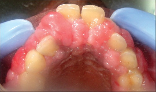
Preoperative view (palatal)
On first appointment, an incisional biopsy was taken for histopathological examination. The report showed densely arranged collagen bundles and numerous fibroblasts in connective tissue stroma, strands of proliferating epithelium and an abundance of inflammatory cells and fluid in the connective tissue core [Figures 3 and 4]. Based on the above mentioned findings, a diagnosis of Grade III[6] GE associated with chronic periodontitis in a nonsyndromic, systemically healthy patient was formulated. After phase I therapy, gingivectomy, a combined approach employing both ledge and wedge technique by scalpel and electrosurgery was planned for the patient. Quadrants 1 and 3 were treated by ledge and wedge technique using scalpel while quadrants 2 and 4 were treated by electrosurgery. After scalpel surgery, the area was thoroughly debrided and gingivoplasty was done to re-contour the tissue. In quadrants 2 and 4, electrosurgery unit (Bonart Co., Lmt.) was utilized to reduce the gingival bulk. Loop electrode was used in “paint-brush” like strokes to trim the excessive gingival overgrowth. A needle electrode was used for cutting. 8–15 s cooling period was given between successive electrode applications to avoid over-heating. Appropriate medication was prescribed. The interval between each surgical procedure was 1-week.
Figure 3.

Histological section under ×10 magnification
Figure 4.

Histological section under ×40 magnification
The patient was placed in a recall program of 1-week, 1-month, 6 months, and 1-year. Mobile teeth upgraded to Grade I by the end of 1-year. The massive GE subsided without recurrence and patient was completely satisfied with the treatment [Figures 5 and 6]. The patient was also assessed on a visual analog scale[7] at 1-week and 1-month recall regarding pain and discomfort experienced after both the treatment protocols. The score 0 was labeled as no pain and score 10 as worst possible pain. Scores 3, 2 were obtained for sites treated with scalpel technique whereas scores 6, 5 were obtained for electrosurgical sites. The convenience of the operator was comparable with both the techniques. Figures 5 and 6 showed the healing after 2 weeks for quadrant 1 and 3, whereas 1-week for quadrant 2 and 4. Delayed healing was observed for the sites treated with electrosurgery.
Figure 5.

Two weeks postoperative (labial)
Figure 6.

Two weeks postoperative (palatal)
DISCUSSION
Idiopathic GE is a rare condition of unknown etiology characterized by slow, progressive enlargement of the gingiva. It can either occur as an isolated disorder or in association with pathoses such as tuberous sclerosis and hypertrichosis.[8] It may also form a part of many syndromes like Zimmermann–Laband syndrome, cross syndrome, and prune belly syndrome.[9]
An imbalance between synthesis and degradation of collagen and/or alterations in fibroblast function and proliferation has been considered for the etiopathogenesis of insulin-like growth factor. The enlargement is confined to fibroblasts in the gingiva without involving the periodontal ligament. It has elucidated that in such cases, the periodontitis and bone resorption is secondary to plaque accumulation due to enlargement.[10] We managed this case comparing scalpel and electrosurgery and also followed the patient for 1-year. Both the procedures were satisfactory, but it should be noted that although reduced bleeding at the electrosurgery site was an advantage, but fumes and burning smell made the patient uncomfortable. Overall, better healing and compliance was observed at the scalpel site as compared to the electrosurgical site.
Regarding the recurrence of this lesion, conflicting studies have been reported in the literature.[11,12] In our case, no recurrence was seen at the end of 1-year follow-up. The well planned professional maintenance protocol might have been the reason for this. The patient was satisfied with esthetic and functional result of the treatment.
CONCLUSION
Since this is a single case study, no consensus among authors related to the mode of treatment could be made but it can be said that scalpel method can be preferred over electrosurgery. Patient education, periodic recall, and proper oral hygiene maintenance reduce and delay the chances of recurrence.
Footnotes
Source of Support: Nil.
Conflicts of Interest: None declared.
REFERENCES
- 1.Pappachan B, Narayan JV, Nayak A. Idiopathic gingival fibromatosis: A neglected case. Indian J Radiol Imaging. 2002;12:335–8. [Google Scholar]
- 2.Dongari-Bagtzoglou A. Research, Science and Therapy Committee, American Academy of Periodontology. Drug-associated gingival enlargement. J Periodontol. 2004;75:1424–31. doi: 10.1902/jop.2004.75.10.1424. [DOI] [PubMed] [Google Scholar]
- 3.Chaturvedi R. Idiopathic gingival fibromatosis associated with generalized aggressive periodontitis: A case report. J Can Dent Assoc. 2009;75:291–5. [PubMed] [Google Scholar]
- 4.Casavecchia P, Uzel MI, Kantarci A, Hasturk H, Dibart S, Hart TC, et al. Hereditary gingival fibromatosis associated with generalized aggressive periodontitis: A case report. J Periodontol. 2004;75:770–8. doi: 10.1902/jop.2004.75.5.770. [DOI] [PubMed] [Google Scholar]
- 5.Coletta RD, Graner E. Hereditary gingival fibromatosis: A systematic review. J Periodontol. 2006;77:753–64. doi: 10.1902/jop.2006.050379. [DOI] [PubMed] [Google Scholar]
- 6.Bökenkamp A, Bohnhorst B, Beier C, Albers N, Offner G, Brodehl J. Nifedipine aggravates cyclosporine A-induced gingival hyperplasia. Pediatr Nephrol. 1994;8:181–5. doi: 10.1007/BF00865474. [DOI] [PubMed] [Google Scholar]
- 7.Huskisson EC. Measurement of pain. Lancet. 1974;2:1127–31. doi: 10.1016/s0140-6736(74)90884-8. [DOI] [PubMed] [Google Scholar]
- 8.Horning GM, Fisher JG, Barker BF, Killoy WJ, Lowe JW. Gingival fibromatosis with hypertrichosis. A case report. J Periodontol. 1985;56:344–7. doi: 10.1902/jop.1985.56.6.344. [DOI] [PubMed] [Google Scholar]
- 9.Harrison M, Odell EW, Agrawal M, Saravanamuttu R, Longhurst P. Gingival fibromatosis with prune-belly syndrome. Oral Surg Oral Med Oral Pathol Oral Radiol Endod. 1998;86:304–7. doi: 10.1016/s1079-2104(98)90176-7. [DOI] [PubMed] [Google Scholar]
- 10.Sapp JP, Eversole LR, Wysocki GP. Contemporary Oral and Maxillofacial Pathology. 2nd ed. London, UK: Mosby; 2004. Connective tissue lesions; pp. 294–7. [Google Scholar]
- 11.Danesh-Meyer MJ, Holborow DW. Familial gingival fibromatosis: A report of two patients. N Z Dent J. 1993;89:119–22. [PubMed] [Google Scholar]
- 12.Cuestas-Carnero R, Bornancini CA. Hereditary generalized gingival fibromatosis associated with hypertrichosis: Report of five cases in one family. J Oral Maxillofac Surg. 1988;46:415–20. doi: 10.1016/0278-2391(88)90229-7. [DOI] [PubMed] [Google Scholar]