SUMMARY
Recent studies have shown that the transcriptional landscape of the pleiomorphic fungus Candida albicans is highly dependent upon growth conditions. Here using a dual RNA-seq approach we identified 299 C. albicans and 72 Streptococcus gordonii genes that were either up- or down-regulated specifically as a result of co-culturing these human oral cavity microorganisms. Seventy five C. albicans genes involved in responses to chemical stimuli, regulation, homeostasis, protein modification and cell cycle were statistically (P ≤0.05) up-regulated, while 36 genes mainly involved in transport and translation were down-regulated. Up-regulation of filamentation-associated TEC1 and FGR42 genes, and of ALS1 adhesin gene, concurred with previous evidence that the C. albicans yeast to hypha transition is promoted by S. gordonii. Increased expression of genes required for arginine biosynthesis in C. albicans was potentially indicative of a novel oxidative stress response. The transcriptional response of S. gordonii to C. albicans was less dramatic, with only eight S. gordonii genes significantly (P ≤0.05) up-regulated ≥ twofold (glpK, rplO, celB, rplN, rplB, rpsE, ciaR, and gat). The expression patterns suggest that signals from S. gordonii cause a positive filamentation response in C. albicans, while S. gordonii appears to be transcriptionally less influenced by C. albicans.
Keywords: Cell wall proteins, adhesin, bacteria-fungi interactions, GPI anchor, RNASeq
INTRODUCTION
Candida albicans is a commensal fungus and opportunistic pathogen found in the human gut, oral cavity and genital tract. It is present in 20-60% humans, depending upon the population studied (Martins et al., 1998). C. albicans can progress from commensal colonization to local invasion, according to subject susceptibility, and then to invasive candidiasis which is associated with high mortality rates (Pfaller et al., 2010; Eggimann et al., 2015). In the oral cavity, C. albicans tends to localize to mucosal surfaces and prostheses e.g. dentures (Zomorodian et al., 2011), but there is also evidence for association with dental caries (de Carvalho et al., 2006) and periodontal disease (Canabarro et al., 2013). C. albicans therefore often coexists with other microorganisms in polymicrobial biofilm communities. There is evidence that this influences the morphogenetic, pathogenic and antifungal susceptibility properties of C. albicans, and therefore such infections may be more difficult to control (Wright et al., 2013).
Biofilm formation in C. albicans is a multistage process (Douglas, 2003) and is dependent upon morphological transitions from yeast to pseudohyphal or hyphal forms. The formation of biofilms involves global changes in gene expression (Garcia-Sanchez et al., 2004) modulated by at least six transcription factors (Nobile et al., 2012). A set of eight genes has been found forming a core filamentation response (Martin et al., 2013). The genes include ALS3, ECE1, HGT2, IHD1 and RBT1, all of which encode cell wall proteins (CWPs).
Recently, RNA sequencing has been utilized to provide transcriptome maps of C. albicans under a range of growth conditions, both in vitro and in vivo (Bruno et al., 2010; Tierney et al., 2012; Grumaz et al., 2013). Such studies have revealed that relatively well established pathways may be affected unexpectedly by the prevailing growth conditions. For example, the arginine biosynthesis genes (e.g. ARG1, ARG3, ARG4) are induced under conditions of mild oxidative stress (0.5 mM hydrogen peroxide, H2O2) such as those which might occur within phagocytes (Jiménez-Lopéz et al., 2013).
In mixed species biofilms there are clearly many opportunities for trans-kingdom signalling to occur (Nobbs & Jenkinson, 2015) and it is therefore important to understand the nature of this communication. Quorum sensing molecules and other metabolites from Gram-negative bacteria such as P. aeruginosa (Hogan et al., 2004) and Burkholderia cenocepacia (Boon et al., 2008) have been shown to block hypha formation in C. albicans. Staphylococcus aureus appears to inhibit filamentation under some conditions (Fox et al., 2013), while the oral bacterium Streptococcus mutans inhibits hypha formation by production of trans-2-decenoic acid (Vilchez et al., 2010) and competence stimulating peptide (Jarosz et al., 2009).
Streptococcus gordonii is found associated with most surfaces in the human oral cavity (Aas et al., 2005) and is one of several oral streptococcal species that have been shown to co-aggregate with C. albicans (Jenkinson et al., 1990). It is suggested that these interactions with bacteria are crucial for C. albicans incorporation into oral cavity biofilms (Jenkinson, 2011), and for the development of polymicrobial communities. More recent studies have shown that one response of C. albicans to the presence of S. gordonii involves promotion of hyphal morphogenesis (Dutton et al., 2014). This could be influenced by cell-cell contact and modulated by secreted metabolites i.e. signalling molecules (Bamford et al., 2009; Jack et al., 2015). The aim of the work described in this paper was to outline the transcriptional responses of C. albicans and S. gordonii in the early stages of their interaction, mimicking the natural biofilm communication processes that occur in the oral cavity. The expression profiles obtained reveal how early recognition responses modulate downstream events involved in dual species, trans-kingdom biofilm development.
METHODS
Growth of microbial cells
C. albicans wild-type strain SC5314 was grown aerobically for 16 h in YPD medium (2% yeast extract, 1% mycological peptone, 2% dextrose) at 37°C, with shaking at 220-rpm. Cells were then harvested by centrifugation (5000 × g for 5 min), washed twice in YPT medium (1 × Difco yeast nitrogen base, 20 mM phosphate buffer pH 7.1, 0.1% Bacto tryptone) by alternate centrifugation (5000 × g for 5 min) and suspension, and finally suspended at optical density 600 nm (OD600) = 1.0 (approximately 1 × 10⁷ cells ml−1) in YPT medium. Aliquots (10 ml; 1 × 10⁷ cells ml−1) were transferred into conical flasks containing YPT medium (90 ml) supplemented with 0.4% glucose (YPT-Glc). The cultures were then incubated at 37°C for 2 h with shaking at 50-rpm to induce hypha formation (Dutton et al., 2014). S. gordonii cells were grown anaerobically for 16 h in 10 ml BHY medium (per litre: 37g Brain Heart infusion broth, 5 g yeast extract) and then harvested by centrifugation (5000 × g for 7 min). The bacterial cells were washed twice with YPT (no glucose) and finally suspended at OD600 = 0.5 (2 × 10⁸ cells ml−1) in YPT-Glc medium.
Several combinations of C. albicans, S. gordonii and growth medium were designed to specifically identify changes in gene expression as a result of co-incubation (Table 1). For the dual-species cultures of S. gordonii and C. albicans, S. gordonii cell suspensions (50 ml; 2 × 10⁸ cells ml−1) were added at 2 h, while for the C. albicans monospecies culture, YPT-Glc medium alone (50 ml) was added. S. gordonii cell suspension (50 ml) was added to pre-warmed (37°C) YPT-Glc medium for 1 h for the S. gordonii monoculture. To prepare C. albicans spent medium, C. albicans cells were removed from the culture medium by centrifugation (5000 × g, 5 min), and the supernatant was vacuum filtered through a 0.45 μm nitrocellulose membrane. The filtered medium was transferred to a sterile glass bottle and warmed to 37°C before S. gordonii suspension (50 ml; 2 × 10⁸ cells ml−1) was added. All cultures were incubated at 37°C with shaking (50-rpm) for a further 1 h.
Table 1.
Composition of microbial cultures utilized to determine transcriptional changes in C. albicans or S. gordonii genes following co-incubation.
| Culture no. | Conditions and components |
|---|---|
| 1 | C. albicans in YPT-Glc, 3 h at 37°C |
| 2 |
C. albicans in YPT-Glc, 2 h at 37°C; + S. gordonii DL1 in YPT- Glc, 1 h at 37°C |
| 3 1 | C. albicans in YPT-Glc, 2 h at 37°C; + YPT-Glc, 1 h at 37°C |
| 4 2 |
C. albicans in YPT-Glc, 2 h at 37°C, spent medium; + S. gordonii in YPT-Glc, 1 h at 37°C |
| 5 3 | S. gordonii in YPT-Glc, 1 h at 37°C |
included to account for effects of adding fresh growth medium in (2)
included to account for effects of C. albicans spent medium on S. gordonii
used to correct for effects of nutritional shift-down in (2)
Cells were harvested by centrifugation (5000 × g, 10 min) in 50 ml-Falcon tubes and all but 5 ml supernatant was aspirated. The cell pellet was suspended in the remaining supernatant, transferred to sterile 15 ml-Falcon tubes and harvested by centrifugation (5000 × g, 5 min). The supernatant was aspirated until only 0.5 ml remained, and this was used to suspend the cell pellet. The cell suspension was frozen into small balls by dropping portions (200 μl) into liquid nitrogen. The balls were stored at −70°C prior to RNA extraction.
RNA extraction
Frozen microbial cell balls were thawed on ice and suspended in ice-cold RLT buffer (Qiagen Ltd., Manchester, UK) containing 2-mercaptoethanol and transferred to a sterile screw cap microfuge tube containing acid-washed Biospec glass beads (0.6 ml). The suspension was mixed with the glass beads and the fungal and bacterial cells were disrupted by alternating shaking (30 s) using a Fast-prep 25 bead beater (MP Biomedicals, Santa Ana, CA) and incubating 1 min on ice (repeated 3 times). The beads were allowed to settle and the supernatant was transferred to a sterile microfuge tube. The disrupted cells were centrifuged (13000 × g, 2 min) and the supernatant transferred to a sterile microfuge tube. An equal volume of 70% ethanol was added and the RNA was extracted and purified using an RNeasy Mini Kit (Qiagen) with the use of an on-column DNAse digestion (Qiagen). The quality of the RNA was checked by formaldehyde agarose-gel electrophoresis. The RNA concentration of each sample was measured spectrophotometrically (Nanodrop 1000, Thermo Scientific, Fisher Scientific UK Ltd, Loughborough, Leics., UK) and stored at −20°C.
Transcriptomic analysis
ERCC RNA Spike-In Control Mix (Ambion, Foster City, CA) was mixed with 2.5 μg RNA. Ribosomal RNA was depleted with a RiboZero Magnetic Gold Kit (Epicentre) and lllumina sequencing libraries were prepared using ScriptSeq v2 (Epicentre, Illumina Inc., Madison, WI) with 10 cycles of PCR amplification. The quality and quantity of each library was determined using a Bioanalyzer and the average (modal) insert size of samples was 400 bp with the spread ranging between 200 bp and 1,000 bp. An equimolar library pool was denatured, diluted to 6.5 pM and clustered on a cBot (Illumina) to create clonal clusters from single molecule DNA templates. One hundred base pair paired-end sequencing was undertaken using HiSeq2500 (Illumina) in high output mode with Truseq v3 reagents. The resulting FASTQ data were then filtered using the fastq-mcf command from the EA-Utils suite to remove adaptor sequences and low quality bases (Aronesty, 2011). The filtered data were then aligned against the reference ERCC transcripts using Bowtie v1.0.0 using the -X 600 flag. SAMtools v0.1.19 was utilized to convert the resulting SAM formatted-file to BAM (Li et al., 2009). The number of reads mapping to each transcript was extracted using the SAMtools idxstats command and used to calculate RPKM values for each ERCC transcript. Log2 values of observed RPKM were then plotted against log2 expected RPKM values and inspected to establish lower limits of detection.
The reads which did not map to the ERCC transcripts were then aligned to a merged FASTA file containing both the Ca21_C_albicans_SC5314 genome (Version 21 from www.candidagenome.org) and the NC_009785 S. gordonii CH1 genome. The Tophat2 v2 2.0.8b program was used with the following parameters: -G -library-type fr-secondstrand -I 10000 -r 50, --mate-std-dev 100 -p 8. The -G parameter was followed by the combined gff file containing annotation for both organisms (Kim et al., 2013). An additional analysis was carried out with the DESeq analysis tool to calculate differential gene expression (Anders & Huber, 2010). The output of Tophat2 was processed using the Bedtools multicov command (Dale et al., 2011). This produces numbers of reads mapping to each annotated gff feature. The gene/read count files were then processed to separate the eukaryote and prokaryote gene features. Each was then analyzed separately using the DESeq package v1.12.1. Default parameters were utilized as outlined in the DESeq manual. DESeq uses a negative binomial model to account for the dispersion of the reads and the variation between replicates (n = 3), and uses a general linear model for comparisons (Love et al., 2014). P-values were calculated from DESeq, and Benjamini-Hochberg adjusted P-values ≤ 0.05 were deemed significant. All transcriptional data have been submitted to the GEO repository and assigned the GEO accession number GSE68477.
Microscopy
Confocal scanning laser microscopy of C. albicans (stained with Calcofluor white) with or without S. gordonii (stained with fluorescein isothiocyanate, FITC) was performed as previously described (Dutton et al., 2014). Transmission electron microscopy (TEM) was performed as follows: cell suspensions were centrifuged (5000 × g, 10 min) and to the pellets was added TEM fixative (4% paraformaldehye, 5% glutaraldehyde, 0. 1M sodium cacodylate buffer pH 7.2, 0.05% Tween 20). Tubes were shaken gently and incubated for 15 min at 22°C. The samples were centrifuged (3000 × g, 2 min), the supernatant carefully removed, and the pellet suspended in TEM fixative (2% paraformaldehye, 2.5% glutaraldehyde; 2 ml) for 16 h at 4°C. The pellets were then washed 3 times in 0.1 M sodium cacodylate buffer (pH 7.2), and set into low melting point 2% agarose. The agarose pellets were cut into ~3 mm sections and incubated for 1 h at 22°C in 1% osmium tetroxide solution. The samples were rinsed in sterile H2O, dehydrated at room temperature using sequential incubations in ethanol (30%, 50%, 75%, 90% and 100%) then propylene oxide, and embedded in Spurr resin. The resin was cut by microtome (RMC Powertome PC) into sections of 80-100 nm using a diamond knife, and sections were collected on copper grids and imaged at 80 kV by TEM.
Scanning Probe Microscopy (SPM) was performed on C. albicans-S. gordonii cultures deposited onto glass cover slips. Cover slips were attached to metal pucks with double sided tape and mounted on a Multimode AFM fitted with a Nanoscope IIIa controller (Veeco, Santa Barbara, CA). Cells were imaged in contact mode using triangular silicon nitride (Si3N4) tips with a nominal spring constant of ~0.06 N m−1 (Veeco). Images were obtained at typical scan rates of 25 μm s−1 and processed using Nanoscope 8 software (Veeco).
RESULTS AND DISCUSSION
C. albicans and S. gordonii form close physical associations
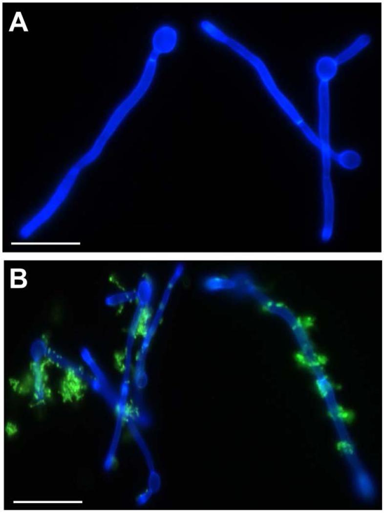
Coaggregation of C. albicans and S. gordonii cells occurs rapidly after mixing of the two cell types (Jenkinson et al., 1990). In order to visualize these interactive events, S. gordonii cells, fluorescently labelled with FITC, were mixed with hyphae-forming C. albicans cells in YPT-Glc medium (see Methods) and incubated for 1 h at 37°C. The associations between the bacteria and fungi were then viewed by confocal scanning laser microscopy (CSLM). After 3 h growth in monoculture, ~50% C. albicans cells had formed hyphae of 20-50 μm in length (Fig. 1A). In the presence of S. gordonii, bacterial cells bound along the lengths of the hyphal filaments (Fig. 1B) and could also be seen to aggregate, forming microcolonies at discrete attachment sites (Fig. 1B). These aggregates must form mainly by recruitment of other streptococci since extensive bacterial cell division does not occur in YPT-Glc medium within the experimental time-frame of 1 h.
Fig. 1.
Light micrograph images of C. albicans SC5314 cells interacting with S. gordonii DL1 cells. S. gordonii cells were fluorescently labelled with fluorescein isothiocyanate (FITC) and incubated with filamentation-induced (2 h at 37°C) C. albicans for 1 h at 37°C with gentle agitation. Calcofluor white (0.3 μg ml−1) was added to fluorescently label C. albicans. Panel A, C. albicans alone; Panel B, C. albicans and S. gordonii (green). Scale bar 5 μm.
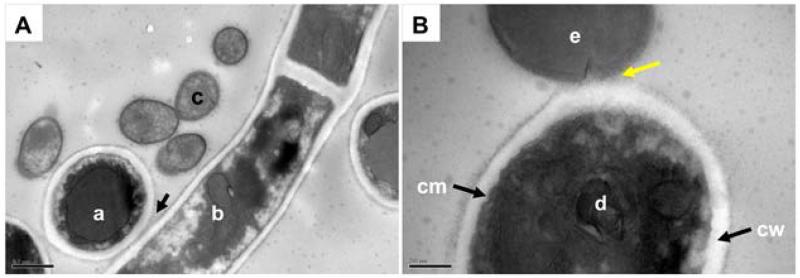
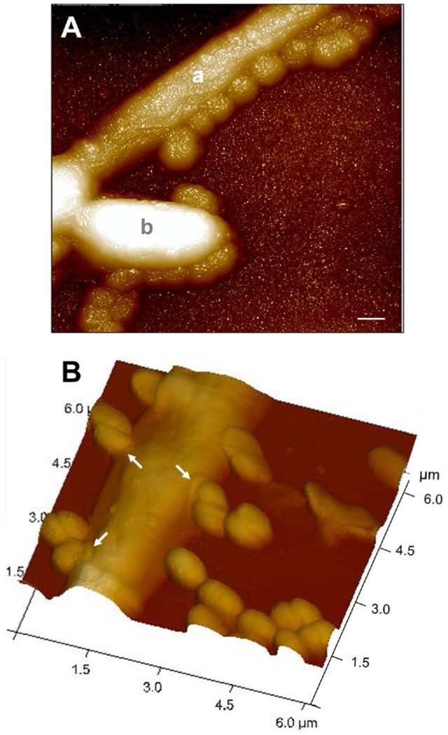
One characteristic of C. albicans hypha formation is that the hyphal filaments clump together. As expected therefore, transmission electron micrographs showed C. albicans hyphal filaments in close contact with each other (Fig. 2A), seemingly connected by networks of fibrillar material. S. gordonii cells were often seen intimately interacting with hyphal filaments, with the cell surfaces in direct contact along a length of about 100 nm (Fig. 2B). There were also streptococci present that were not interacting with hyphae (Fig. 2B). The closeness of the bacterium-fungus interaction was further shown by scanning probe microscopy. In scans of wet mounts, streptococcal and C. albicans cell surfaces are apparently coalesced (Fig. 3A), while under dried conditions there appears to be a structural difference on the hyphal cell surface at the point of contact with bacteria (Fig. 3B) which could suggest some form of fungal cell wall remodelling. It is these close associations between the two cell types that led us to hypothesize that recognition signals (contact or diffusible) could be relayed through cell wall sensors to modulate gene expression in response to the other microorganism.
Fig. 2.
Transmission electron microscope (TEM) images of C. albicans SC5314 cells interacting with S. gordonii DL1. Filamentation-induced C. albicans cells in YPT-Glc were incubated with S. gordonii cells for 1 h at 37°C. Cells were fixed, resin embedded and sectioned (see Experimental procedures) for visualization by TEM. Panel A, vertical cross section of a C. albicans hyphal filament (a) shows cell wall in close contact (arrowed) with the cell wall of a longitudinally-sectioned hyphal filament (b). Smaller streptococcal cells (c) can clearly be seen nearby expressing surface fibrillar structures. Panel B, shows the cell membrane (cm) and cell wall (cw) of a vertical section of a C. albicans hyphal filament (d) with the cell wall physically associated with the outer wall of an S. gordonii cell (e). The interaction is occurring at the newly forming septum of the streptococcal cell and the fibrils appear to interdigitate with the material on the C. albicans cell surface (yellow arrowed). Scale bars: A, 0.5 μm; B, 200 nm.
Fig. 3.
Scanning Probe Microscope (SPM) images of C. albicans SC5314 hypha-forming cells interacting with S. gordonii DL1. Filamentation-induced C. albicans cells in YPT-Glc medium were incubated with S. gordonii cells for 1 h at 37°C. Cells were then deposited onto glass cover slips and imaged in contact mode as described in Experimental procedures. Panel A, undried specimen showing hyphal filament (a) with smaller streptococcal cells attached along its length. A budding pseudohypha (b) appears also to have streptococci attached. Panel B, dried specimen showing numerous streptococcal cells in close physical contact with a C. albicans hyphal filament. At the point of contact there is an annular modification visible on the C. albicans cell surface (arrowed), implying a hyphal cell surface structural response. Note that quite often the streptococcal cell septum region was involved in binding hyphae (see also Fig. 2B). Scale bar 0.5 μm.
Transcriptional landscape studies
Following suspension of C. albicans cells in YPT-Glc medium, and incubation for 2 h at 37°C, fungal cells were undergoing early-stage hyphal morphogenesis (see above). At this point the C. albicans cells were allowed to proceed for a further 1 h, either in the presence or absence of S. gordonii, or in the presence of spent C. albicans culture medium (to correct for metabolic effects), and RNA was then prepared. Transcriptomic data therefore represent the response of C. albicans to the presence of S. gordonii for 1 h, and they take into account also the transcriptional effects of nutritional shift-down and C. albicans culture medium on S. gordonii cells (see Table 1). Under these conditions, physical trans-kingdom interactions occur, and chemical signals would carry on being exchanged between the two microorganisms since both C. albicans and S. gordonii continue to metabolize within this medium (Bamford et al., 2009).
Illumina sequencing from the combined C. albicans and S. gordonii samples yielded 258,347,928 raw reads (Table S1). A total of 6,653 open reading frames (features) from all the samples were functionally annotated to the haploid assembly 21 of the C. albicans genome (van het Hoog et al., 2007) using the Candida Genome Database (CGD) (www.candidagenome.org), while 2,051 open reading frames were matched to the S. gordonii database for annotation, visualization and integrated discovery (DAVID version 6.7 - http://david.abcc.ncifcrf.gov) (Huang et al., 2007; Sherman et al., 2007). The total numbers of candidal and streptococcal reads for all three sample replicates were calculated, and the % distribution of candidal and bacterial reads were calculated for the three combined replicates for each sample (Table S2).
When C. albicans and S. gordonii were co-cultured in YPT-Glc medium, the overall ratio of reads was 37.7% C. albicans to 62.7% S. gordonii. This showed there was a suitable distribution of C. albicans and S. gordonii reads with no undue bias towards one single organism. It should be noted that features can include any sequence belonging to the genome, including reads that are not translated into proteins. This explains why there were a small number of reads (0.09% and 0.03%) from the S. gordonii samples that matched to the C. albicans genome. These relate to regions of the genome with some homology that are similar in the two organisms e.g. tRNA or mitochondrial RNA.
One technical consideration was if there might be a bias towards long or short transcripts. To check this, the gene (orf) coordinates from the 6,653 orfs in the CGD were used to prepare a dataset of the lengths of every orf. The normalized expression reads (tag counts) for every gene were then plotted against gene length for the entire C. albicans genome. The plot was compared with a corresponding graph of normalized expression reads versus gene length for the up- and down-regulated C. albicans genes in the presence of S. gordonii. These data, presented in Fig.S1, show that there was no shift in length distribution between total C. albicans orfs and differentially regulated genes, and no obvious change in range of distribution. Therefore it was concluded that the expression data were not biased by gene length.
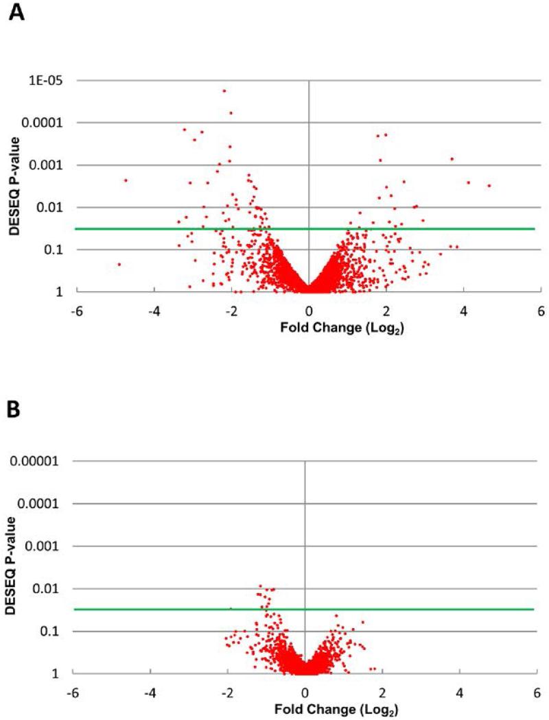
The Illumina sequencing data from co-incubated cultures of C. albicans and S. gordonii were compared to the data obtained from C. albicans grown with only the addition of growth medium (YPT-Glc) minus S. gordonii. This was to rule out the likelihood that any changes in gene expression were caused by the addition of the extra growth medium after 2 h rather than an effect caused by S. gordonii cells. Volcano plots of P-value vs. mean fold change in gene expression (Fig. 4) derived from analysis of the Illumina data (by the statistical DESeq package v1.12.1 with default parameters) showed that when C. albicans and S. gordonii were co-cultured in YPT-Glc medium the expression levels of a large number of C. albicans genes were significantly (P ≤0.05) up- or down-regulated by at least a twofold change (Fig. 4A, Table 2). On the other hand, only one S. gordonii gene was significantly (P ≤0.05) decreased in expression when the bacteria were incubated with C. albicans. S. gordonii gene expression significantly increased only slightly overall, with the majority of increases around twofold or less (Fig. 4B). Statistical analysis using DESeq v1.12.1 also showed the total number of genes with altered expression (either up- or down-regulated) was much higher in C. albicans (299 genes) compared with S. gordonii (72 genes).
Fig. 4.
Distribution of differentially regulated genes of C. albicans and S. gordonii following co-incubation for 1 h at 37°C. RNA was extracted and gene transcriptional levels were determined following Ilumina HISeq2500 sequencing. The transcriptional profiles were constructed and analyzed using the statistical software DESeq. Volcano plots of P-value vs. mean fold change in gene expression were constructed for C. albicans genes when incubated with S. gordonii (A) and S. gordonii genes when incubated with C. albicans (B). Green horizontal lines represent P = 0.05.
Table 2.
List of 111 Candida albicans genes significantly (P ≤0.05) differentially expressed following co-incubation of C. albicans filamentation-induced cells with S. gordonii for 1 h at 37°C in YPT-Glc medium.
| ORF | Gene name |
Fold change (log2)1 |
Description2 | Total abundance3 (log2) |
P- value4 |
|---|---|---|---|---|---|
| orf19.3537 | 5.47 | Putative sulfiredoxin; biofilm-induced gene; regulated by Tsa1p, Tsa1Bp in minimal medium at 37°C | −3.310 | <0.001 | |
| orf19.7469 | ARG1 | 4.73 | Argininosuccinate synthase; arginine biosynthesis; regulated by Gcn4p, Rim101p; induced by amino acid starvation (3-AT) and benomyl treatment; stationary phase enriched protein; repressed in alkalinizing medium; planktonic growth-induced |
−2.256 | 0.002 |
| orf19.3107 | 3.65 | Ortholog of Candida dubliniensis CD36 : CD36_46490, Pichia stipitis Pignal : PICST_33598, Candida tropicalis MYA-3404 : CTRG_03758 and Candida albicans WO-1 : CAWG_03126 |
−3.325 | <0.001 | |
| orf19.4630 | CPA1 | 3.36 | Putative carbamoyl-phosphate synthase subunit; alkaline down-regulated; transcription is up-regulated in both intermediate and mature biofilms |
−2.646 | 0.022 |
| orf19.2285 | 3.22 | Increased transcription is observed upon benomyl treatment | −3.729 | <0.001 | |
| orf19.3221 | CPA2 | 3.17 | Putative arginine-specific carbamoylphosphate synthetase; protein enriched in stationary phase yeast cultures; transcription is up-regulated in both intermediate and mature biofilms |
−1.987 | 0.017 |
| orf19.4789 | 3.13 | Has domain(s) with predicted metal ion binding activity | −3.971 | 0.048 | |
| orf19.2165 | 3.08 | Predicted ORF in Assemblies 19, 20 and 21; induced by nitric oxide | −2.750 | <0.001 | |
| orf19.5610 | ARG3 | 3.07 | Putative ornithine carbamoyltransferase; Gcn4p-regulated; Hap43p-induced gene; repressed in alkalinizing medium |
−3.019 | 0.003 |
| orf19.6689 | ARG4 | 3.03 | Argininosuccinate lyase, catalyzes the final step in the arginine biosynthesis pathway; alkaline down- regulated; late-stage biofilm-induced |
−2.027 | 0.042 |
| orf19.2593 | BIO2 | 2.95 | Putative biotin synthase; transcriptionally up-regulated in high iron; transcription down-regulated by treatment with ciclopiroxolamine; up-regulated in clinical isolates from HIV+ patients with oral candidiasis; Hap43p-repressed |
−3.257 | <0.001 |
| orf19.176 | OPT4 | 2.76 | Oligopeptide transporter; detected at germ tube plasma membrane; transcriptionally induced upon phagocytosis by macrophage; fungal-specific (no human or murine homolog); Hap43p-repressed; merged with orf19.2292 in Assembly 20 |
−3.061 | <0.001 |
| orf19.3770 | ARG8 | 2.73 | Putative acetylornithine aminotransferase; Gcn2p-, Gcn4p-regulated; transcription is up-regulated in both intermediate and mature biofilms |
−2.480 | 0.028 |
| orf19.6229 | CAT1 | 2.71 | Catalase; resistance to oxidative stress, neutrophils, peroxide; role in virulence; regulated by iron, ciclopirox, fluconazole, carbon source, pH, Rim101p, Ssn6p, Hog1p, Hap43p, Sfu1p, Sef1p, farnesol, core stress response |
−1.845 | 0.010 |
| orf19.6899 | 2.65 | Putative oxidoreductase; mutation confers hypersensitivity to toxic ergosterol analog | −3.382 | 0.017 | |
| orf19.4788 | ARG5,6 | 2.61 | Arginine biosynthetic enzyme activities; in S. cerevisiae, processed into distinct polypeptides with acetylglutamate kinase (Arg6p) activity and acetylglutamate-phosphate reductase (Arg5p) activity; Gcn4p regulated; alkaline down-regulated |
−2.088 | 0.003 |
| orf19.3902 | 2.40 | Predicted ORF in Assemblies 19, 20 and 21; decreased transcription is observed upon fluphenazine treatment or in an azole-resistant strain that over-expresses CDR1 and CDR2 |
−3.078 | 0.036 | |
| orf19.3395 | 2.36 | Predicted membrane transporter, member of the drug:proton antiporter (12 spanner) (DHA1) family, major facilitator superfamily; induced by nitric oxide, oxidative stress, α-pheromone; fungal-specific; Hap43p-repressed |
−3.151 | 0.001 | |
| orf19.5094 | BUL1 | 2.31 | Protein not essential for viability; macrophage/pseudohyphal-induced; similar to S. cerevisiae Bul1p, which may be involved in selection of substrates for ubiquitination |
−2.361 | 0.001 |
| orf19.4335 | TNA1 | 2.29 | Putative nicotinic acid transporter; fungal-specific (no human or murine homolog); detected at germ tube plasma membrane by mass spectrometry; transcriptionally induced upon phagocytosis by macrophage |
−3.902 | 0.042 |
| orf19.7042 | 2.25 | Increased transcription is observed upon benomyl treatment or in an azole-resistant strain that over- expresses MDR1; induced by nitric oxide |
−2.697 | 0.036 | |
| orf19.4773 | AOX2 | 2.22 | Alternative oxidase; induced by antimycin A, some oxidants; growth- and carbon-source-regulated; one of two isoforms (Aox1p and Aox2p); involved in cyanide-resistant respiratory pathway that is absent from S. cerevisiae; Hap43p-repressed |
−2.949 | 0.014 |
| orf19.4290 | TRR1 | 2.19 | Thioredoxin reductase; regulated by Tsa1p/Tsa1Bp, Hap43p; induced by nitric oxide, peroxide; oxidative stress-induced via Cap1p; up-regulated by human neutrophils; fungal-specific (no human/murine homolog); stationary phase enriched protein |
−1.407 | <0.001 |
| orf19.125 | EBP1 | 2.15 | NADPH oxidoreductase; interacts with phenolic substrates such as 17β-estradiol; possible role in estrogen response; induced by oxidative, weak acid stress, nitric oxide, benomyl, GlcNAc; activated by Cap1p, Mnl1p; Sko1p; Hap43p-repressed |
−2.803 | 0.021 |
| orf19.6500 | ECM42 | 2.10 | Putative ornithine acetyltransferase; fungal-specific (no human or murine homolog); Gcn2p-, Gcn4p- regulated; clade-specific gene expression; possibly an essential gene, disruptants not obtained by UAU1 method |
−2.809 | 0.009 |
| orf19.113 | CIP1 | 2.07 | Possible oxidoreductase; transcription induced by cadmium but not by other heavy metals, heat shock, yeast-hyphal switch, oxidative stress (via Cap1p), or macrophage interaction; stationary phase enriched protein |
−3.055 | 0.019 |
| orf19.4689 | PGA57 | 2.05 | Putative GPI-anchored protein; Hap43p-induced gene | −3.348 | <0.001 |
| orf19.3131 | OYE32 | 2.04 | NAD(P)H oxidoreductase family protein; induced by nitric oxide, amphotericin B, oxidative stress (via Cap1p); increased transcription associated with MDR1 over-expression or benomyl treatment; macrophage-down-regulated protein abundance |
−2.821 | 0.0003 |
| orf19.5741 | ALS1 | 2.01 | Adhesin; ALS family of cell-surface glycoproteins; adhesion, virulence roles; immunoprotective; band at hyphal base; amyloid domain; biofilm-induced; Rfg1p, Ssk1p; strain background affects expression; N- term binds fucose-containing glycans |
−0.998 | <0.001 |
| orf19.3981 | MAL31 | 1.98 | Putative high-affinity maltose transporter; transcription is up-regulated in clinical isolates from HIV+ patients with oral candidiasis; alkaline up-regulated |
−2.581 | 0.005 |
| orf19.5908 | TEC1 | 1.97 | TEA/ATTS transcription factor; involved in white cell pheromone response, regulates hypha-specific genes, biofilm formation; regulates BCR1; transcription regulated by Cph2 in some conditions; alkaline- and biofilm-induced |
−2.348 | 0.050 |
| orf19.2970 | LYS2 | 1.96 | Large subunit of heterodimeric α-aminoadipate reductase; enzyme of lysine biosynthesis; contains predicted binding sites for AMP and α-aminoadipate; feedback inhibited by lysine or thialysine; regulated by Gcn2p and Gcn4p |
−1.794 | 0.030 |
| orf19.4873 | 1.89 | Biofilm-induced gene; expression is regulated upon white-opaque switching | −3.192 | 0.009 | |
| orf19.2724 | 1.88 | Hap43p-repressed gene; late-stage biofilm induced | −2.217 | 0.007 | |
| orf19.4274 | PUT1 | 1.83 | Putative proline oxidase; alkaline up-regulated by Rim101p; biofilm-induced | −2.623 | 0.011 |
| orf19.3803 | MNN22 | 1.57 | Putative Golgi α-1,2-mannosyltransferase; regulated by Tsa1p, Tsa1Bp in minimal media at 37°C; Hog1p-induced; induced by nitric oxide; down-regulated in core stress response; planktonic growth- induced gene |
−2.198 | 0.002 |
| orf19.2833 | PGA34 | 1.55 | Putative GPI-anchored protein; transcription is repressed in response to α-pheromone in Spider M medium; late-stage biofilm-induced gene; induced in oropharyngeal candidiasis |
−3.811 | 0.035 |
| CaalfMr16 | RRNL | 1.55 | Mitochondrial ribosomal RNA of the large ribosomal subunit | 0.022 | 0.002 |
| orf19.4513 | 1.53 | Protein not essential for viability | −2.036 | 0.013 | |
| orf19.701 | CFL11 | 1.51 | Protein similar to ferric reductase Fre10p; flucytosine repressed; possibly adherence-induced; possibly an essential gene, disruptants not obtained by UAU1 method |
−3.182 | 0.008 |
| orf19.2262 | 1.50 | Protein similar to quinone oxidoreductases; increased transcription upon benomyl treatment; induced by nitric oxide; oxidative stress-induced via Cap1p; stationary-phase enriched protein |
−2.494 | 0.002 | |
| orf19.276 | 1.48 | Plasma membrane-associated protein; increased transcription is observed in an azole-resistant strain that over-expresses MDR1; Hap43p-repressed gene |
−2.959 | 0.035 | |
| orf19.7637 | YHB4 | 1.46 | Protein related to flavohemoglobins; not required for wild-type nitric oxide resistance; has predicted globin, FAD-binding, and NAD(P)-binding domains but lacks some conserved residues of flavohemoglobins; Hap43p-repressed gene |
−2.416 | 0.007 |
| orf19.1651 | 1.44 | Ortholog of Candida dubliniensis CD36 : CD36_81960, Candida tropicalis MYA-3404 : CTRG_02378 and Candida albicans WO-1 : CAWG_02525 |
−3.581 | 0.045 | |
| orf19.2020 | HGT6 | 1.43 | Putative high-affinity major facilitator superfamily glucose transporter; 20 family members; 12 probable membrane-spanning segments; core stress response, fluconazole-induced; biofilm-induced; induced in oropharyngeal candidiasis |
−1.355 | 0.033 |
| orf19.7417 | TSA1 | 1.43 | TSA/alkyl hydroperoxide peroxidase C (AhPC) family protein; similar to thiol-dependent peroxidases of oxidative stress signaling; antigenic; hyphal surface, nucleus; yeast-form nucleus, cytoplasm; biofilm, phagocytosis, peroxide induced |
−1.232 | 0.034 |
| orf19.7398 | 1.42 | Hap43p-induced gene; decreased expression in response to prostaglandins | −1.227 | 0.031 | |
| orf19.4482 | IFI3 | 1.42 | Predicted ORF in Assemblies 19, 20 and 21; constitutive expression independent of MTL or white- opaque status |
−2.289 | 0.003 |
| orf19.85 | GPX2 | 1.42 | Similar to glutathione peroxidase; expression greater in high iron; alkaline up-regulated by Rim101p; transcriptionally induced by α-factor or interaction with macrophage; regulated by Efg1p; caspofungin repressed |
−2.053 | 0.006 |
| orf19.2809 | CTN3 | 1.41 | Predicted peroxisomal carnitine acetyl transferase; Ura+ deletion strain has no obvious metabolic, hyphal, or virulence defects; transcription induced by macrophage engulfment, hyphal growth, starvation, or non- fermentable carbon sources |
−2.913 | 0.034 |
| orf19.7244 | 1.40 | Putative fumarylacetoacetate hydrolase; biofilm-induced gene; induced by nitric oxide independent of Yhb1p; regulated by Sef1p, Sfu1p, and Hap43p |
−2.277 | 0.043 | |
| orf19.6501 | 1.40 | Ortholog of Candida dubliniensis CD36 : CD36_71960, Spathaspora passalidarum NRRL Y-27907 : SPAPADRAFT_63786, Candida tropicalis MYA-3404 : CTRG_05049 and Candida albicans WO-1 : CAWG_05564 |
−3.862 | 0.043 | |
| orf19.5760 | IHD1 | 1.39 | Putative GPI-anchored protein; alkaline up-regulated; greater transcription in hyphal form than yeast form; regulated by Nrg1p, Rfg1p, Tup1p; regulated by Tsa1p, Tsa1Bp in minimal media at 37°C; induced in oropharyngeal candidiasis |
−1.770 | 0.016 |
| orf19.5140 | 1.38 | Ortholog of Candida dubliniensis CD36 : CD36_72790 and Candida albicans WO-1 : CAWG_05648 | −3.507 | 0.017 | |
| orf19.1037 | 1.37 | Ortholog of Candida tenuis NRRL Y-1498 : CANTEDRAFT_114303, Candida dubliniensis CD36 : CD36_03460, Pichia stipitis Pignal : PICST_63037 and Spathaspora passalidarum NRRL Y-27907 : SPAPADRAFT_136773 |
−1.996 | 0.004 | |
| orf19.84 | CAN3 | 1.36 | Hap43p-repressed gene; expression is regulated upon white-opaque switching | −2.433 | 0.010 |
| orf19.4370 | 1.35 | Predicted ORF in Assemblies 19, 20 and 21; induced by nitric oxide; oxidative stress-induced via Cap1p; fungal-specific (no human or murine homolog) |
−2.730 | 0.010 | |
| orf19.1363 | 1.34 | Putative protein of unknown function; late-stage biofilm-induced gene; Plc1p-regulated; transcriptionally activated by Mnl1p under weak acid stress |
−2.494 | 0.011 | |
| orf19.3142 | 1.34 | Ortholog of C. parapsilosis CDC317 : CPAR2_501210, Candida tenuis NRRL Y-1498 : CANTEDRAFT_97195, Debaryomyces hansenii CBS767 : DEHA2E08800g and Candida dubliniensis CD36 : CD36_46140 |
−3.388 | 0.045 | |
| orf19.238 | CCP1 | 1.30 | Similar to cytochrome-c peroxidase N terminus; negatively regulated by Rim101p or alkaline pH; transcription induced by interaction with macrophage or low iron; oxygen-induced activity; regulated by Sef1p, Sfu1p, and Hap43p |
−1.804 | 0.045 |
| orf19.3122.2 | 1.29 | Ortholog(s) have role in exocytosis, filamentous growth, mRNA export from nucleus, proteasomal ubiquitin-dependent protein catabolic process, proteasome assembly, regulation of cell cycle |
−2.692 | 0.010 | |
| orf19.6994 | BAT22 | 1.26 | Putative branched chain amino acid aminotransferase; regulated by Gcn4p; induced by farnesol treatment, GlcNAc, amino acid starvation (3-aminotriazole treatment); present in exponential and stationary growth phase yeast cultures |
−2.006 | 0.028 |
| orf19.5762 | PGA61 | 1.26 | Putative GPI-anchored protein | −3.810 | 0.044 |
| orf19.699 | 1.25 | Biofilm-induced gene | −2.757 | 0.035 | |
| orf19.690 | PLB2 | 1.25 | Putative phospholipase B; conserved catalytic region; 6 putative N-glycosylation motifs; predicted secretion signal; no GPI anchor predicted; fungal-specific (no human or murine homolog) |
−3.009 | 0.017 |
| orf19.3443 | OYE2 | 1.24 | Putative NAPDH dehydrogenase; fungal-specific (no human or murine homolog); induced by nitric oxide | −2.530 | 0.011 |
| orf19.4653 | 1.24 | Transcriptionally regulated by iron; expression greater in low iron; similar to GPI-linked cell-wall proteins | −2.937 | 0.034 | |
| orf19.3528 | 1.21 | Ortholog(s) have cytosol, nucleus localization | −3.001 | 0.023 | |
| orf19.847 | YIM1 | 1.20 | Protein similar to protease of mitochondrial inner membrane; increased transcription is observed upon benomyl treatment; macrophage-down-regulated gene |
−2.193 | 0.019 |
| orf19.1077 | ATM1 | 1.15 | Member of MDR subfamily of ABC family; similar to S. cerevisiae ABC transporter, Atm1p; transcriptionally regulated by iron; expression greater in low iron; induced by nitric oxide independent of Yhb1p |
−2.405 | 0.030 |
| CaalfMr17 | RRNS | 1.13 | Mitochondrial ribosomal RNA of the small ribosomal subunit | 0.567 | 0.014 |
| orf19.1701 | RKI1 | 1.11 | Ortholog(s) have ribose-5-phosphate isomerase activity, role in pentose-phosphate shunt, pyridoxine biosynthetic process and cytoplasm, nucleus localization |
−2.653 | 0.038 |
| orf19.6947 | GTT11 | 1.10 | Glutathione S-transferase, localized to ER; induced in exponentially growing cells, under oxidative stress; induced by nitric oxide |
−2.348 | 0.019 |
| orf19.4147 | GLR1 | 1.03 | Glutathione reductase; up-regulated by human neutrophils; oxidative stress-induced regulation via Cap1p; over-expression correlates with multidrug resistance in a cap1 mutant, farnesol induced; stationary phase enriched protein |
−1.585 | 0.029 |
| orf19.7567 | 1.03 | Predicted ORF in Assemblies 19, 20 and 21; transcription is induced in response to α-pheromone in Spider M medium |
−2.655 | 0.043 | |
| orf19.2179.2 | RPS10 | −0.99 | Ribosomal protein S10; down-regulated in the presence of human whole blood or polymorphonuclear (PMN) cells |
−1.674 | 0.041 |
| orf19.54 | RHD1 | −1.01 | Putative β-mannosyltransferase, required for the addition of β-mannose to the acid-labile fraction of cell wall phosphopeptidomannan; member of a 9-gene family; transcriptionally regulated on yeast-hyphal and white-opaque switches |
−1.871 | 0.034 |
| orf19.1860.1 | −1.03 | Has domain(s) with predicted aminopeptidase activity and role in proteolysis | −1.689 | 0.041 | |
| orf19.5839 | PDR17 | −1.08 | Fungal-specific protein (no human or murine homolog); role in sensitivity to fluconazole, specifically | −2.608 | 0.023 |
| orf19.1264 | CFL2 | −1.08 | Putative oxidoreductase, iron utilization; regulated by Sfu1p, Sef1p, Hap43p, Nrg1p, Tup1p, Rim101p; alkaline, low iron, fluphenazine, ciclopirox olamine, flucytosine, fluconazole, biofilm induced; caspofungin, amphotericin B repressed |
−1.512 | 0.047 |
| orf19.252 | −1.09 | Putative protein of unknown function; Hap43p-repressed gene; S. cerevisiae ortholog FMP37 localizes to mitochondria |
−2.420 | 0.047 | |
| orf19.4151 | SPO1 | −1.25 | Protein similar to phospholipase B; fungal-specific (no human or murine homolog) | −3.236 | 0.042 |
| orf19.5673 | OPT7 | −1.25 | Putative oligopeptide transporter; possibly transports GSH or related compounds; induced by biofilm formation; Hog1p-induced; expression of OPT6, -7, or -8 does not suppress defect of mutant lacking OPT1-3; fungal-specific; Hap43p-repressed |
−2.516 | 0.049 |
| orf19.1258 | −1.30 | Putative adhesin-like protein; regulated by Tsa1p, Tsa1Bp in minimal media at 37°C; transcription is induced in response to alpha pheromone in Spider M medium; clade-associated gene expression; Hap43p-induced gene |
−2.537 | 0.030 | |
| orf19.5806 | ALD5 | −1.32 | NAD-aldehyde dehydrogenase; decreased expression in fluconazole-resistant isolate, or in hyphae; biofilm induced; fluconazole-down-regulated; protein abundance is affected by URA3 expression in the CAI-4 strain; stationary phase enriched |
−0.500 | 0.014 |
| snR61 | −1.40 | C/D box small nucleolar RNA (snoRNA) | −3.152 | 0.045 | |
| orf19.5986 | THI4 | −1.48 | Thiamine biosynthetic enzyme precursor; repressed during the mating process; fungal-specific (no human or murine homolog); stationary phase enriched protein; planktonic growth-induced gene |
−3.220 | 0.046 |
| orf19.2619 | PHO113 | −1.48 | Putative constitutive acid phosphatase; transcription is negatively regulated by Rim101p; DTT- extractable; N-glycosylated; possibly an essential gene, disruptants not obtained by UAU1 method |
−2.760 | 0.036 |
| orf19.1608 | −1.61 | Ortholog(s) have cytosol, nucleus localization | −2.858 | 0.032 | |
| orf19.984 | PHO8 | −1.66 | Putative repressible vacuolar alkaline phosphatase; transcription is positively regulated by Rim101p; regulated by Tsa1p, Tsa1Bp in minimal media at 37°C; possibly adherence-induced |
−3.745 | 0.023 |
| orf19.1263 | CFL1 | −1.78 | Protein similar to ferric reductase Fre10p; possible functional homolog of S. cerevisiae Fre1p (reports differ); transcription is negatively regulated by Sfu1p, copper, amphotericin B, caspofungin; induced by ciclopirox olamine |
−1.393 | <0.001 |
| orf19.6570 | NUP | −1.82 | Nucleoside permease; adenosine and guanosine are substrates, whereas cytidine, adenine, guanine, uridine, uracil are not; similar to a nucleoside permease of S. pombe; possibly processed by Kex2p |
−2.942 | 0.006 |
| orf19.3934 | CAR1 | −1.85 | Arginase involved in arginine catabolism; transcription regulated by Nrg1p, Mig1p, and Tup1p; colony morphology-related regulation by Ssn6p; alkaline up-regulated; protein decreased in stationary phase; biofilm-induced; sumoylation target |
−1.998 | <0.001 |
| tE(UUC)4 | tE(UUC)4 | −1.92 | tRNA-Glu, predicted by tRNAscan-SE; UUC anticodon | −3.276 | 0.046 |
| orf19.4211 | FET3 | −1.98 | Multicopper oxidase; required for growth in low-iron; required for prostaglandin E2 production; functional homolog of S. cerevisiae Fet3p; ketoconazole, caspofungin, amphotericin B repressed; regulated by Sef1p, Sfu1p, and Hap43p |
−0.909 | <0.001 |
| RDN18 | RDN18 | −2.01 | 18S ribosomal RNA; component of the small (40S) ribosomal subunit; encoded in about 55 copies of the rDNA repeat on Chromosome R |
−0.717 | 0.003 |
| tI(AAU)2 | tI(AAU)2 | −2.07 | tRNA-Ile, predicted by tRNAscan-SE; AAU anticodon | −3.416 | 0.022 |
| snR57b | −2.12 | C/D box small nucleolar RNA (snoRNA) | −3.210 | 0.005 | |
| tE(UUC)6 | tE(UUC)6 | −2.12 | tRNA-Glu, predicted by tRNAscan-SE; UUC anticodon | −3.135 | 0.050 |
| orf19.3152 | AMO2 | −2.21 | Protein similar to A. niger predicted peroxisomal copper amino oxidase; mutation confers hypersensitivity to toxic ergosterol analog; induced upon biofilm formation |
−1.986 | 0.011 |
| orf19.6520 | −2.22 | Putative allantoate permease; fungal-specific (no human or murine homolog) | −3.636 | 0.042 | |
| tE(UUC)3 | tE(UUC)3 | −2.24 | tRNA-Glu, predicted by tRNAscan-SE; UUC anticodon | −3.269 | 0.028 |
| snR69 | −2.40 | C/D box small nucleolar RNA (snoRNA) | −3.517 | 0.025 | |
| orf19.2652 | TEF4 | −2.46 | Putative translation elongation factor; genes encoding ribosomal subunits, translation factors, and tRNA synthetases are down-regulated upon phagocytosis by murine macrophage |
−3.230 | 0.003 |
| orf19.7071 | FGR2 | −2.73 | Protein similar to phosphate transporters; transposon mutation affects filamentous growth; expression is regulated upon white-opaque switching |
−3.598 | 0.010 |
| snR67 | −2.78 | C/D box small nucleolar RNA (snoRNA) | −3.508 | 0.009 | |
| orf19.2197 | −2.95 | Has domain(s) with predicted N,N-dimethylaniline monooxygenase activity, NADP binding, flavin adenine dinucleotide binding activity and role in oxidation-reduction process |
−3.766 | 0.020 | |
| orf19.4599 | PHO89 | −3.70 | Putative phosphate permease; expression is regulated upon white-opaque switching; alkaline up- regulated by Rim101p; induced upon biofilm formation; possibly adherence-induced |
−2.523 | 0.001 |
| orf19.1344 | −4.12 | Predicted ORF in Assemblies 19, 20 and 21; fluconazole-induced | −2.533 | 0.003 | |
| tL(UAA)3 | tL(UAA)3 | −4.66 | tRNA-Leu, predicted by tRNAscan-SE; UAA anticodon | −3.785 | 0.003 |
| xi-Rb | Inf | Long terminal repeat (LTR); about 387 bp long, 5 copies per genome | −4.329 | 0.029 |
Positive values, up-regulated; negative values, down-regulated (log2 ≥1.0 equivalent to ≥ twofold linear change)
Description details from the CGD (www.candidagenome.org)
Abundance reads represent the fractional expression of all targets in the genome
P-values were calculated from DESeq and adjusted P-values ≤ 0.050 were considered to be significant
Table 2 shows all genes identified as significantly (P ≤0.05) up- or down-regulated by DESeq in C. albicans when hypha-forming cells were incubated with S. gordonii for 1 h. Eighteen out of 75 genes up-regulated ≥ twofold are annotated as being associated with stress responses (core, oxidative, acid, macrophage up-regulated), while 15 genes are annotated as being up-regulated in biofilm formation (Table 2). Only one up-regulated gene (TSA1) was associated with both stress and biofilm formation, suggesting that at least two response pathways were being activated. These results, together with the volcano plots, indicated that when C. albicans and S. gordonii were co-incubated there was a much larger overall effect on C. albicans compared to a relatively small effect on S. gordonii.
GO Biological Processes
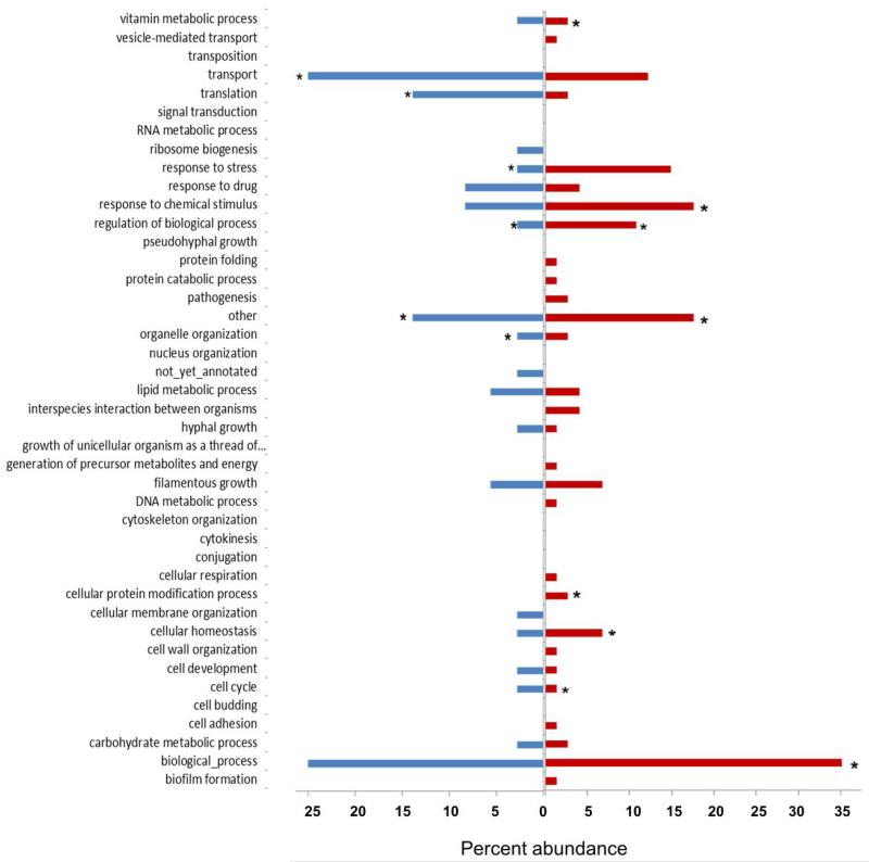
Within the Gene Ontology (GO) category for Biological Processes, from 152 C. albicans up-regulated genes (Table 2), 75 were significant (P ≤0.05), while 36 of the 147 down-regulated C. albicans genes were found to be significantly (P ≤0.05) down-regulated. The genes with significant differential gene expression were assigned to 41 GO Slim categories (Fig. 5). The numbers of genes assigned to biological process, other and chemical stimulus (the most abundant) were all found to be significantly (P ≤0.05) affected (Fig. 5). From the C. albicans down-regulated genes the terms biological process and transport were the most abundant (25%) and the terms other and translation were the next highest in abundance (13.9%). The genes assigned to transport were found to be significantly (P ≤0.05) affected (Fig. 5).
Fig. 5.
Distribution of significantly up- and down-regulated C. albicans genes in the Gene Ontology (GO) category Biological Process following C. albicans co-incubation with S. gordonii. Transcriptional profiles were analyzed with DESEQ. There were 75 significantly up-regulated genes, and 36 significantly down-regulated genes, which were assigned as a percentage to 41 identified level-2 GO-categories associated with the GO namespace Biological Process. *Denotes significance (P ≤0.05) based on hypergeometric distribution test. Red, up-regulated; blue, down-regulated.
GO Cellular Components
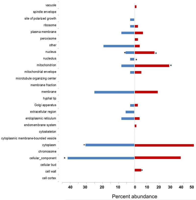
Cellular Components describe locations, at the levels of subcellular and macromolecular complexes. For GO category Cellular Components, 75 up-regulated C. albicans genes were assigned to 27 Slim categories (Fig. 6). From the up-regulated C. albicans genes the term cytoplasm (48.6%) was the most abundant with the terms cellular component (37.8%) and mitochondrion (28.4%) showing the next highest abundance. Gene ontology terms associated with the nucleus, mitochondrion, and cell wall were all found to be significantly (P ≤0.05) enriched in the up-regulated gene sets (Fig. 6). For the down-regulated C. albicans genes, GO Slim terms assigned to cytoplasm and cellular components were found to be significantly (P ≤0.05) affected (Fig. 6).
Fig. 6.
Distribution of significantly up- and down-regulated C. albicans genes in the Gene Ontology (GO) category Cellular Component following C. albicans co-incubation with S. gordonii. Transcriptional profiles were analyzed with DESeq. There were 75 significantly up-regulated, and 36 significantly down-regulated genes, which were assigned as a percentage to 27 level-2 GO-categories associated with the GO namespace Cellular Component. *Denotes significance (P ≤0.05) based on hypergeometric distribution test.
GO Molecular Functions
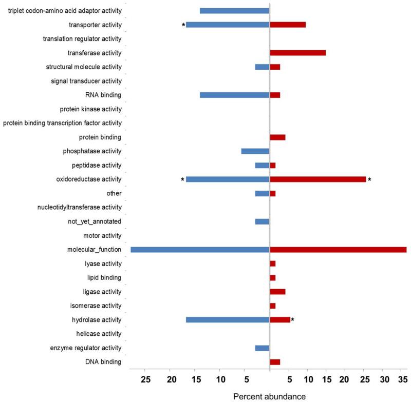
Molecular Functions describe activities, such as catalytic or binding activities that occur at the molecular level. For the GO category Molecular Function, the 75 significantly up-regulated C. albicans genes (from a total of 152 up-regulated genes) (Fig. 7) and 36 significantly down-regulated C. albicans genes were assigned to 26 GO Slim categories. From the up-regulated C. albicans genes, the term molecular function (36.5%) was the most abundant, with the terms oxidoreductase activity (25.7%) and transferase activity (14.9%) showing the next highest abundance. The number of genes assigned to oxidoreductase activity and hydrolase activity were found to be significantly (P ≤0.05) affected (Fig. 7). From the C. albicans down-regulated genes the term molecular function (27.8%) was the most abundant, with oxidoreductase activity (16.0%) and transporter activity (16.7%) significantly (P≤0.05) affected.
Fig. 7.
Distribution of significantly up- or down-regulated C. albicans genes in the Gene Ontology (GO) category Molecular Function following C. albicans co-incubation with S. gordonii. Transcriptional profiles were analyzed by the statistical software DESeq. There were 75 significantly up-regulated genes and 36 significantly down-regulated genes which were assigned as a percentage to 26 identified level-2 GO-categories associated with the GO namespace Molecular Function. *Denotes significance (P ≤0.05) based on hypergeometric distribution test.
Colonization and pathogenesis genes
From 211 genes associated with pathogenesis by CGD, only 10 genes were shown to be up- or down-regulated by ≥ twofold (Table 3). The genes ALS1, CAT1 and TEC1 are strongly associated with the transition from yeast to hyphae during filamentous growth as well as pathogenesis. The up-regulation of TEC1 in this study tends to suggest that S. gordonii stimulation of C. albicans hyphal growth might occur through the Cyr1/cAMP signalling pathway (Sudbery, 2011).
Table 3.
Mean fold changes in transcription levels of genes associated with C. albicans filamentous growth and pathogenesis following co-incubation with S. gordonii.
| Gene* | Filamentous growth (G) or pathogenesis (P) |
Mean fold change (log2)1 |
Protein identity or function2 | Total abundance (log10)3 |
P-value4 |
|---|---|---|---|---|---|
| FGR42 | G | 2.94 | Protein lacking an orthologue in S. cerevisiae; transposon mutation affects filamentous growth |
−1.941 | 0.181 |
| CAT1 | G/P | 2.71 | Catalase; resistance to oxidative stress | −1.845 | 0.010 * |
| ALS1 | G/P | 2.00 | Cell surface glycoprotein ALS family; biofilm formation, germ tube induction; expressed at infection of human buccal epithelial cells; GPI-modified; induced by ketoconazole, low iron and at cell wall regeneration; regulated by Sfu1 |
−0.998 | <0.001* |
| TEC1 | G/P | 1.97 | TEA/ATTS transcription factor; involved in white cell pheromone response, regulates hypha-specific genes, biofilm formation; regulates BCR1; transcription regulated by Cph2 in some conditions; alkaline- and biofilm-induced |
−2.348 | 0.050 * |
| CUP9 | G | 1.55 | Gene up-regulated in clinical isolates from HIV patients with oral candidiasis; transcription reduced in yeast-hyphal switch |
−1.331 | 0.205 |
| CAT8 | G | 1.52 | Predicted zinc finger protein B not essential for viability |
−2.687 | 0.152 |
| GAL10 | G | 1.49 | UDP-glucose 4-epimerase required for galactose utilization |
−1.941 | 0.269 |
| HSP21 | G/P | 1.46 | Small heat shock protein, stress response and virulence |
−2.800 | 0.374 |
| TSA1 | G | 1.43 | TSA/alkyl hydroperoxide peroxidase C (AhPC) family protein; similar to thiol- dependent peroxidases of oxidative stress signaling; antigenic; hyphal surface; peroxide induced |
−1.232 | 0.034 * |
| CTN3 | G | 1.41 | Predicted peroxisomal carnitine acetyl transferase |
−2.913 | 0.034 * |
| MDR1 | P | 1.38 | Stationary phase enriched protein; Gcn4-regulated; |
−2.454 | 0.220 |
| YHB1 | G/P | 1.36 | Member of MDR subfamily of ABC family; similar to S. cerevisiae ABC transporter Atm1; transcriptionally regulated by iron; expression greater in low iron; induced by nitric oxide independent of Yhb1 |
−1.881 | 0.365 |
| VPS20 | G | 1.15 | ESCRT III complex protein with a role in multivesicular body (MVB) trafficking; required for processing of Rim8 |
−2.608 | 0.078 |
| NRG2 | G | 1.15 | Protein similar to S. cerevisiae Nrg2 transcription factor, which regulates invasive growth in S. cerevisiae; transposon mutation affects filamentous growth |
−4.503 | 0.598 |
| PHHB | G | 1.04 | Transposon mutation affects filamentous growth; late stage biofilm- induced gene |
−2.771 | 0.413 |
| ICL1 | P | 1.03 | Isocitrate lyase enzyme of glyoxylate cycle |
−2.034 | 0.176 |
| TTR1 | P | 1.02 | Putative glutaredoxin; described as a glutathione reductase; up-regulated in the presence of human neutrophils, and upon benomyl treatment; alkaline down-regulated; regulated by Gcn2 and Gcn4; required for virulence in mouse model |
−2.719 | 0.475 |
| GAP1 | G | 1.00 | Amino acid permease | −2.191 | 0.215 |
| CHT2 | G | −1.05 | GPI-linked chitinase required for normal filamentous growth |
−1.457 | 0.498 |
| SOD5 | P | −1.26 | Copper- and zinc-containing superoxide dismutase; member of a gene family that includes SOD1, SOD4, SOD5, and SOD6; gene may contain an intron; Hap43-repressed; biofilm-induced |
−2.507 | 0.329 |
| FGR12 | G | −1.41 | Protein lacking an orthologue in S. cerevisiae; transposon mutation affects filamentous growth; biofilm induced gene |
−4.642 | 0.590 |
| LIP2 | P | −1.69 | Secreted lipase member of a differentially expressed lipase gene family |
−4.418 | 0.486 |
| FGR2 | G | − 2.73 | Transcription factor with zinc cluster DNA-binding motif |
−3.598 | 0.010 * |
Positive values, up-regulated; negative values, down-regulated (log2 ≥1.0 equivalent to ≥ twofold linear change)
Information from CGD (www.candidagenome.org)
Abundance reads represent the fractional expression of all targets in the genome. The most highly-expressed genes on this Table are therefore (in order): ALS1, TSA1, CUP9 and CHT2
P-values were calculated from DESeq and adjusted P-values ≤ 0.05 were considered to be significant
Bold type significant change P ≤0.05
For successful colonization and pathogenesis, once C. albicans blastospores adhere to host surfaces they rapidly begin to produce hyphal filaments. Hyphae physically penetrate host endothelial and epithelial cells. Aided by a large array of hyphal surface proteins they also create stronger attachments to the host surfaces. Overall there were 18 genes associated with filamentous growth whose expression was affected (six significantly) when C. albicans was challenged with S. gordonii (Table 3). FRG42, ALS1, CAT1, and TEC1 each showed a mean > four-fold increase. ALS1 and TSA1 transcripts were of highest abundance (Table 3). Tec1 and Fgr42 proteins are both filamentous growth regulators (Sudbery, 2011), so are involved in the switch from yeast form to hyphal form, suggesting that the gene expression network associated with the yeast to hypha transition was stimulated by the presence of S. gordonii in the culture. This is supported by evidence for growth synergy of streptococci and C. albicans in biofilms (Bamford et al., 2009) and morphological observations of more extensive hyphal filament formation with S. gordonii present (Dutton et al., 2014).
Oxidative stress related genes
Up-regulation of CAT1 (catalase) during the yeast-hypha transition might be consistent with reports that reactive oxygen species (ROS) levels are increased under hypha-forming conditions (Schroter et al., 2000). During the transition of yeast to hyphal filament, the cell wall must go through a considerable conformational change, involving restructuring, which ultimately requires a greater energy input from the cell. The energy comes from oxidative phosphorylation in mitochondria, which generate ROS as a by-product. The ROS will attack and inhibit the functions of proteins, lipids and DNA if not decomposed by Cat1 and other anti-oxidant enzymes. So an increased energy output during hypha formation will generate a need for an increase in anti-oxidants.
HSP21, one of the up-regulated genes in Table 3, encodes a multifunctional heat shock protein, with a role in both stress adaptation and virulence in C. albicans (Mayer et al., 2012). Hsp21 modulates thermal stress by fine tuning homeostasis of compatible solutes and activation of the Cek1 pathway, which has previously been shown to be influenced by S. gordonii (Bamford et al., 2009). Hsp21 also mediates adaptation to oxidative stress, while an hsp21Δ/Δ mutant forms shorter hyphae than the wild-type and is strongly attenuated in virulence in vivo (Mayer et al., 2012).
Numerous C. albicans anti-oxidant genes in addition to CAT1 responded to the presence of S. gordonii including ORF19.3537 a putative sulfiredoxin, two oxidoreductases (ORF19.2262 and CIP1), cytochrome c peroxidase (CCP1), glutathione reductase (GLR1), glutathione-S-transferase (GTT11), glutathione peroxidase (GPX2), thioredoxin reductase (TRR1) and the thiol-specific antioxidant protein (TSA1). TSA1, TRR1, CAT1, GLR1 and CPA2 transcripts were of high abundance (Table 2). However, not all known oxidative stress response genes were up-regulated e.g. TRX1, SOD1, GRX2, GPX31-33 (da Silva Dantas et al., 2015) suggesting a more specific type of response to S. gordonii. Many of the antioxidant genes are reported to be regulated by Cap1 (Wang et al., 2006) and their products are up-regulated upon H2O2 treatment of C. albicans (Kusch et al., 2007). Overall, protection against H2O2 potentially confers added resistance to macrophages and cells of the innate immune system that utilize oxidative stress to kill C. albicans.
ARG1, ARG3, ARG4, ARG5,6, CPA1, and CPA2 are all involved with arginine biosynthesis are also greatly up-regulated in C. albicans with S. gordonii present. Conversely CAR1, an arginase, is strongly down-regulated suggesting that increased arginine production is important for the response of C. albicans to S. gordonii. Since the ARG genes are up-regulated in the presence of H2O2 (Jiménez-Lopéz et al., 2013) these effects could be in direct response to the production of H2O2 by S. gordonii (Liu et al., 2011). The importance of arginine biosynthesis is further strengthened by the up-regulation of PUT1, a putative proline oxidase that is expected to convert L-proline into Δ1-pyrroline-5-carboxylate and glutamate-γ-semialdehyde that act as the main precursors for arginine production and polyamine production. In Saccharomyces cerevisiae, exposure to H2O2 and freeze-thaw stress also leads to an accumulation of arginine (Almeida et al., 2007; Momose et al., 2010), with supplementary arginine conferring resistance to oxidative and thermal stress (Nishimura et al., 2010).
Covalently linked cell wall proteins (CWPs)
The C. albicans cell wall consists of an internal scaffold of (β-1,3)- and (β-1,6)-linked glucan and chitin, to which an outer protein coat is attached (Klis et al., 2001; Ruiz-Herrera et al., 2006). The protein coat is thought to contain about 20 different polypeptides attached to the cell wall by covalent bonds linking proteins to the inner glucan/chitin skeleton (Klis et al., 2009). These cell wall proteins (CWPs) have been associated with many functions including host cell adhesion and invasion, biofilm formation, cell-cell and intergeneric aggregation, and enzymatic functions such as superoxide dismutases and yapsin-like aspartic proteases (Monod et al., 1998; Martchenko et al., 2004; Krysan et al., 2005). Because of the diversity of their roles, it is not surprising that expression of CWP-encoding genes can vary enormously, not only with mode of growth, but also with environmental signals and input from distinct signalling pathways, triggered by changing environmental conditions (e.g. temperature, pH, N-acetyl-D-glucosamine etc.). This analysis investigated 36 experimentally-validated covalently-linked CWPs of C. albicans (Klis et al., 2009) and how their gene expression profiles changed when C. albicans cells were co-incubated with S. gordonii (Table S3).
Thirteen out of 18 C. albicans CWP genes showed ≥ twofold up-regulation of gene expression, while five were ≥ twofold down-regulated (Table 4). Chi-squared tests showed that cell wall protein genes PGA57, ALS1, PGA34, PHA36 (IHD1), PGA61, and ORF19.4653 were all significantly up-regulated (P ≤0.05). PGA10 was up-regulated > four-fold, though this was not considered significant at P ≤0.05. Pga10 (Rbt51) belongs to a sub-set of fungal proteins with an eight cysteine residues domain CFEM (Common in several Fungal Extracellular Membrane proteins). Pga10/Rbt51 along with other proteins Rbt5 and Wap1/Csa1, each contain CFEM domains, which play key roles during biofilm formation (Perez et al., 2006).
Table 4.
Mean fold changes in expression of C. albicans genes associated with adherence, or encoding covalently-linked cell wall proteins or GPI-modified proteins, following co-incubation with S. gordonii.
| Gene* | Association1 | Mean fold change (log2)2 |
Total abundance (log10)3 |
P-value4 |
|---|---|---|---|---|
| PGA10 | CL, GPI | 2.23 | −3.196 | 0.147 |
| PGA37 | GPI | 2.20 | −3.680 | 0.054 |
| PGA57 | GPI | 2.05 | −3.348 | <0.001* |
| ALS1 | CL, GPI, Adh | 2.01 | −0.998 | <0.001* |
| TEC1 | Adh | 1.97 | −2.348 | 0.050 * |
| HIS4 | Adh | 1.58 | −1.663 | 0.160 |
| PGA34 | GPI | 1.55 | −3.811 | 0.035 * |
| PGA60 | GPI | 1.49 | −3.813 | 0.083 |
| PGA36/IHD1 | GPI | 1.39 | −1.172 | 0.016 * |
| EAP1 | CL, Adh | 1.39 | −3.295 | 0.062 |
| PGA61 | GPI | 1.26 | −3.810 | 0.044 * |
| ORF19.4653 | GPI | 1.24 | −2.256 | 0.034 * |
| HYR1 | CL, GPI, Adh | 1.10 | −2.300 | 0.253 |
| CHT2 | CL, GPI | −1.05 | −1.457 | 0.498 |
| PGA3/SOD5 | CL, GPI | −1.26 | −2.507 | 0.329 |
| PGA16 | GPI | −1.42 | −5.187 | 0.959 |
| CSA1 | CL, GPI | −1.43 | −1.638 | 0.280 |
| PGA39 | GPI | −1.56 | −4.541 | 0.487 |
Adh, adhesion; CL, covalently linked; GPI, Glycophosphatidylinositol-modified
Positive values, up-regulated; negative values, down-regulated (log2 ≥1.0 equivalent to ≥ twofold linear change)
Abundance reads represent the fractional expression of all targets in the genome
P-values were calculated from DESeq and adjusted P-values ≤ 0.05 were considered to be significant
Bold type significant change P ≤0.05
The up-regulation of expression of HYR1 is most likely to be associated with an increase in the extent or rate of hypha formation. Studies have shown HYR1 is induced specifically in response to hyphal development when morphogenesis is stimulated by growth conditions such as serum, temperature elevation, pH and the addition of N-acetyl-D-glucosamine (Bailey et al., 1996). The findings suggest that the just over twofold increase in expression of HYR1 could be associated with yeast to hypha transition, as shown by Spiering et al. (2010), stimulated by the addition of S. gordonii to the culture.
GPI-modified proteins
Glycophosphatidylinositol (GPI)-modified proteins all share conserved features of an N-terminal signal sequence and C-terminus tethered to the cell wall or cell membrane by a preformed GPI anchor. In silico predictions suggest that there are 115 genes encoding GPI-modified proteins in C. albicans (Richard & Plaine, 2007). Several lists of C. albicans GPI-modified proteins (GpiPs) published in the literature (De Groot et al., 2003; Garcera et al., 2003; Eisenhaber et al., 2004) have been amalgamated and refined (Richard & Plaine, 2007) to avoid duplications and allow for differences in the algorithms used to define GpiPs.
The functions of the majority of these GpiPs (66%) still remain unknown. The others can be assigned to functions related to cell wall biosynthesis or remodelling, and cell-cell adhesion and interactions. Since the compilation of this list of GpiPs, 45 knock-out mutants of computer predicted GpiPs have been screened for their roles in cell wall structure (Plaine et al., 2008). Deletion mutants that result in cell wall modifications and reduced caspofungin sensitivity included DFG5, PHR1, PGA4 and PGA62.
In our data, from the 115 predicted GpiPs, only 16 had a change in expression > twofold in the presence of S. gordonii (Table S4). A small number of genes (five) were down-regulated while 11 genes showed up-regulation (Table S4), six of which were significant (P <0.05). These were ALS1, ORF19.4653, PGA34, PGA36, PGA57 and PGA61 (Table 4). The CGD provides information on PGA34 (role in host infection) and PGA36 (induced during hyphal development), while ORF19.4653, PGA57 and PGA61 are currently uncharacterized but clearly respond transcriptionally to S. gordonii.
Adhesins
C. albicans cells within the oral cavity must avoid being washed away by the continuous flow of saliva, therefore adhesion to a multitude of host surfaces including epithelial cells, teeth, and dentures is of paramount importance. C. albicans possesses numerous potential adhesins (Zordan and Cormack, 2012) and this part of the study investigated 23 proteins which have been linked to cell adhesion in the CGD.
When C. albicans was co-incubated with S. gordonii, 19 out of the 23 C. albicans cell wall protein genes associated with adhesion showed up-regulation of gene expression, while four were seen to be down-regulated (these were all less than twofold changes). Both ALS1 and TEC1 showed the highest mean fold change in up-regulated adhesion-associated genes with significant four-fold changes (P ≤0.05) (Table 4). ALS1 expression is known to be associated with hypha formation and, on the emergence of the germ tube, Als1 is the first member of the Als family to be expressed. Interestingly, the expressed protein is localized at the neck of the growing hypha (Fu et al., 2002). Differential expression patterns of the ALS genes have shown that there is a major spike in ALS1 expression when cells are inoculated into fresh medium, a procedure which also triggers hyphal growth, indicating that Als1 might have a regulatory role as well as an adhesin function. The up-regulation of TEC1, a TEA/ATTS transcription factor, is consistent with its reported role in regulating hypha formation and virulence (Schweizer et al., 2000).
EAP1 (2.6-fold increase), HIS4 (three-fold increase) and HYR1 (2.1-fold increase) were also seen to be up-regulated. Eap1 is a glucan-cross-linked cell wall-localized protein that has been reported to be required for C. albicans to form robust biofilms on polystyrene surfaces (Nobbs et al., 2010) and in central venous catheters (Li et al., 2007) under shear flow in vitro and in vivo. Although expressed in both yeast and hyphal cells, it has been suggested that Eap1 protein expression is not directly associated with hypha formation (Li and Palecek, 2003). However, there are contradicting reports where transcriptional profiling studies of the yeast to hypha transition reveal a twofold increase in Eap1 at 6 h (Nantel et al., 2002) similar to the approximately three-fold up-regulation seen here. Interestingly, studies of S. cerevisiae strains expressing Eap1 have confirmed that Eap1 is able to bind S. gordonii in planktonic culture (Grubb et al., 2009). This indicates that Eap1, along with a number of other adhesins such as Als3 and Hwp1, promotes trans-kingdom interactions with other microorganisms to aid successful colonization and the formation of polymicrobial communities (Nobbs et al., 2010; Xu et al., 2014). Although EAP1 expression was up-regulated, expression of the genes encoding other C. albicans adhesins ALS3 and HWP1 was not over the time-course of the experiment.
S. gordonii transcriptional response to C. albicans
A total of 72 S. gordonii genes were either up- or down-regulated specifically in response to co-culturing with C. albicans (Table 5). Eighteen S. gordonii genes were significantly up-regulated including ≥ twofold increases in glpK, rplO, celB, rplN, rplB, rpsE, ciaR, and gat (Table 5). Glycerol kinase (GlpK) allows glycerol to be utilized as a carbon source. Notably, a highly up-regulated gene in C. albicans biofilms was RHR2 encoding the glycerol biosynthetic enzyme glycerol-3-phosphatase. Glycerol is five times more abundant in C. albicans biofilm cells (Desai et al., 2013) so it seems possible that S. gordonii may respond to glycerol production or secretion by C. albicans hypha-forming cells by utilizing this as an alternate carbon and energy source.
Table 5.
Mean fold changes in expression of S. gordonii genes following co-incubation with C. albicans.
| Gene* | Mean fold change (log2)1 |
Description2 | Total abundance (log10)3 |
P-value4 |
|---|---|---|---|---|
| glpK | 1.92 | Glycerol kinase | −1.612 | 0.030 * |
| SGO_0149 | 1.91 | Hypothetical protein | −2.812 | 0.135 |
| SGO_0092 | 1.85 | Hypothetical protein | 0.000 | 0.181 |
| SGO_0093 | 1.81 | Hypothetical protein | 0.000 | 0.123 |
| SGO_0631 | 1.78 | α-glyerophosphae oxidase | −0.955 | 0.101 |
| SGO_1071 | 1.67 | Hypothetical protein | −0.961 | 0.146 |
| adk | 1.56 | Adenylate kinase | −1.587 | 0.100 |
| xpt | 1.49 | Xanthine phosphoribosyltransferase | −1.144 | 0.318 |
| rpoA | 1.48 | DNA-directed RNA polymerase | −0.282 | 0.133 |
| SGO_2091 | 1.37 | Hypothetical proein | −2.210 | 0.257 |
| SGO_2079 | 1.29 | Hypothetical protein | 0.000 | 0.690 |
| rplR | 1.26 | 50S ribosomal protein | −0.776 | 0.064 |
| SGO_1136 | 1.25 | Esterase superfamily protein | −2.636 | 0.119 |
| SGO_1581 | 1.25 | MtN3/saliva family protein | −2.511 | 0.091 |
| rpsS | 1.24 | 30S ribosomal protein S19 | −1.088 | 0.069 |
| rplO | 1.21 | Ribosomal protein | −1.086 | 0.014 * |
| trpA-1 | 1.18 | Tryptophan synthase subunit α | 0.000 | 0.193 |
| celB | 1.15 | Cellobiose metabolism operon | −1.538 | 0.014 * |
| rplN | 1.15 | Ribosomal protein L14 | −0.918 | 0.009 * |
| rplB | 1.11 | Ribosomal protein L2 | −0.599 | 0.027 * |
| SGO_0630 | 1.05 | Glycerol uptake facilitator protein | −1.642 | 0.256 |
| rpsE | 1.02 | Ribosomal protein S5 | −0.666 | 0.016 * |
| ciaR | 1.00 | Transcription regulator | −0.885 | 0.032 * |
|
SGO_1261
(gat) |
1.00 | Glutamine amidotransferase | −1.765 | 0.044 * |
| rpmD | 0.98 | Ribosomal protein L30 | −0.961 | 0.011 * |
| celA | 0.98 | Cellobiose metabolism operon | −0.738 | 0.026 * |
| SGO_0571 | 0.97 | Hypothetical protein | 0.000 | 0.041 * |
| rplX | 0.94 | Ribosomal protein L24 | −1.020 | 0.023 * |
| secY | 0.92 | Preprotein translocase subunit | −1.095 | 0.018 * |
| SGO_1625 | 0.88 | Acetoin utilization | −2.511 | 0.045 * |
| SGO_1579 | 0.85 | Transcription antiterminator (BglG family) |
−0.745 | 0.011 * |
| celD | 0.80 | Cellobiose metabolism operon | −0.775 | 0.010 * |
| rplF | 0.80 | Ribosomal protein L6 | −1.062 | 0.039 * |
| celC | 0.67 | Cellobiose metabolism operon | −0.989 | 0.043 * |
| SGO_0991 | −0.31 | Hypothetical protein | −2.812 | 1.000 |
| SGO_0924 | −0.53 | HsdD | 0.000 | 0.921 |
| sodA | −0.57 | Superoxide dismutase | −0.818 | 0.760 |
| SGO_1461 | −0.62 | Stress response transcriptional regulator |
−2.511 | 0.630 |
| SGO_0378 | −0.64 | Hypthothetical protein | −0.567 | 0.783 |
| rpmF | −0.73 | Ribosomal protein L32 | −1.409 | 0.650 |
| rpmG | −0.79 | Ribosomal protein L33 | −2.636 | 0.403 |
| glgP-2 | − 0.82 | Maltodextrin phosphorylase | −0.913 | 0.043 * |
| trx-2 | −0.83 | Thioredoxin 2 | −2.113 | 0.201 |
| gloA | −0.83 | Lactolylglutathione lyase | −1.454 | 0.347 |
| hutU | −0.85 | Urocanate hydratase | −2.414 | 0.235 |
| SGO_0584 | −0.85 | Acetyltransferase | −1.591 | 0.073 |
| SGO_2084 | −0.86 | Quinone family NAD(P)H deydrogenase | 0.000 | 0.406 |
| ldh | −0.89 | Lactate dehydrogenase | −0.791 | 0.111 |
| SGO_1457 | −0.90 | Hypothetical protein | −2.210 | 0.367 |
| SGO_1300 | −0.90 | NADPH-dependant reductase | −2.210 | 0.465 |
| pyrB | −0.91 | Aspartate carbomyl transferase | −1.260 | 0.295 |
| gtfG | −0.91 | Glucosyltransferase | −0.943 | 0.151 |
| SGO_1301 | −0.93 | Hypothetical protein | −2.812 | 0.322 |
| SGO_0194 | −0.94 | Hypothetical protein | −1.958 | 0.265 |
| ftcD | −0.96 | Glutamate formiminotransferase | 0.000 | 0.323 |
| SGO_0233 | −0.98 | Lipoprotein | 0.000 | 0.083 |
| SGO_1740 | −0.99 | Integral membrane protein | −1.834 | 0.575 |
| bta | −1.01 | Bacteriocin transport accessory protein | −1.334 | 0.300 |
| SGO_1508 | −1.01 | Uncharacterized | 0.000 | 0.650 |
| secE | −1.04 | Preprotein translocase subunit | −2.636 | 0.113 |
| mscL | −1.10 | Mechanosensitive channel protein | −1.312 | 0.196 |
| SGO_1256 | −1.13 | PyrK(dihydroorotate dehydrogenase) | −2.812 | 0.238 |
| SGO_1108 | −1.15 | Uracil permease | −0.812 | 0.187 |
| SGO_1106 | −1.16 | Hypthothetical protein | −1.958 | 0.384 |
| trx-1 | −1.17 | Thioredoxin 1 | −0.957 | 0.164 |
| SGO_1107 | −1.18 | PyrR (pyrimidine regulatory protein) | −2.812 | 0.182 |
| glmS | −1.25 | Glucosamine fructose-6-phosphate aminotransferase |
−1.001 | 0.090 |
| pyrd | −1.29 | Dihydroorotate dehydrogenase | −3.113 | 0.202 |
| carB | −1.42 | Carbamyl phosphate synthetase | −0.291 | 0.207 |
| pyrE | −1.49 | Orotate phosphoribosyl transferase | −2.636 | 0.228 |
| SGO_0296 | −1.50 | Integral membrane protein | 0.000 | 0.061 |
| SGO_1105 | −1.54 | Hypothetical protein | −1.958 | 0.257 |
Positive values, up-regulated; negative values, down-regulated (log2 ≥1.0 equivalent to ≥ twofold linear change)
Description details from the S. gordonii CH1 annotated genome sequence
Abundance reads represent the fractional expression of all targets in the genome
P-values were calculated from DESeq and adjusted P-values ≤ 0.05 were considered to be significant
Bold type significant change P ≤0.05
CiaR is a response regulator of competence for DNA uptake (Mascher et al., 2003) and biofilm formation (Blanchette-Cain et al., 2013) in Streptococcus pneumoniae. We have shown recently that S. gordonii competence-development is involved in formation of dual species biofilms with C. albicans (Jack et al., 2015), and so CiaR may be a factor in this process. cel genes annotated as encoding components of cellobiose metabolism were up-regulated, perhaps indicating that S. gordonii is responding to the presence of C. albicans cell wall glucans. Up-regulation of rplO, rplN, rplB, and rpsE, all involved in translation, may be an artefact of ribosomal depletion. Only glgP-2 (maltodextran phosphorylase) was significantly down-regulated (Table 5). Regions of the S. gordonii genome containing ORFs SGO_1105 to SGO_1108 showed coordinated down-regulation, and are all implicated in pyrimidine biosynthesis. Overall, these results suggest that S. gordonii was transcriptionally much less reactive to the presence of C. albicans for 1 h in mixed culture, while the effects of CiaR up-regulation in S. gordonii are a topic of future investigation.
Summary and conclusions
This work describes the responses of C. albicans and S. gordonii to each other at the transcriptional level. The experiments were undertaken under growth conditions that induce filamentation in C. albicans. The results presented suggest that S. gordonii has a range of significant effects on the biological processes occurring within C. albicans during the early phase of co-culture, with a large number of genes affected in C. albicans in comparison to a smaller number in S. gordonii. Genes involved in responses to chemical stimuli, regulation, homeostasis, protein modification and cell cycle were up-regulated, while genes involved in transport and translation were down-regulated. These patterns suggest that C. albicans was responding positively to signals produced by S. gordonii. Mitochondrial genes were up-regulated together with genes encoding cell wall proteins, suggesting triggering of metabolic functions.
On the other hand, down-regulation of triplet codon-amino acid adaptor and transporter activities suggest modulation of the rate of protein synthesis. Oxidoreductase activities were up- and down-regulated, while hydrolase activity genes were up-regulated, suggesting perhaps that new macromolecular substrates e.g. polysaccharide, peptidoglycan etc. were now available for metabolism by C. albicans. Overall the data are consistent with C. albicans not being growth-inhibited in the presence of S. gordonii, unlike the inhibitory effects of some other bacteria (e.g. P. aeruginosa) on growth and hyphal development (Hogan & Kolter, 2004; Fox et al., 2013). There was some evidence of up-regulation of genes that might be linked with growth-stimulatory effects, supporting previous observations from biofilm experiments (Bamford et al., 2009; Dutton et al., 2014; Xu et al., 2014).
The major changes in expression of morphogenesis-related genes in response to S. gordonii were upregulation of TEC1, ALS1, and CAT1. Notably, Tec1 is a hyphal-development activator (Nobile & Mitchell, 2005) that regulates expression of cell wall protein genes, but probably not ALS1 (Nobile & Mitchell, 2006). ALS1 also appeared in the up-regulated genes encoding covalently-linked CWPs and GPI-modified proteins. Clearly TEC1, ALS1 and anti-oxidant genes seem to be major players in the response of C. albicans to S. gordonii. The Als1 protein is not thought to be a component of the hyphal cell wall, but appears at the initial site of hyphal filament growth from the mother cell (Coleman et al., 2012). ALS1 has been shown to be one of the genes that is first up-regulated following adhesion to a surface (Garcia-Sanchez et al., 2004) that then leads onto biofilm formation. Further work is required to establish the factors affecting expression of ALS1 and if it is involved in regulation as well as adhesion. Of the GPI-modified proteins, six genes (including ALS1) encoding these were significantly (P ≤0.05) up-regulated in the presence of S. gordonii. The functions of the other five PGA (Protein with Glycosylphosphatidylinositol Anchor) genes are unknown. Further studies could help to identify the functions of the proteins encoded by these genes and they may reveal new factors for adhesion, biofilm formation and pathogenesis. In summary, these transcriptional data findings seem to correlate with the biological data indicating that S. gordonii promotes hyphal development and grows synergistically with C. albicans. They also identify a number of new target genes for further study of their roles in development of interkingdom biofilm communities.
Supplementary Material
ACKNOWLEDGMENTS
We thank Massimo Micaroni for help with electron microscopy and Jane Brittan for technical assistance. The support of NIH (NIDCR), Bethesda, USA to HFJ and RJL is gratefully acknowledged (R01DE016690).
REFERENCES
- Aas JA, Paster BJ, Stokes LN, Olsen I, Dewhirst FE. Defining the normal bacerial flora of the oral cavity. J Clin Microbiol. 2005;43:5721–5732. doi: 10.1128/JCM.43.11.5721-5732.2005. [DOI] [PMC free article] [PubMed] [Google Scholar]
- Almeida B, Buttner S, Ohlmeier S, et al. NO-mediated apoptosis in yeast. J Cell Sci. 2007;120:3279–3288. doi: 10.1242/jcs.010926. [DOI] [PubMed] [Google Scholar]
- Anders S, Huber W. Differential expression analysis for sequence count data. Genome Biol. 2010;11:R106. doi: 10.1186/gb-2010-11-10-r106. [DOI] [PMC free article] [PubMed] [Google Scholar]
- Aronesty E. ea-utils: Command-line tools for processing biological sequencing data. 2011. [Google Scholar]
- Bailey DA, Feldmann PJ, Bovey M, Gow NA, Brown AJ. The Candida albicans HYR1 gene, which is activated in response to hyphal development, belongs to a gene family encoding yeast cell wall proteins. J Bacteriol. 1996;178:5353–5360. doi: 10.1128/jb.178.18.5353-5360.1996. [DOI] [PMC free article] [PubMed] [Google Scholar]
- Bamford CV, d’Mello A, Nobbs AH, Dutton LC, Vickerman MM, Jenkinson HF. Streptococcus gordonii modulates Candida albicans biofilm formation through intergeneric communication. Infect Immun. 2009;77:3696–3704. doi: 10.1128/IAI.00438-09. [DOI] [PMC free article] [PubMed] [Google Scholar]
- Blanchette-Cain K, Hinojosa CA, Akula Suresh Babu R, et al. Streptococcus pneumoniae biofilm formation is strain dependent, multifactorial, and associated with reduced invasiveness and immunoreactivity during colonization. mBio. 2013;4:e00745–13. doi: 10.1128/mBio.00745-13. [DOI] [PMC free article] [PubMed] [Google Scholar]
- Boon C, Deng Y, Wang LH, et al. A novel DSF-like signal from Burkholderia cenocepacia interferes with Candida albicans morphological transition. ISME J. 2008;2:27–36. doi: 10.1038/ismej.2007.76. [DOI] [PubMed] [Google Scholar]
- Bruno VM, Wang Z, Marjani SL, et al. Comprehensive annotation of the transcriptome of the human fungal pathogen Candida albicans using RNA-seq. Genome Res. 2010;20:1451–1458. doi: 10.1101/gr.109553.110. [DOI] [PMC free article] [PubMed] [Google Scholar]
- Canabarro A, Valle C, Farias MR, Santos FB, Lazera M, Wanke B. Association of subgingival colonization of Candida albicans and other yeasts with severity of chronic periodontitis. J Periodontal Res. 2013;48:428–432. doi: 10.1111/jre.12022. [DOI] [PubMed] [Google Scholar]
- Coleman DA, Oh SH, Manfra-Maretta SL, Hoyer LL. A monoclonal antibody specific for Candida albicans Als4 demonstrates overlapping localization of Als family proteins on the fungal cell surface and highlights differences between Als localization in vitro and in vivo. FEMS Immunol Med Microbiol. 2012;64:321–333. doi: 10.1111/j.1574-695X.2011.00914.x. [DOI] [PMC free article] [PubMed] [Google Scholar]
- Dale RK, Pedersen BS, Quinlan AR. Pybedtools: a flexible Python library for manipulating genomic datasets and annotations. Bioinformatics. 2011;27:3423–3424. doi: 10.1093/bioinformatics/btr539. [DOI] [PMC free article] [PubMed] [Google Scholar]
- da Silva Dantas A, Day A, Ikeh M, Kos I, Achan B, Quinn J. Oxidative stress responses in the human fungal pathogen, Candida albicans. Biomolecules. 2015;5:142–165. doi: 10.3390/biom5010142. [DOI] [PMC free article] [PubMed] [Google Scholar]
- de Carvalho FG, Silva DS, Hebling J, Spolidorio LC, Spolidorio DM. Presence of mutans streptococci and Candida spp. Indental plaque/dentine of carious teeth and early childhood caries. Arch Oral Biol. 2006;51:1024–1028. doi: 10.1016/j.archoralbio.2006.06.001. [DOI] [PubMed] [Google Scholar]
- De Groot PW, Hellingwerf KJ, Klis FM. Genome-wide identification of fungal GPI proteins. Yeast. 2003;20:781–796. doi: 10.1002/yea.1007. [DOI] [PubMed] [Google Scholar]
- Desai JV, Bruno VM, Ganguly S, et al. Regulatory role of glycerol in Candida albicans biofilm formation. mBio. 2013;4:e00637–00612. doi: 10.1128/mBio.00637-12. [DOI] [PMC free article] [PubMed] [Google Scholar]
- Douglas LJ. Candida biofilms and their role in infection. Trends Microbiol. 2003;11:30–36. doi: 10.1016/s0966-842x(02)00002-1. [DOI] [PubMed] [Google Scholar]
- Dutton LC, Nobbs AH, Jepson K, et al. O-mannosylation in Candida albicans enables development of interkingdom biofilm communities. mBio. 2014;5:e00911. doi: 10.1128/mBio.00911-14. [DOI] [PMC free article] [PubMed] [Google Scholar]
- Eggimann P, Que YA, Revelly JP, Pagani JL. Preventing invasive candida infections. Where could we do better? J Hosp Infect. 2015;89:302–308. doi: 10.1016/j.jhin.2014.11.006. [DOI] [PubMed] [Google Scholar]
- Eisenhaber B, Schneider G, Wildpaner M, Eisenhaber F. A sensitive predictor for potential GPI lipid modification sites in fungal protein sequences and its application to genome-wide studies for Aspergillus nidulans, Candida albicans, Neurospora crassa, Saccharomyces cerevisiae and Schizosaccharomyces pombe. J Mol Biol. 2004;337:243–253. doi: 10.1016/j.jmb.2004.01.025. [DOI] [PubMed] [Google Scholar]
- Fox SJ, Shelton BT, Kruppa MD. Characterization of genetic determinants that modulate Candida albicans filamentation in the presence of bacteria. PLoS One. 2013;8:e71939. doi: 10.1371/journal.pone.0071939. [DOI] [PMC free article] [PubMed] [Google Scholar]
- Fu Y, Ibrahim AS, Sheppard DC, et al. Candida albicans Als1p: an adhesin that is a downstream effector of the EFG1 filamentation pathway. Mol Microbiol. 2002;44:61–72. doi: 10.1046/j.1365-2958.2002.02873.x. [DOI] [PubMed] [Google Scholar]
- Garcera A, Martinez AI, Castillo L, Elorza MV, Sentandreu R, Valentin E. Identification and study of a Candida albicans protein homologous to Saccharomyces cerevisiae Ssr1p, an internal cell-wall protein. Microbiology. 2003;149:2137–2145. doi: 10.1099/mic.0.26301-0. [DOI] [PubMed] [Google Scholar]
- Garcia-Sanchez S, Aubert S, Iraqui I, Janbon G, Ghigo JM, d’Enfert C. Candida albicans biofilms: a developmental state associated with specific and stable gene expression patterns. Eukaryot Cell. 2004;3:536–545. doi: 10.1128/EC.3.2.536-545.2004. [DOI] [PMC free article] [PubMed] [Google Scholar]
- Grubb SE, Murdoch C, Sudbery PE, Saville SP, Lopez-Ribot JL, Thornhill MH. Adhesion of Candida albicans to endothelial cells under physiological conditions of flow. Infect Immun. 2009;77:3872–3878. doi: 10.1128/IAI.00518-09. [DOI] [PMC free article] [PubMed] [Google Scholar]
- Grumaz C, Lorenz S, Stevens P, et al. Species and condition specific adaptation of the transcriptional landscapes in Candida albicans and Candida dubliniensis. BMC Genomics. 2013;14:212. doi: 10.1186/1471-2164-14-212. [DOI] [PMC free article] [PubMed] [Google Scholar]
- Hogan DA, Vik A, Kolter R. A Pseudomonas aeruginosa quorum-sensing molecule influences Candida albicans morphology. Mol Microbiol. 2004;54:1212–1223. doi: 10.1111/j.1365-2958.2004.04349.x. [DOI] [PubMed] [Google Scholar]
- Huang DW, Sherman BT, Tan Q, et al. The DAVID Gene Functional Classification Tool: a novel biological module-centric algorithm to functionally analyze large gene lists. Genome Biol. 2007;8:R183. doi: 10.1186/gb-2007-8-9-r183. [DOI] [PMC free article] [PubMed] [Google Scholar]
- Jack AA, Daniels DE, Jepson MA, et al. Streptococcus gordonii comCDE (competence) operon modulates biofilm formation with Candida albicans. Microbiology. 2015;161:411–421. doi: 10.1099/mic.0.000010. [DOI] [PMC free article] [PubMed] [Google Scholar]
- Jarosz LM, Deng DM, Van Der Mei HC, Crielaard W, Krom BP. Streptococcus mutans competence-stimulating peptide inhibits Candida albicans hypha formation. Eukaryot Cell. 2009;8:1658–1664. doi: 10.1128/EC.00070-09. [DOI] [PMC free article] [PubMed] [Google Scholar]
- Jenkinson HF. Beyond the oral microbiome. Environ Microbiol. 2011;13:3077–3087. doi: 10.1111/j.1462-2920.2011.02573.x. [DOI] [PubMed] [Google Scholar]
- Jenkinson HF, Lala HC, Shepherd MG. Coaggregation of Streptococcus sanguis and other streptococci with Candida albicans. Infect Immun. 1990;58:1429–1436. doi: 10.1128/iai.58.5.1429-1436.1990. [DOI] [PMC free article] [PubMed] [Google Scholar]
- Jiménéz-Lopéz C, Collette JR, Brothers KM, et al. Candida albicans induces arginine biosynthesis genes in response to host-derived reactive oxygen species. Eukaryot Cell. 2013;12:91–100. doi: 10.1128/EC.00290-12. [DOI] [PMC free article] [PubMed] [Google Scholar]
- Kim D, Pertea G, Trapnell C, Pimentel H, Kelley R, Salzberg SL. TopHat2: accurate alignment of transcriptomes in the presence of insertions, deletions and gene fusions. Genome Biol. 2013;14:R36. doi: 10.1186/gb-2013-14-4-r36. [DOI] [PMC free article] [PubMed] [Google Scholar]
- Klis FM, De Groot P, Hellingwerf K. Molecular organization of the cell wall of Candida albicans. Med Mycol. 2001;39(Suppl 1):1–8. [PubMed] [Google Scholar]
- Klis FM, Sosinska GJ, De Groot PW, Brul S. Covalently linked cell wall proteins of Candida albicans and their role in fitness and virulence. FEMS Yeast Res. 2009;9:1013–1028. doi: 10.1111/j.1567-1364.2009.00541.x. [DOI] [PubMed] [Google Scholar]
- Krysan DJ, Ting EL, Abeijon C, Kroos L, Fuller RS. Yapsins are a family of aspartyl proteases required for cell wall integrity in Saccharomyces cerevisiae. Eukaryot Cell. 2005;4:1364–1374. doi: 10.1128/EC.4.8.1364-1374.2005. [DOI] [PMC free article] [PubMed] [Google Scholar]
- Kusch H, Engelmann S, Albrecht D, Morschhäuser J, Hecker M. Proteomic analysis of the oxidative stress response in Candida albicans. Proteomics. 2007;7:686–697. doi: 10.1002/pmic.200600575. [DOI] [PubMed] [Google Scholar]
- Li F, alecek SP. EAP1, a Candida albicans gene involved in binding human epithelial cells. Eukaryot Cell. 2003;2:1266–1273. doi: 10.1128/EC.2.6.1266-1273.2003. [DOI] [PMC free article] [PubMed] [Google Scholar]
- Li F, Svarovsky MJ, Karlsson AJ, et al. Eap1p, an adhesin that mediates Candida albicans biofilm formation in vitro and in vivo. Eukaryot Cell. 2007;6:931–939. doi: 10.1128/EC.00049-07. [DOI] [PMC free article] [PubMed] [Google Scholar]
- Li H, Handsaker B, Wysoker A, et al. The Sequence Alignment/Map format and SAMtools. Bioinformatics. 2009;25:2078–2079. doi: 10.1093/bioinformatics/btp352. [DOI] [PMC free article] [PubMed] [Google Scholar]
- Liu X, Ramsey MM, Chen X, Koley D, Whiteley M, Bard AJ. Real-time mapping of a hydrogen peroxide concentration profile across a polymicrobial bacterial biofilm using scanning electrochemical microscopy. Proc Natl Acad Sci USA. 2011;108:2668–2673. doi: 10.1073/pnas.1018391108. [DOI] [PMC free article] [PubMed] [Google Scholar]
- Love MI, Huber W, Anders S. Moderated extimation of fold change and dispersion for RNA-seq data with DESeq2. Genome Biol. 2014;15:550. doi: 10.1186/s13059-014-0550-8. [DOI] [PMC free article] [PubMed] [Google Scholar]
- Martchenko M, Alarco AM, Harcus D, Whiteway M. Superoxide dismutases in Candida albicans: transcriptional regulation and functional characterization of the hyphal-induced SOD5 gene. Mol Biol Cell. 2004;15:456–467. doi: 10.1091/mbc.E03-03-0179. [DOI] [PMC free article] [PubMed] [Google Scholar]
- Martin R, Albrecht-Eckardt D, Brunke S, Hube B, Hunniger K, Kurzai O. A core filamentation response network in Candida albicans is restricted to eight genes. PLoS One. 2013;8:e58613. doi: 10.1371/journal.pone.0058613. [DOI] [PMC free article] [PubMed] [Google Scholar]
- Martins MD, Lozano-Chiu M, Rex JH. Declining rates of oropharyngeal candidiasis and carriage of Candida albicans associated with trends towards reduced rates of carriage of fluconazole-resistant C. albicans in human immunodeficiency virus-infected patients. Clin Infect Dis. 1998;27:1291–1294. doi: 10.1086/515006. [DOI] [PubMed] [Google Scholar]
- Mascher T, Zähner D, Merai M, Balmelle N, de Saizieu AB, Hakenbeck R. The Streptococcus pneumoniae cia regulon: CiaR target sites and transcription profile analysis. J Bacteriol. 2003;185:60–70. doi: 10.1128/JB.185.1.60-70.2003. [DOI] [PMC free article] [PubMed] [Google Scholar]
- Mayer FL, Wilson D, Jacobsen ID, et al. Small but crucial: the novel small heat shock protein Hsp21 mediates stress adaptation and virulence in Candida albicans. PLoS One. 2012;7:e38584. doi: 10.1371/journal.pone.0038584. [DOI] [PMC free article] [PubMed] [Google Scholar]
- Momose Y, Matsumoto R, Maruyama A, Yamaoka M. Comparative analysis of transcriptional responses to the cryoprotectants, dimethyl sulfoxide and trehalose, which confer tolerance to freeze-thaw stress in Saccharomyces cerevisiae. Cryobiology. 2010;60:245–261. doi: 10.1016/j.cryobiol.2010.01.001. [DOI] [PubMed] [Google Scholar]
- Monod M, Hube B, Hess D, Sanglard D. Differential regulation of SAP8 and SAP9, which encode two new members of the secreted aspartic proteinase family in Candida albicans. Microbiology. 1998;144:2731–2737. doi: 10.1099/00221287-144-10-2731. [DOI] [PubMed] [Google Scholar]
- Nantel A, Dignard D, Bachewich C, et al. Transcription profiling of Candida albicans cells undergoing the yeast-to-hyphal transition. Mol Biol Cell. 2002;13:3452–3465. doi: 10.1091/mbc.E02-05-0272. [DOI] [PMC free article] [PubMed] [Google Scholar]
- Nishimura A, Kotani T, Sasano Y, Takagi H. An antioxidative mechanism mediated by the yeast N-acetyltransferase Mpr1: oxidative stress-induced arginine synthesis and its physiological role. FEMS Yeast Res. 2010;10:687–698. doi: 10.1111/j.1567-1364.2010.00650.x. [DOI] [PubMed] [Google Scholar]
- Nobbs AH, Jenkinson HF. Interkingdom networking within the oral microbiome. Microbes Infect. 2015 doi: 10.1016/j.micinf.2015.03.008. doi:10.1016/j.micinf.2015.03.008 (Epub ahead of print) [DOI] [PMC free article] [PubMed] [Google Scholar]
- Nobbs AH, Vickerman MM, Jenkinson HF. Heterologous expression of Candida albicans cell wall-associated adhesins in Saccharomyces cerevisiae Reveals differential specificities in adherence and biofilm formation and in binding oral Streptococcus gordonii. Eukaryot Cell. 2010;9:1622–1634. doi: 10.1128/EC.00103-10. [DOI] [PMC free article] [PubMed] [Google Scholar]
- Nobile CJ, Fox EP, Nett JE, et al. A recently evolved transcriptional network controls biofilm development in Candida albicans. Cell. 2012;148:126–138. doi: 10.1016/j.cell.2011.10.048. [DOI] [PMC free article] [PubMed] [Google Scholar]
- Nobile CJ, Mitchell AP. Regulation of cell-surface genes and biofilm formation by the C. albicans transcription factor Bcr1p. Curr Biol. 2005;15:1150–1155. doi: 10.1016/j.cub.2005.05.047. [DOI] [PubMed] [Google Scholar]
- Nobile CJ, Mitchell AP. Genetics and genomics of Candida albicans biofilm formation. Cell Microbiol. 2006;8:1382–1391. doi: 10.1111/j.1462-5822.2006.00761.x. [DOI] [PubMed] [Google Scholar]
- Perez A, Pedros B, Murgui A, Casanova M, Lopez-Ribot JL, Martinez JP. Biofilm formation by Candida albicans mutants for genes coding fungal proteins exhibiting the eight-cysteine-containing CFEM domain. FEMS Yeast Res. 2006;6:1074–1084. doi: 10.1111/j.1567-1364.2006.00131.x. [DOI] [PubMed] [Google Scholar]
- Pfaller MA, Castanheira M, Messer SA, Moet GJ, Jones RN. Variation in Candida spp. Distribution and antifungal resistance rates among bloodstream infection isolates by patient age: report from the SENTRY Antimicrobial Surveillance Program (2008-2009) Diagn Microbiol Infect Dis. 2010;68:278–283. doi: 10.1016/j.diagmicrobio.2010.06.015. [DOI] [PubMed] [Google Scholar]
- Plaine A, Walker L, Da Costa G, et al. Functional anlaysis of Candida albicans GPI-anchored proteins: roles in cell wall integrity and caspofungin sensitivity. Fungal Genet Biol. 2008;45:1404–1414. doi: 10.1016/j.fgb.2008.08.003. [DOI] [PMC free article] [PubMed] [Google Scholar]
- Richard ML, Plaine A. Comprehensive analysis of glycosylphosphatidylinositol-anchored proteins in Candida albicans. Eukaryot Cell. 2007;6:119–133. doi: 10.1128/EC.00297-06. [DOI] [PMC free article] [PubMed] [Google Scholar]
- Ruiz-Herrera J, Elorza MV, Valentin E, Sentandreu R. Molecular organization of the cell wall of Candida albicans and its relation to pathogenicity. FEMS Yeast Res. 2006;6:14–29. doi: 10.1111/j.1567-1364.2005.00017.x. [DOI] [PubMed] [Google Scholar]
- Schroter C, Hipler UC, Wilmer A, Kunkel W, Wollina U. Generation of reactive oxygen species by Candida albicans in relation to morphogenesis. Arch Dermatol Res. 2000;292:260–264. doi: 10.1007/s004030050484. [DOI] [PubMed] [Google Scholar]
- Schweizer A, Rupp S, Taylor BN, Rollinghoff M, Schroppel K. The TEA/ATTS transcription factor CaTec1p regulates hyphal development and virulence in Candida albicans. Mol Microbiol. 2000;38:435–445. doi: 10.1046/j.1365-2958.2000.02132.x. [DOI] [PubMed] [Google Scholar]
- Sherman BT, Huang DW, Tan Q, et al. DAVID Knowledgebase: a gene-centered database integrating heterogeneous gene annotation resources to facilitate high-throughput gene functional analysis. BMC Bioinformatics. 2007;8:426. doi: 10.1186/1471-2105-8-426. [DOI] [PMC free article] [PubMed] [Google Scholar]
- Spiering MJ, Moran GP, Chauvel M, et al. Comparative transcript profiling of Candida albicans and Candida dubliniensis Identifies SFL2, a C. albicans gene required for virulence in a reconstituted epithelial infection model. Eukaryotic Cell. 2010;9:251–265. doi: 10.1128/EC.00291-09. [DOI] [PMC free article] [PubMed] [Google Scholar]
- Sudbery PE. Growth of Candida albicans hyphae. Nat Rev Microbiol. 2011;9:737–748. doi: 10.1038/nrmicro2636. [DOI] [PubMed] [Google Scholar]
- Tierney L, Linde J, Muller S, et al. An interspecies regulatory network inferred from simultaneous RNA-seq of Candida albicans invading innate immune cells. Front Microbiol. 2012;3:85. doi: 10.3389/fmicb.2012.00085. [DOI] [PMC free article] [PubMed] [Google Scholar]
- van het Hoog M, Rast TJ, Martchenko M, et al. Assembly of the Candida albicans genome into sixteen supercontigs aligned on the eight chromosomes. Genome Biol. 2007;8:R52. doi: 10.1186/gb-2007-8-4-r52. [DOI] [PMC free article] [PubMed] [Google Scholar]
- Vilchez R, Lemme A, Ballhausen B, et al. Streptococcus mutans inhibits Candida albicans hyphal formation by the fatty acid signaling molecule trans-2-decenoic acid (SDSF) Chembiochem. 2010;11:1552–1562. doi: 10.1002/cbic.201000086. [DOI] [PubMed] [Google Scholar]
- Wang Y, Cao YY, Jia XM, et al. Cap1p is involved in multiple pathways of oxidative stress response in Candida albicans. Free Radic Biol Med. 2006;40:1201–1209. doi: 10.1016/j.freeradbiomed.2005.11.019. [DOI] [PubMed] [Google Scholar]
- Wright CJ, Burns LH, Jack AA, et al. Microbial interactions in building of communities. Mol Oral Microbiol. 2013;28:83–101. doi: 10.1111/omi.12012. [DOI] [PMC free article] [PubMed] [Google Scholar]
- Xu H, Jenkinson HF, Dongari-Bagtzoglou A. Innocent until proven guilty: mechanisms and roles of Streptococcus-Candida interactions in oral health and disease. Mol Oral Microbiol. 2014;29:99–116. doi: 10.1111/omi.12049. [DOI] [PMC free article] [PubMed] [Google Scholar]
- Zomorodian K, Haghighi NN, Rajaee N, et al. Assessment of Candida species colonization and denture-related stomatitis in complete denture wearers. Med Mycol. 2011;49:208–211. doi: 10.3109/13693786.2010.507605. [DOI] [PubMed] [Google Scholar]
- Zordan R, Cormack B. Adhesins in opportunistic fungal pathogens. In: Calderone RA, Clancy CJ, editors. Candida and Candidosis. ASM Press; Washington D.C.: 2012. pp. 243–259. [Google Scholar]
Associated Data
This section collects any data citations, data availability statements, or supplementary materials included in this article.


