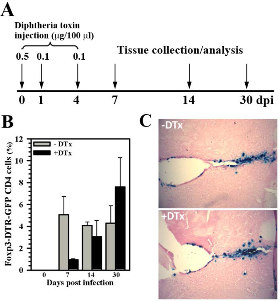
Figure 2. DTx treatment depletes Tregs from the brain but still allows establishment of viral infection.
(A) Foxp3-DTR-GFP transgenic mice were injected with diphtheria toxin (DTx) at 0 (0.5 µg), 1 (0.1 µg), and 4 (0.1 µg) dpi to deplete Treg cells during the acute phase of viral brain infection. (B) To quantify Treg depletion from the brain, leukocytes were isolated from untreated (−DTx) as well as DTx-treated (+DTx), MCMV-infected mice and stained for flow cytometry to detect Foxp3-GFP expression within the CD4+ subpopulation of brain-infiltrating T lymphocytes (CD45+CD11blow). Pooled data are presented as mean ± SEM of two separate experiments using six animals per treatment group/time point. (C) Establishment of viral brain infection with or without DTx treatment was confirmed using X-gal (blue) staining to detect β-gal expression from the recombinant MCMV viral genome at 4 dpi.

