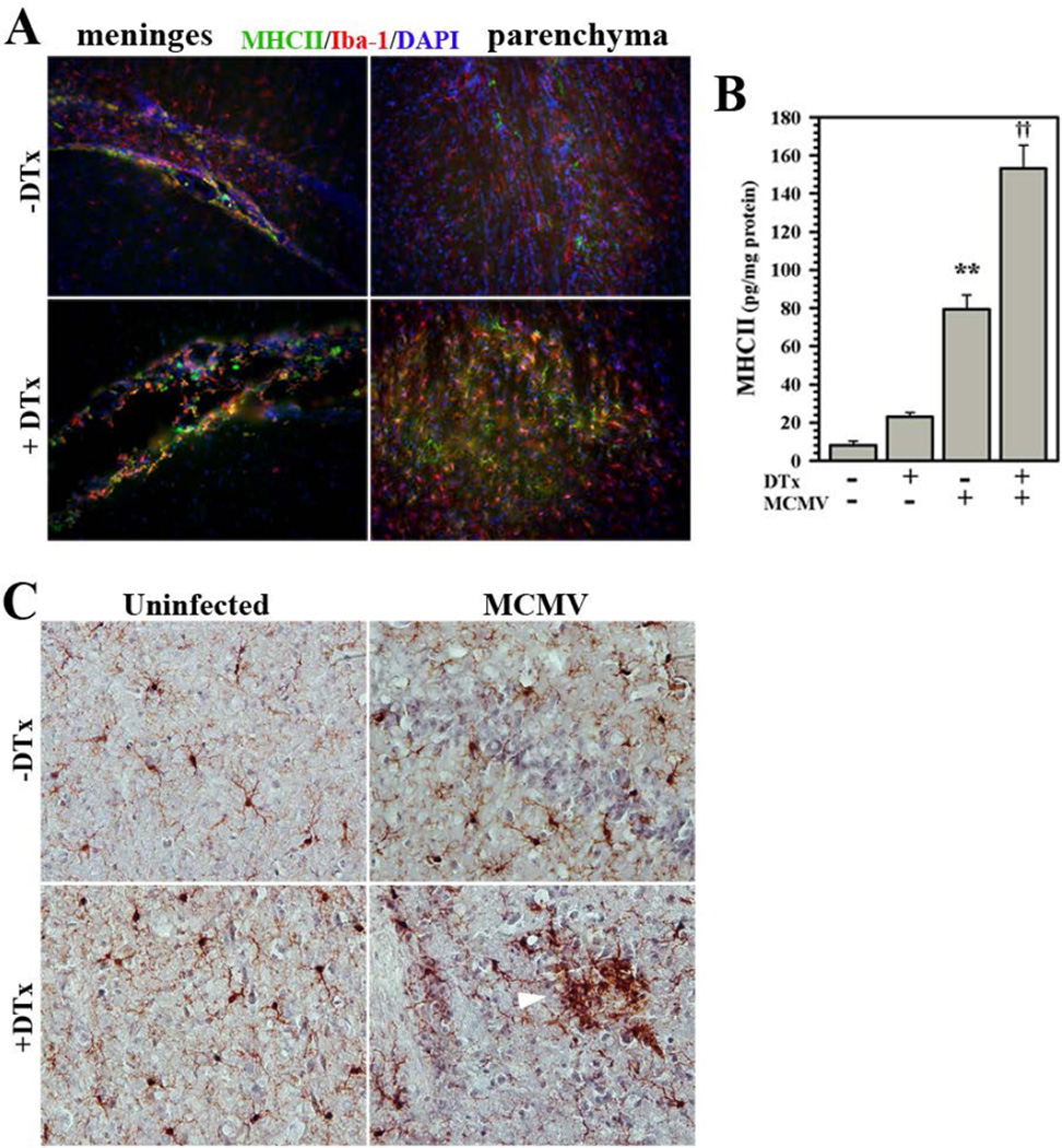
Figure 5. Immunohistochemical staining of chronic reactive microglia.
Microglia chronically express MHC Class II and PD-L1 following MCMV brain infection. (A) Infected Foxp3-DTR-GFP transgenic mice with (+DTx) and without (−DTx) DTx treatment were perfused and brains were cryosectioned for immunohistochemistry. Co-labeling of MHC II (green), as an indicator of cell reactivity, and the microglial cell marker Iba-1 (red) was observed in the meninges and brain parenchyma at 40 dpi. (B) Elevated MHC Class II protein levels in chronically-infected brains with or without Treg depletion (±DTx) were quantified using ELISA. Pooled data presented are derived from three to nine animals/group at 40 dpi. (C) Brain sections from infected animals ±DTx treatment stained for Iba-1 at 24 dpi displayed microglial nodules with reactive morphology (white arrow). **P ≤ 0.01 vs. uninfected. ††P ≤ 0.01 vs. MCMV.

