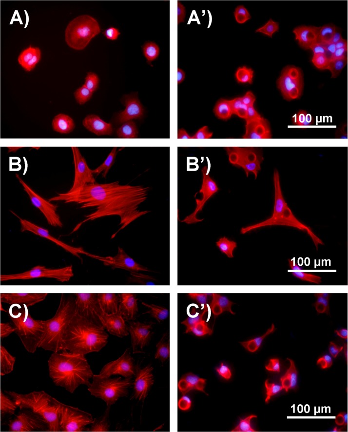
FIG. 3.
Comparison of cell morphology and cytoskeleton after 24-h culture on the flat and micropillar substrates. (a) Fluorescence image of HaCaT cells cultured on the flat substrate. (a') Fluorescence image of HaCaT cells cultured on the d18s18-μm micropillar substrate. (b) Fluorescence image of ESF-1 cells cultured on the flat substrate. (b') Fluorescence image of ESF-1 cells cultured on the d18s18-μm micropillar substrate. (c) Fluorescence image of HUVEC cells cultured on the flat substrate. (c') Fluorescence image of HUVEC cells cultured on the d18s18-μm micropillar substrate. Cells were stained for actin filament (red) and nuclei (blue).

