Abstract
Background
Airborne fiber size has been shown to be an important factor relative to adverse lung effects of asbestos and suggested in animal studies of carbon nanotubes and nanofibers (CNT/CNF).
Materials and Methods
The International Standards Organization (ISO) transmission electron microscopy (TEM) method for asbestos was modified to increase the statistical precision of fiber size determinations, improve efficiency, and reduce analysis costs. Comparisons of the fiber size distributions and exposure indices by laboratory and counting method were performed.
Results
No significant differences in size distributions by the ISO and modified ISO methods were observed. Small but statistically-significant inter-lab differences in the proportion of fibers in some size bins were found, but these differences had little impact on the summary exposure indices. The modified ISO method produced slightly more precise estimates of the long fiber fraction (>15 μm).
Conclusions
The modified ISO method may be useful for estimating size-specific structure exposures, including CNT/CNF, for risk assessment research.
Keywords: asbestos, carbon nanotubes, carbon nanofibers, transmission electron microscopy (TEM), exposure estimation
Introduction
Epidemiological evidence has established a causal relationship between exposure to asbestos and various adverse health outcomes, including asbestosis, lung cancer, and mesothelioma. These demonstrated adverse health effects of asbestos have focused attention on the potential occupational health hazards of other elongated particles which have dimensions similar to asbestos fibers, such as synthetic vitreous fibers [NIOSH, 2011] and engineered carbon nanotubes (CNT) and carbon nanofibers (CNF) [Donaldson et al., 2006; Schulte et al., 2012; NIOSH, 2013].
CNT are allotropes of carbon with a fibrous morphology and are classified as single-walled (SWCNT) or multi-walled (MWCNT). SWCNT are typically 0.4–3 nm in diameter and are composed of a single cylindrical sheet of graphene whereas MWCNT range from 2 to 200 nm in diameter and consist of several concentric, coaxial rolled up graphene sheets [Murray et. al, 2012]. CNF are composed of stacked graphene with diameters ranging from 70 to 200 nm with lengths ranging from 10 to 100 μm [Murray et. al, 2012]. Of the various forms of asbestos, chrysotile may be most similar in dimensions to CNT and CNF, as the vast proportion of textile workplace chrysotile structures were <250 nm (<0.25 μm) in diameter [Dement et al., 2008; and fig. 1 of Stayner et al., 2008]. Additionally, both chrysotile and CNT/CNF have curved and twisted fibers and form complex clusters. Most airborne chrysotile structures in this study were <5 μm in length, although some structures were up to 40 mm or more in length [fig.1 of Stayner et al., 2008].
The toxicity and adverse health effects of asbestos, CNT/CNF, and other elongated particles and fibers is likely a complex function of particle physical, chemical, and surface properties. The epidemiology and toxicology data for asbestos suggests that fiber dimensional characteristics as well as surface area are important determinants of toxic effects. Biologically-based exposure indices have been proposed based on fiber length and width size fractions, although the specific length and diameter cut-offs vary among these proposed indices [Pott et al., 1974; Stanton et al., 1981; Lippmann, 1988; Berman et al., 1995; Quinn et al., 2000]. In a review of potential health effects of asbestos fibers of various lengths, Dodson et al. [2003] concluded that asbestos fibers of all lengths induce pathological responses. Recent epidemiology studies of US chrysotile textile workers also found that cumulative exposure to airborne fibers of all sizes, including those shorter than 5 μm were significantly associated with asbestosis or lung cancer mortality. The strongest associations were observed with fibers less than 0.25 μm in diameter (for either asbestosis or lung cancer) and with fibers longer than 10 μm (for lung cancer) [Stayner et al., 2008; Loomis et al., 2010, 2012]. One limitation of these investigations was the high degree of correlation between the size-specific cumulative exposure measures, which limited the ability to identify precise fiber dimensions predicting asbestos-related lung disease. This was due to the high overlapping size distributions of the airborne structures sampled in different jobs in the facility; although some jobs had higher proportions of certain fiber dimensions, the distributions were generally wide and included some proportion of most fiber sizes.
Despite these fiber-specific effects, the current risk assessments for occupational asbestos exposures are based on measures of airborne fiber concentrations by phase contrast microscopy (PCM) which quantifies fibers >5 μm in length and >0.25 μm in width [OSHA, 1994; Stayner et al., 1997]. Challenges in developing transmission electron microscopy (TEM)-based risk estimates include the absence of a statistically independent TEM-based fiber exposure metric due to difficulties correlating among the various size-specific cumulative exposuremeasures [Stayner et al., 2008]. While PCM fiber exposures integrated over a working lifetime are predictive of lung cancer and asbestosis risk, exposure-response relationships using this exposure metric vary greatly by industry [Dement and Wallingford, 1990; Hodgson and Darnton, 2000]. This may be due to problems inherent with the PCM method as well as the exposure metric (i.e., only fibers >5 μm are counted). The PCM has a limit of resolution of approximately 0.2–0.3 μm which means that thin airborne asbestos particles (i.e., less than approximately 0.25 μm in diameter) will not be included in the PCM fiber count (even though many of these thinner fibers are longer than 5 μm). Although the thinnest structures (<0.25 μm) comprise the highest proportion of past workplace airborne chrysotile exposures [Dement et al., 2008] and are the most predictive of asbestosis and lung cancer mortality in US textile workers [Stayner et al., 2008; Loomis et al., 2012], these structures are not currently included in the occupational exposure limits for chrysotile and other asbestos minerals (based on PCM methods) [NIOSH, 2011]. In addition, PCM would not be a reasonable technique to use to estimate airborne exposures to CNT/CNF as PCM does not detect structures less than approximately 250 nm in diameter. Electron microscopy methods (including TEM and scanning electron microscopy, SEM) have not yet been established for characterizing these nanomaterials.
Several recent epidemiological studies have investigated the role of fiber dimensions on mortality from lung cancer or asbestosis after occupational exposure to chrysotile among US textile workers [Dement et al., 2008, 2011; Stayner et al., 2008; Loomis et al., 2012]. These studies used TEM analysis of archived membrane filter samples to obtain bivariate (diameter and length) airborne fiber size data [Dement et al., 2008]. The bivariate fiber size distributions were applied to historical estimates of exposure by PCM to develop a fiber size-specific job-exposure matrix (JEM) [Dement et al., 2008, 2009, 2011] for use in the exposure-response analyses [Stayner et al., 2008; Loomis et al., 2010, 2012]. The TEM methods applied to obtain those estimated bivariate fiber size distributions were evaluated in an inter-lab and inter-method comparison of fiber counts within bivariate size bins in the current study. A sensitivity analysis was performed to evaluate the influence of laboratory or method-specific differences in the fiber size distribution estimates and the various fiber size-specific exposure indices derived from PCM concentrations. Those methods and analyses are reported in this paper.
This paper also discusses characteristics of CNT/CNF and the potential application of the modified ISO TEM method [Kuempel et al., 2004] in the measurement and characterization of airborne CNT /CNF exposures. Specific modifications to the International Organization for Standardization (ISO) (1995) direct-transfer method for asbestos were made to increase the statistical precision of the bivariate (length and width) size determinations and to improve the efficiency and reduce costs of the analysis. The modified ISO TEM methoddescribed in this paper is designed for use in research studies in exposure estimation and epidemiology, and is not designed for routine workplace and environmental exposure monitoring. However, this modified method may be useful for the refinement or further development of standard TEM methods for asbestos, as supported bythe statistical evaluation of the methods described in this paper. Additionally, the modified method may be relevant to the development of standard methods for counting and sizing other airborne fibrous materials such as CNT/CNF for research studies and may provide the basis for development of routine monitoring methods.
Materials and Methods
TEM Methods
All of the recent TEM analyses for the recent US chrysotile textile workerstudies [Dement et al., 2008, 2009, 2011; Stayner et al., 2008; Loomis et al., 2010, 2012] were conducted in a single laboratory using a modified TEM fiber counting and sizing protocol based on the ISO, Direct-Transfer Method (ISO 10312, 1995-01-01) [ISO, 1995; Kuempel et al., 2004] The primary objective of those TEM analyses was to estimate bivariate size distributions by plant and operation [Dement et al., 2008, 2009, 2011]; therefore, modifications of the ISO methods were needed to increase the statistical precision of the bivariate size determinations. The complete modified ISO TEM protocol is available in the online supplemental materials and the following is a summary of the method modifications:
A minimum aspect ratio of 3:1 was used to define fibers and structures for consistency with PCM methods. ISO allows use of either 3:1 or 5:1.
In order to accommodate sizing of the large number of fibers needed, diameter and length were recorded into discrete interval categories (rather than precise measurements of each fiber dimension) to increase the efficiency and reduce the cost of the analyses.
The ISO stopping rule for dispersed matrices and dispersed clusters was not used and all visible fibers and fiber bundles within these structures were enumerated and sized. This enhancement allowed better resolution of the true diameter/length distribution. Even with this modification, complete enumeration of fibers and fiber bundles within these complex structures is sometimes impossible.
- The ISO method employs stopping rules of 100 primary structures in the all-sizes count and 100 structures in the PCM-equivalent count (>5 μm in length, >0.25 μm in diameter). In order to increase the count of the less-prevalent longer fibers (>15 μm) and thereby achieve greater statistical precision of the bivariate size distributions, three separate analyses were performed on each sample based on fiber length and without limitation based on fiber diameter. These analyses consisted of counting all structures >0.5 μm, >5 μm, and > 15 μm. The stopping rules (i.e., minimum number of primary structures to be sized and recorded) used were:
- All structures: 50
- Structures longer than 5 μm: 80
- Structures longer than 15 μm: 50
The primary laboratory conducting the TEM analyses implemented quality control procedures that included replicate sample analyses. The current study was undertaken to further evaluate the modification of the ISO protocol with the following objectives:
To evaluate the intra-laboratory variability in the bivariate diameter/length distributions using the modified ISO TEM protocol.
To evaluate the inter-laboratory variability in the bivariate diameter/length distributions using the modified ISO TEM protocol in two different laboratories.
To evaluate the inter-method variability in the bivariate diameter/length distributions using the modified ISO TEM and the standard ISO 10312 protocols within the same laboratory.
To evaluate the sensitivity of the TEM-based fiber-size specific exposure estimates to variability in the estimates of the bivariate fiber size distributions due to inter-laboratory and inter-method sources.
Sample Selection
Membrane filter samples used for these analyses were a subset of the 86 filters used to develop the fiber size-specific JEM for an epidemiological study of US asbestos textile workers [Dement et al., 2008; Stayner et al., 2008]. Resources did not allow replicate analyses of all 86 samples by TEM in a second laboratory; therefore, a 10% stratified random sample of the membrane filters was selected. Samples were selected from textile exposure zones (departments) shown to have differences in fiber length, that is, fiber preparation (Zone 1) and light weaving (Zone 9) [Dement et al., 2008]. Three samples which were heavily-loaded/overloaded or did not have complete TEM analysis were eliminated a priori, leaving four samples from Zone 1 and five samples from Zone 9 for study. A random order was established for analyses by a second laboratory.
Sample Analyses
The TEM grids for the nine selected samples were exchanged between the primary laboratory and the second laboratory; however, instability of the TEM preparations necessitated new TEM grid preparations by the second laboratory for all samples. The second laboratory conducted TEM analyses of each sample using the modification of ISO method 10312, as described above. In order to provide a comparison with results using the unmodified ISO method, the second laboratory also analyzed all samples by ISO 10312 using the ISO optional counting procedure for structures longer than 5 μm with enumeration of all fiber diameters. All data were recorded in Excel files using the data field format developed for the modified ISO TEM method.
Data Reduction and Statistical Analyses
The primary statistical analyses evaluated the degree of agreement between the two laboratories and the two TEM protocols with regard to the bivariate length/diameter distributions. Initial tests of overall agreement were followed by more detailed analyses by bivariate diameter/length category in order to explore any systematic differences.
Data reduction procedures developed for the TEM fiber size-specific JEM were used [Dement et al., 2008]. For each TEM counting stratum (all structures, structures >5 μm, and structures >15 μm) the samples within each exposure zone for each laboratory were combined based on pooling the fibers counted. The TEM fiber size data were reduced to a matrix containing 24 categories for fibers and fiber bundles for each combination of laboratory and exposure zone (fiber diameters [D] of <0.25, 0.25–1, 1–3, and >3 μm and fiber lengths [L] of <1.5, 1.5–3, 3–5, 5–15, 15–40, and >40 μm). Diameter/length combinations not satisfying a minimum aspect ratio of fiber length to diameter (L/D) of 3-to-1 were eliminated, resulting in 20 cells for all analyses.
Intra-Laboratory Evaluation of Modified ISO Protocol
Intra-laboratory agreement in bivariate diameter/length distributions obtained using the modified ISO protocol was first evaluated. For these analyses we selected all primary laboratory quality control samples that included fiber counts on the same filter for the all structure and the >5 μm strata. One repeated analysis was available for each quality control sample. The primary laboratory QC analyses for the >15 μm stratum used different filter preparations as the >15 μm stratum was added later in the protocol development; therefore, the >15 μm data were not used in this evaluation. The bivariate diameter/length data (given the total fiber count) within each counting stratum for each TEM sample was assumed to follow a multinomial distribution. In order to combine the counting strata to arrive at an overall distribution for statistical tests of agreement, the fiber counts for the all lengths stratum was used for counts of fibers <5 μm in length, which accounts for the vast majority of fibers counted in this stratum, and the >5 μm stratum was used to estimate the multinomial distribution for all fibers 5 μm in length or longer. A likelihood ratio chi-square statistic for comparing the data from each quality control sample to the corresponding original sample was computed. These likelihood ratio chi-square values were then summed across all five samples with 90 degrees of freedom as each sample contributed 7+11 = 18 degrees of freedom to the sum.
Inter-Laboratory Evaluation of Modified ISO Protocol
Overall agreement in the bivariate diameter/length fiber size distributions between the two laboratories using the modified ISO TEM protocol was evaluated by summing the likelihood ratio chi-squares by laboratory within each counting stratum and each exposure zone thus allowing for exposure zone heterogeneity. For each evaluation the data were assumed to have been sampled from a multinomial distribution within each stratum and zone and the likelihood ratio chi-squares were summed over the strata and zones. The analysis for the all lengths stratum was limited to fibers <5 μm in length, as previously described. Similarly, the counts based on the lengths >5 μm were limited to lengths <15 μm and the sum was compared to a chi-squared distribution with 34 degrees of freedom.
Inter-Method Comparison (Within and Between Laboratories)
A similar analysis was conducted to compare the modified ISO TEM protocol to the ISO 10312 protocol within the same laboratory. Although the ISO protocol includes only two strata (all lengths and >5 μm) this comparison of the two methods was based on likelihood ratio chi-square values which partitioned the ISO >5 μm stratum into two strata (5-15 μm, and > 15 μm) in order to match the modified ISO TEM method. Again, the chi-square values were summed across the strata and zones and compared to a chi-squared distribution with 33 degrees of freedom, where one degree of freedom was subtracted due to a lack of fiber counts by either method from the bivariate interval [1.0<D<3.0 μm] × [3.0<L<5.0 μm].
For both the inter-laboratory and inter-method comparisons, likelihood ratio chi-square values for each combination of fiber diameter and length were calculated and tabulated to allow closer scrutiny of fiber size categories contributing to the overall evaluation of laboratories and methods.
Evaluation of Bivariate Fiber Size Distributions and Variability (by Method and Laboratory)
In addition to tests of overall agreement by laboratory and TEM method, we conducted additional analyses to investigate any systematic differences within bivariate diameter/length categories. Within each TEM counting stratum (all structures, structures >5 μm, and structures >15 μm), the fiber counts were combined and the proportional distribution by diameter/length category calculated directly as a simple proportion. Bivariate fiber size distributions were developed for each combination of plant exposure zone, laboratory, and TEM method by weighting the proportions associated with the TEM counting strata by their conditional probabilities, and bootstrapping was used to estimate statistical variability for the proportion of airborne fibers in each diameter/length category, as previously described [Dement et al., 2008]. The distribution of estimates for cell proportions from a minimum of 4,000 replications was used to construct approximate 95% confidence intervals using the bootstrap percentile method [Mooney and Duval, 1993]. Using a similar nonparametric bootstrapping approach, we calculated inter-laboratory and inter-method differences in the mean cell proportions by cell in the diameter/length matrices as well as approximate 95% confidence intervals for these differences.
Sensitivity Analyses in Estimation of Previously Proposed Exposure Indices
The bivariate fiber size distributions determined using the modified ISO TEM protocol were applied to historical estimates of exposure by PCM to develop a fiber size-specific JEM [Dement et al., 2008, 2009, 2011] for the exposure- response analyses [Stayner et al., 2008; Loomis et al., 2010, 2012]. Worker exposures (f/cc-years) to any desired airborne fiber size fraction for a given time period, exposure zone, and job were derived using the following expression:
TEM Size-Specific Exposure (f/cc) = (PCM) × [(F)/(PCM)] where PCM is the phase contrast fiber concentration for given time period, exposure zone, and job [Dement et al., 1980, 1983], F is the fraction of all TEM fibers in the exposure zone fitting the definition for the exposure metric based on TEM fiber size data, and FPCM is the fraction of all airborne fibers measured by TEM which would be counted by PCM (>0.25 μm in diameter and >5.0 μm in length) for the exposure zone.
The ratio (F/FPCM) is the PCM to TEM factor for a given cell in the bivariate diameter/length distribution that, when multiplied by the PCM concentration, results in fiber size-specific exposure estimates. Differences in the TEM bivariate size distributions by method and laboratory impacts both parameters (F and FPCM) used to convert PCM to TEM size-specific exposures.
The final analysis undertaken was an evaluation of the sensitivity of several previously proposed TEM-based fiber-size specific exposure indices to variability in the estimates of the bivariate fiber size distributions (and parameters F and FPCM) due to inter-laboratory and inter-method sources. The exposure indices were selected from those used by Stayner et al. [2008] and are summarized in Table I. These three exposure indices serve the purpose of the current paper, which is to evaluate the utility of TEM methods to estimate airborne fiber size distributions for use in size-specific exposure estimation. The primary objective of the current study was to compare TEM methods; therefore, fiber length categories incorporated into the modified ISO method for the three counting strata were used for statistical analyses. Some of the exposure indices used fiber size cuts slightly different from those available in the current data; therefore, these indices were approximated by selecting the closest size category, as shown in Table I. Two additional indices were considered but not included in these analyses. The exposure index suggested by Berman [Berman et al., 1995] specified regression-model derived weights for fibers with diameters <0.25 μm in two length categories (5.0–40.0 μm and >40 μm). The proposed weight constantsaccount for relative disease potency derived from analyses of animal data. The Berman index also includes fibers >5.0 μm in diameter; however fibers >5.0 μm diameter have a very low probability of respiratory deposition [Timbrell, 1982; Baron, 1996] and were extremely rare in the current TEM data. The Berman index, when applied to the current data, thus reduces approximately to the Stanton index in the absence of the weights. Multiplication by these weights does not impact our evaluation of the ability of the TEM methods to estimate size fractions; therefore the Berman index was not included in the current analyses. We also considered the exposure index proposed by Quinn et al. [Quinn et al., 2000]; however, our approximation of the Stanton index is equal to the Quinn index minus one. For this reason, we did not include the Quinn index in this evaluation. In order to explore differences in exposure indices by plant operation, results for Zones 1 and 9 were summarized separately.
Table I. Fiber Exposure Indices Evaluated.
| Fiber exposure index | Fiber exposure index fiber dimensions | Fiber dimensions used for simulations |
|---|---|---|
| Stanton [1981] | Diameter <0.25 μm, Length >8.0 μm, Aspect Ratio≥3:1 | Diameter <0.25 μm, Length ≥5.0 μm, Aspect Ratio≥3:1 |
| Pott [1974] | Diameter <1.0 μm, Length >3.0 μm, Aspect Ratio≥5:1 | Diameter <1.0 μm, Length >3.0 μm, Aspect Ratio≥3:1a |
| Modified Lippman [1990]b | Diameter >0.15 μm, Length >10.0 μm, Aspect Ratio≥3:1 | Diameter >0.25 μm, Length ≥15.0 μm, Aspect Ratio≥3:1 |
An aspect ratio of 3-to-1 was used in the JEM. Approximately 25% of fibers had aspect ratios <5:1.
Lippmann index included fibers >10 μm; however,the closest category in the current data was 15.0 μm.
Using inter-laboratory and inter-method TEM bivariate size data, we calculated fiber concentrations for the exposure indices shown in Table I assuming an equivalent PCM exposure of 1.0 f/cc. For these analyses 4,000 bootstrap samples were generated in the same manner as previously described [Dement et al., 2008] to estimate statistical variability for the proportion of airborne fibers in each diameter/length category. Results of each of the 4,000 bootstrap samples were used to calculate each of the desired fiber exposure indices by laboratory and method which were summarized using univariate statistics (mean and approximate 95% confidence intervals using the bootstrap percentile method) and plotted to observe the degree of dispersion, overlap, and separation of the four indices by zone, laboratory, and method.
Results
Detailed summaries of the bivariate size data by zone, laboratory, and method are provided in Supplementary Appendix I.
Intra-Laboratory Comparison of the Modified ISO Method
Results of the overall statistical analysis of the primary laboratory quality control samples failed to provide evidence of statistically significant differences in the bivariate diameter/length distributions between the original analyses and the quality control analyses (χ2 =45.15, df = 90). However, the primary laboratory quality control analyses used the same grid openings for the original and the recounts; therefore, statistical independence cannot be assumed and its violation may have contributed to the observation of what would otherwise be a surprisingly small value of the χ2 statistic and the covariation of fiber counts within TEM grids was not considered. Although subject to substantial limitations this analysis suggests that the modified ISO method results in consistent fiber size distributions when counting the same TEM grids. No further analyses of these samples were undertaken.
Inter-Laboratory Comparison of the Modified ISO Method
The inter-laboratory comparison of the primary laboratory and the second laboratory using the modified ISO TEM method resulted in the overall likelihood ratio chi-square values as shown in Table II (Zones 1 and 9 combined). All comparisons were based on nine samples and counts of over 100 structures by each laboratory. This overall evaluation found statistically significant differences in the bivariate diameter/length distributions between the two laboratories analyses using the modified ISO TEM method. Table I indicates that these statistical differences are largely driven by differences in the relative distribution of fibers in the <5 μm stratum, which also contains the largest number of fibers enumerated. This conclusion is supported by the detailed likelihood ratio chi-square values by cell shown in Supplementary Appendix II which shows that the overall likelihood ratio chi-square for the <5 μm stratum is largely driven by a few cells with high values.
Table II. Overall Inter-Laboratory Comparison Usingthe Modified ISO TEM Method.
| Zone | Stratum | Likelihood ratio χ2 | DF | LR P-value |
|---|---|---|---|---|
| 1 | <5 μm | 28.6325 | 7 | 0.0002 |
| 1 | 5–14 μm | 6.6353 | 3 | 0.0845 |
| 1 | ≥15 μm | 4.4928 | 7 | 0.7216 |
| 9 | <5 μm | 11.3307 | 7 | 0.1248 |
| 9 | 5–14 μm | 6.0999 | 3 | 0.1069 |
| 9 | ≥15 μm | 9.0586 | 7 | 0.2484 |
| Overall LR χ2 | 66.2498 | 34 | 0.0008 |
Results of the detailed analyses of the bivariate diameter/length distributions, comparing data between laboratories by zone are presented in Supplementary Appendix III. These tables compare distributions by zone and diameter/length cells which were found to be statistically different based on the bootstrap analyses are indicated. No significant differences were observed for Zone 9 and for Zone 1 only six of the 20 cells found significant differences by laboratory. The actual differences in the values for many of the statistically different size categories are small and the overall pattern suggests that the second laboratory estimates of the proportion of fibers longer than 3 μm are less than those obtained by the primary laboratory for fibers <1.0 μm in diameter.
Inter-Method Comparison (Modified ISO and ISO TEM Protocols)
For comparison of TEM protocols, data from the second laboratory on the same nine samples using the modified ISO 10312 method and unmodified ISO method for all size fibers were used (the primary laboratory used the modified protocol only). The inter-method comparison resulted in the overall evaluation (Zones 1 and 9 combined) based on likelihood ratio chi-square values as shown in Table III. The overall bivariate diameter/length distributions were not statistically different when comparing modified ISO and ISO methods, although the <5 μm was significant in Zone 9. Appendix II of the supplemental materials shows the detailed likelihood ratio chi-square values by fiber diameter and length category.
Table III. Overall Comparison of the Modified ISO and ISO TEM Methods Second Laboratory Lab Data.
| Zone | Stratum | Likelihood Ratio (χ2) | DF | LR P-value |
|---|---|---|---|---|
| 1 | <5 μm | 10.2788 | 7 | 0.1733 |
| 1 | 5–14 μm | 1.5754 | 3 | 0.6636 |
| 1 | ≥15 μm | 4.8742 | 7 | 0.6753 |
| 9 | <5 μm | 13.6676 | 6 | 0.0336 |
| 9 | 5–14 μm | 2.1396 | 3 | 0.5439 |
| 9 | ≥15 μm | 4.8146 | 7 | 0.6826 |
| Overall LR χ2 | 37.3502 | 33 | 0.2759 |
Note: The ISO >5 μm stratum was partitioned into 5–14 μm and a ≥15 μm stratum for these analyses.
More detailed analyses of inter-method differences in fiber size distributions by exposure zone are presented in Supplementary Appendix IV. For Zone 1 only two cells with significant differences were observed and only one cell was found to be significantly different for Zone 9. Overall, results for individual cells in the bivariate diameter/length matrix were found to be remarkably similar by the two methods and a systematic pattern of differences in estimated cell proportions was not apparent.
Sensitivity Analyses for Estimating Fiber Exposure Indices
Univariate summary statistics for the fiber exposure indices evaluated in the sensitivity analyses are presented in Tables IV and V. As expected, these data show size-specific concentrations to vary by exposure zone for the same exposure index (due to the difference in the zones’ fiber length distributions [Dement et al., 2008]). The inter-laboratory data by the modified ISO method found the 95% confidence intervals for each exposure index to overlap within each exposure zone (Table IV). Table V also shows that the inter-method data using the modified ISO and ISO protocols in the second laboratory produced overlapping 95% confidence intervals for each exposure index within each zone. These results suggest that the modified ISO method produces mean exposure indices in reasonable agreement with those produced by the ISO method with good reproducibility between laboratories.
Table IV. Modified ISO TEM Size-Specific Exposures Using Primary and Second Laboratory Data: Estimated TEM (f/cc) Exposure for a Measured PCM Exposure of 1.0 f/cc*.
| Fiber Exp. Index | Approximate fiber dimensions used for simulations | Zone 1 | Zone 9 | ||
|---|---|---|---|---|---|
|
|
|
||||
| Primary laboratory | Second laboratory | Primary laboratory | Second laboratory | ||
| Stanton | Dia. <0.25 μm | Mean: 0.88 | Mean: 1.15 | Mean: 1.87 | Mean: 1.47 |
| Length ≥5.0 μm | Range: 0.60–1.29 | Range: 0.83–1.78 | Range: 1.22–3.02 | Range: 0.97–2.50 | |
| Aspect Ratio≥3:1 | Std Dev: 0.088 | Std Dev: 0.110 | Std Dev: 0.216 | Std Dev: 0.171 | |
| 95% CI: 0.71–1.06 | 95% CI: 0.94–1.38 | 95% CI: 1.48–2.33 | 95% CI: 1.17–1.84 | ||
| Pott | Dia. <1.0 μm | Mean: 2.93 | Mean: 3.04 | Mean: 4.20 | Mean: 3.69 |
| Length >3.0 μm | Range: 2.18–4.07 | Range: 2.14–4.53 | Range: 2.82–6.78 | Range: 2.65–5.74 | |
| Aspect Ratio≥ 3:1 | Std Dev: 0.255 | Std Dev: 0.318 | Std Dev: 0.442 | Std Dev: 0.402 | |
| 95% CI: 2.48–3.48 | 95% CI: 2.48–3.75 | 95% CI: 3.42–5.18 | 95% CI: 2.99–4.55 | ||
| Modified Lippmann | Diameter>0.15 μm | Mean: 0.26 | Mean: 0.25 | Mean: 0.24 | Mean: 0.24 |
| Length >15. 0 μm | Range: 0.17–0.37 | Range: 0.16–0.36 | Range: 0.13–0.41 | Range: 0.12–0.44 | |
| Aspect Ratio≥3:1 | Std Dev: 0.030 | Std Dev: 0.029 | Std Dev: 0.037 | Std Dev: 0.037 | |
| 95% CI: 0.20–0.32 | 95% CI: 0.19–0.31 | 95% CI: 0.16–0.31 | 95% CI: 0.17–0.32 | ||
Predicted size-specific exposures for each exposure index based on a measured PCM concentration of 1.0 f/cc. Approximate 95% confidence intervals were calculated using the bootstrap percentile method.
Table V. Modified ISO and ISO TEM Size-Specific Exposures Indices Using Second Laboratory Data: Estimated TEM (f/cc) Exposure for a Measured PCM Exposure of 1.0 f/cc*.
| Fiber exp. Index | Approximate fiber dimensions used for simulations | Zone 1 | Zone 9 | ||
|---|---|---|---|---|---|
|
|
|
||||
| ISO method | Modified ISO method | ISO method | Modified ISO method | ||
| Stanton | Dia. <0.25 μm | Mean: 1.00 | Mean: 1.15 | Mean: 1.49 | Mean: 1.47 |
| Length ≥5.0 μm | Range: 0.71–1.33 | Range: 0.82–1.62 | Range: 1.10–2.01 | Range: 0.97–2.50 | |
| Aspect Ratio ≥3:1 | Std Dev: 0.084 | Std Dev: 0.110 | Std Dev: 0.139 | Std Dev: 0.171 | |
| 95% CI: 0.85–1.16 | 95% CI: 0.95–1.37 | 95% CI: 1.24–1.78 | 95% CI: 1.17–1.84 | ||
| Pott | Dia. <1.0 μm | Mean: 3.33 | Mean: 3.04 | Mean: 3.93 | Mean: 3.69 |
| Length >3.0 μm | Range: 2.43–5.12 | Range: 2.14–4.53 | Range: 2.99–5.23 | Range: 2.65–5.74 | |
| Aspect Ratio ≥3:1 | Std Dev: 0.333 | Std Dev: 0.318 | Std Dev: 0.322 | Std Dev: 0.402 | |
| 95% CI: 2.75–4.05 | 95% CI: 2.48–3.75 | 95% CI: 3.35–4.63 | 95% CI: 2.99–4.55 | ||
| Modified Lippmann | Diameter >0.15 μm | Mean: 0.23 | Mean: 0.25 | Mean: 0.30 | Mean: 0.24 |
| Length >15.0 μm | Range: 0.14–0.33 | Range: 0.16–0.36 | Range: 0.20–0.42 | Range: 0.12–0.44 | |
| Aspect Ratio ≥3:1 | Std Dev: 0.026 | Std Dev: 0.029 | Std Dev: 0.034 | Std Dev: 0.037 | |
| 95% CI: 0.18–0.28 | 95% CI: 0.19–0.31 | 95% CI: 0.23–0.37 | 95% CI: 0.17–0.32 | ||
Predicted size-specific exposures for each exposure index based on a measured PCM concentration of 1.0 f/cc. Approximate 95% confidence intervals were calculated using the bootstrap percentile method.
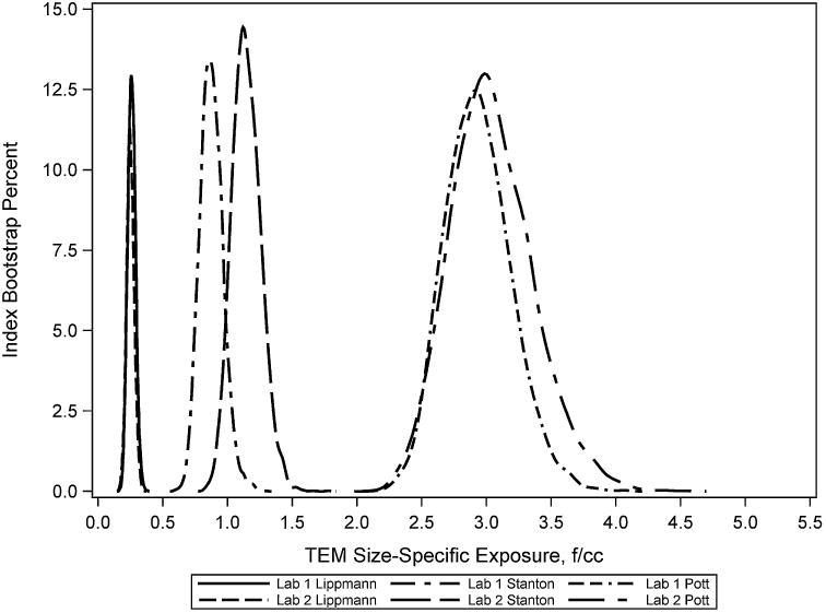
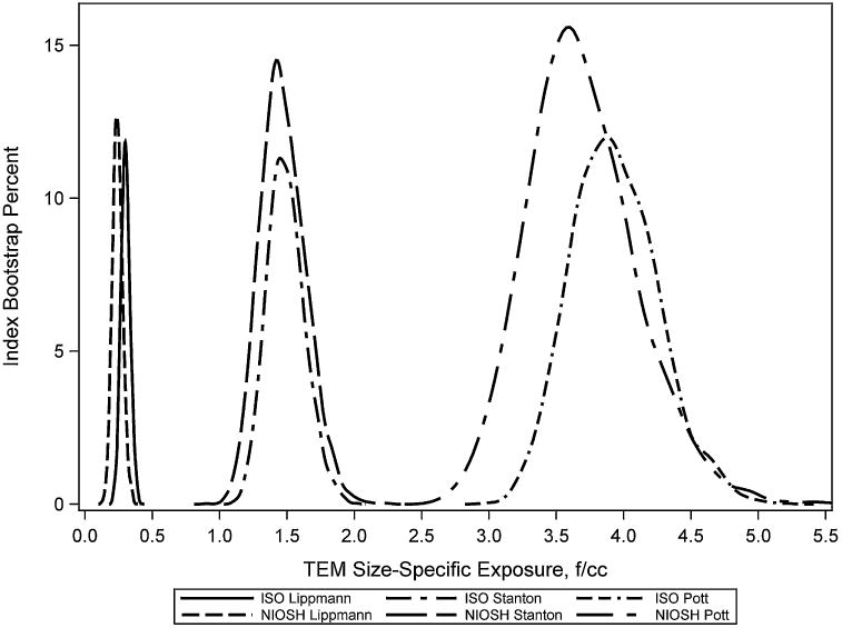
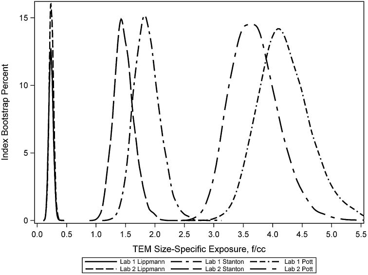
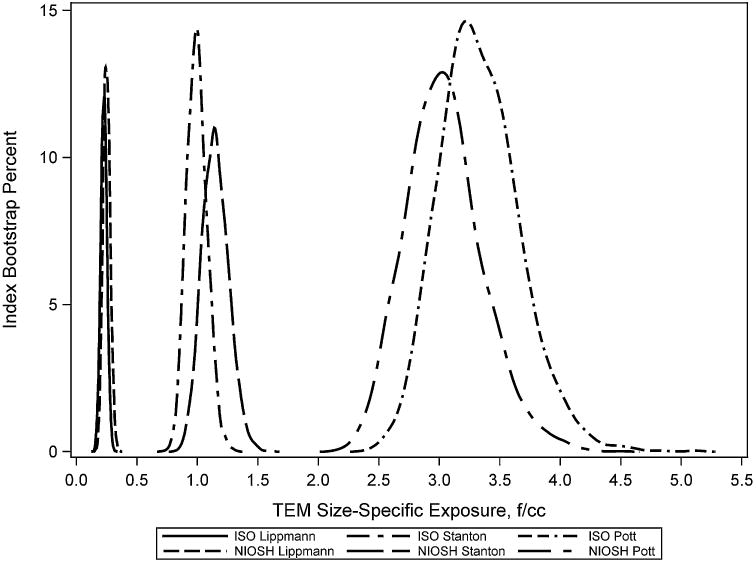
Distributions of size-specific concentrations for the fiber exposures indices are presented in Figures 1–4. Several observations are noteworthy. First, size-specific concentrations for the three indices were very different within each of the exposure zones, as expected, given the differences in the fiber size categories for each of the exposure indices (Table I). Secondly, use of the modified ISO method by the primary laboratory and second laboratory produced distributions of exposure indices that overlapped and were similar with regard to their shape. Lastly, use of either the modified ISOorthe ISO method within the same laboratory produced overlapping exposure indices with similar shapes and distributions. The 95% confidence intervals for the mean difference in size-specific concentrations from the bootstrap results (data not shown) found no statistically significance differences in the mean values for the four exposure indices (P > 0.05) for the inter-laboratory or the inter-method comparisons.
Figure 1.

Size-specific exposures for a PCM exposure of 1.0 f/cc—Zone 1 comparison by laboratory using modified ISO methods.
Figure 4.

Size-specific exposures for a PCM exposure of 1.0 f/cc—Laboratory 2, Zone 9 data by ISO and modified ISO methods.
Discussion
Quantitative comparisons of the fiber size distribution estimates by laboratory and method showed reasonable C While this study is based on a limited number of samples, the following findings are relevant to fiber-size specific exposure estimation using TEM methods:
An analysis of the primary laboratory quality control samples (for comparing initial and repeat analyses) suggested that the modified ISO method produces consistent results on the same TEM grids when used by an experienced analyst.
While the overall statistical test comparing inter-laboratory results using the modified ISO TEM method produced statistically significant results, most differences were relatively small in magnitude. These differences had only moderate effects on fiber exposure indices that combine cells of the bivariate diameter/length matrix and give greater weight to longer fibers. Additionally, some of the observed differences in results between the primary laboratory and the second laboratory using the modified ISO TEM method can be attributed to the need to prepare additional TEM grids for analyses by the second laboratory, thus adding an additional source of variability. The second laboratory had substantially less experience with the modified ISO protocol which may have had an effect.
The inter-method comparison of the modified ISO and ISO TEM protocols (within the laboratory that evaluated both methods) found no statistically significant differences in the bivariate diameter/length distributions. These results provide support for the ability of the modified ISO method to produce bivariate fiber size data comparable to the ISO method at reduced analytical costs.
The modified ISO and ISO TEM methods each produce bivariate size data useful in constructing reasonably consistent fiber size-specific exposure indices within the size fractions evaluated by both methods. Three indices proposed in the literature were investigated and found to vary considerably by textile exposure zone, which is as expected given the differences in the fiber size categories in these exposure indices (Table I). Standard deviations for the three summary exposure indices studied were generally slightly larger using the modified ISO method based on the inter-method data (Table V); however, the secondary laboratory had much less experience in using the modified ISO method.
The modified ISO TEM method, which is based on modifications to the ISO direct transfer method, produces results comparable to the ISO method while allowing more analytical resources to be focused on assessment of the long fiber fraction, thought to be more important for asbestos health risk assessments. The modified ISO method counts more long fibers and produces estimates of the fraction of very long fibers with slightly reduced variability compared to the ISO method (Supplementary Appendix I). These results suggest that the choice of either the modified ISO method or the ISO method with the supplemental count of fibers >5 μm appears to be largely governed by the required analytical time and associated costs. Fewer samples within a given exposure zone will result in sparse counts of the longest fibers by the ISO method, and this would favor use of the modified ISO TEM method to enhance the counts for longer fibers and increase statistical precision. An additional consideration for use of the modified ISO method, which places structures into predetermined diameter and length size bins, is the state of knowledge concerning critical size ranges relative to health effects. Use of the modified binning method is preferred when the approximate critical size ranges are reasonably well defined; however, the modified binning method may not be the most appropriate choice during initial exploratory analyses where maximum flexibility in defining critical size ranges during subsequent data analyses is the objective. In these instances, use of the modified ISO method with direct recording of individual structure diameter and length but retaining all other aspects of the modified ISO TEM method including the three counting strata and procedures for enumerating fibers in clusters and matrices would be preferred.
Evaluation of Electron Microscopy Methods for Airborne Exposure Estimation of CNT/CNF
Aerosols of CNF and CNT consist of mixtures of individual nano-sized particles as well as micron-sized agglomerates with complex structures and irregular shapes [Chen et al., 2012]. Murray et al. [2012] generated aerosols of SWCNT for inhalation studies and observed both dispersed SWCNT and well as agglomerates of SWCNT. Agglomerated structures were composed of SWCNT and bundled into “ropes” ranging from 65–150 nm and these authors estimated that a SWCNT bundle ∼65 nm in diameter contained ∼300 SWCNT. Chen et al. [2012] also noted the tendency for aerosolized MWCNT to form agglomerated structures and to combine into bundled rope configurations, creating significant difficulties for particle count-based exposure assessments. Chen et al. [2012] further described airborne MWCNT as containing particles of many different shape configurations including single smooth nanotubes with various aspect ratios, bundled nanotubes, nanotube nodules (fiber-like particles having nano-sized “nodules” attached), and fiber-like agglomerates having nanotubes and/or compact particles attached.
The role of the CNT/CNF morphology on health hazard potential is not well understood, but their high aspect ratio and ability to persist in the lung has resulted in findings of adverse pulmonary effects in experimental animal studies [NIOSH, 2013]. Acute and sub-chronic experimental animal studies with SWCNT, CNF, or asbestos have shown inflammation and early onset of pulmonary fibrosis [Shvedova et al., 2005, 2008; Kisin et al., 2011; Delorme et al., 2012; Murray et al., 2012]. Morphometric analysis has also indicated that well-dispersed SWCNT structures are more potent than agglomerated structures in causing pulmonary fibrosis [Mercer et al., 2008]. These findings, along with the epidemiological evidence of greater respiratory health hazard of the thinner and longer asbestos structures, provide the health basis for evaluating the feasibility of developing a more sensitive, structure-based particle count measurement method for CNT and CNF. Current occupational exposure limits for CNT/CNF are based on mass concentration, as evidenced by the NIOSH REL for respirable CNT/CNF of 1μg/m3 (8 hr time weighted average concentration) [NIOSH, 2013].
An additional motivation for developing count-based (vs. mass-based) sampling methods for CNT/CNF is the observation that even at the relatively low mass concentration at the NIOSH REL for CNT/CNF (1μg/m3) [Johnson et al., 2010; NIOSH, 2013], the number of airborne CNT and CNF structures by TEM can be relatively high in some workplaces (e.g., compared to an equivalent number concentration to the asbestos REL of 0.1 fiber/cc (structures >5 μm in length; 3:1 aspect ratio; measured byPCM) [Dahm et al., 2012; Schulte et al., 2012]. The NIOSH REL for CNT/CNF is set at the limit of quantification (LOQ) for the airborne sampling and analytical method for elemental carbon, and residual risks of early-stage adverse lung effects are estimated at working lifetime exposures to the REL [NIOSH, 2013].
While there is a need for count-based methods for CNT/CNF, PCM is not a sufficiently sensitive method for CNT/CNF due to its inability to resolve small diameter structures. TEM has the resolution needed to detect and enumerate CNT/CNF; however, use of TEM for routine exposure monitoring may be infeasible due to instrument availability and associated costs. Rather, TEM methods might be used in conjunction with CNT/CNF exposure assessments based on mass concentrations to characterize airborne aerosols by source and industrial process. Correlation of TEM size-specific concentrations with respirable mass concentrations might be used to generate TEM size-specific exposure measurements. It is unlikely that the same filters could be used for both determinations; however, parallel samples could be collected. However, the degree of correlation is likely to be low and highly varied by CNT/CNF source and process. This combined approach may be useful for research (e.g., in developing exposure estimates for an epidemiology study) but currently would be problematic as a routine sampling method for compliance.
While there is currently no widely accepted method for microscopic analyses of CNT/CNF, several studies have adapted modified versions of TEM-based NIOSH method 7402 for assessing CNT/CNF aerosols and worker exposures [Han et al., 2008; Chen et al., 2012; Dahm et al., 2012]. Electron microscope methods for these studies have included TEM, scanning transmission electron microscopy (STEM), and field emission scanning electron microscopy (FE-SEM). NIOSH method 7402 is primarily intended as a means of asbestos fiber identification to supplement PCM fiber concentration measurements (which does not provide fiber identification). The unmodified 7402 TEM method for asbestos specifies that structures with a diameter >0.25 μm that meet the definition of a PCM fiber (i.e., aspect ratio ≥3-to-1 and longer than 5 μm) be counted, with length and diameter recorded. The ratio of asbestos to non-asbestos fibers by TEM is then used to adjust PCM measurements for expression of airborne asbestos fiber concentrations.
Clearly, fiber counting rules incorporated in NIOSH method 7402 are inadequate for characterizing total asbestos or CNT/CNF aerosols as only fibers >0.25 μm in diameter >5 μm in length are enumerated. Additionally, the unmodified NIOSH method 7402 definitions of structures to be counted are inadequate for characterization of complex structures observed in both asbestos and CNT/CNF aerosols. Several CNT/CNF studies have modified NIOSH method 7402 particle counting rules to include all tubes and fiber structures with aspect ratios greater than 3-to-1 [Bello et al., 2008, 2009, 2010; Han et al., 2008; Lee et al., 2010]. However, these studies did not further define structures by morphology and did not attempt to enumerate fibers and fiber bundles contained within more complex structures.
Dahm et al. [2012] used a modified version of NIOSH method 7402 to count and characterize CNT structures on membrane filter samples collected from numerous workplaces air samples and reported concentrations as CNT structures per cubic centimeter. In that modified NIOSH 7402 method [Dahm et al., 2012], no minimum length or diameter cutoffs for structure counting were employed, and CNT/CNF structure morphology definitions followed the EPA Asbestos Hazard Emergency Response Act (AHERA) method [USEPA, 1987]. Primary structures are defined in the AHERA TEM protocol as fibers, bundles, clusters, and matrices, similar to the ISO TEM and modified ISO TEM methods [ISO, 1995; Kuempel et al., 2004]; however, the AHERA protocol does not require enumeration of fibers and fiber bundles contained within more complex structures as required by the ISO method.
Chen et al. [2012] used a modification of NIOSH TEM method 7402 [NIOSH, 1994] for quantitatively describing MWCNT aerosols using field emission scanning electron microscopy (FE-SEM) rather than TEM. Particle diameter in these MWCNT aerosols ranged from 15 to >500 nm and particle length ranged from 0.2 to 15 μm. The count median fiber diameter was estimatedto be 100.3 nm with a geometric standard deviation of 1.73 and the count median fiber length was 3.04 μm with a geometric standard deviation of 2.23. These authors devised a classification system which visually separated MWCNT particles into two categories, fiber-like particles and isometric particles. Particles having a shape with dimensions approximately the same in all directions were classified as isometric particles and particles with an aspect ratio of 3 or greater in two dimensions were classified as fibers. This approach allowed particles in each category to be differentiated, sized, and counted by electron microscopy using a range of magnifications (2,000 to 40,000×). During the counting process, the number of primary components (including individual nanotubes of various aspect ratios, nano-sized nodules, and/or micron-sized compact particles) in each complex particle structure was also measured when possible. Enumeration of the numbers of nanotubes was difficult for heavily agglomerated samples, so their numbers were estimated based on the cross-sectional area of the agglomerate and the average area of slightly agglomerated particles whose nanotubes could be counted.
Standardized TEM Method Development
The ASTM-International subcommittee D22.07 has initiated a work group to develop a TEM method for determining airborne CNT concentrations in ambient and indoor atmospheres [ASTM Work Item WK28561, 2011] based on their TEM method for counting asbestos in air [ASTM D6281–09, 2009] and specific identifying criteria for CNT [Millette et al., 2009]. The work group is discussing a number of questions including the minimum length for structure enumeration, the morphological categories, and what information about structure diameter and length should be recorded.
Clearly, much additional work is needed to further develop and evaluate TEM sizing and structure counting methods for evaluating CNT and CNF inhalation exposures. Differing levels of detail may be needed depending on the intended use of the data. For example, “routine” exposure monitoring of already well characterized CNT and CNF environments (where the primary objective is to determine structure concentrations), methods such as the draft ASTM or the modified NIOSH method 7402 (which record less detail concerning particle size as well as composition of complex structures) could be used. However, toxicology and epidemiological studies evaluating potential adverse health effects of carbon nanotubes and nanofibers require more detailed determinations of particle concentrations and airborne size characteristics.
The modification of the ISO method for asbestos described in this paper may be applicable to the characterization of CNT/CNF morphology and airborne size characteristics for research studies in exposure estimation, epidemiology, and toxicology. Primary advantages of the modified ISO TEM method include: 1) well defined morphological definitions and counting rules for primary structures, 2) enumeration of primary structures as well as components of complex structures, 3) measurements of both particle diameter and length, allowing determinations of size-specific exposures, and 4) stratified particle counting based on particle length, which increases statistical accuracy for less prevalent size categories. However, the heterogeneity of CNT/CNF structures may require additional modifications to the modified ISO TEM method described in this paper. For example, the modified method uses three strata of fiber length based typical asbestos size determinations (<5 μm, 5–15 μm, and >15 μm). These length categories may need modification to reflect the range of typical CNT and CNF lengths, which can often be <5 μm. The modified method also places fibers into pre-determined bins by length and diameter, and these may need additional modification for CNTs and CNFs. Alternatively, length and diameter of each recorded CNT and CNF could be recorded directly and bins for determination of size-specific exposures determined in the analysis phase. While recording of individual structure diameter and length would likely increase analytical time and costs, this approach would allow flexibility in defining size-specific exposures and may be the preferred approach until risk-based CNT/CNF fiber dimensions are more clearly defined.
One potential disadvantage of the modified ISO method is the inability to directly calculate aspect ratios for each recorded structure due to placement of structures into size bins. However, estimates of nominal aspect ratios for structures within each combination of fiber diameter and length are possible and should be sufficient to place structures into meaningful categories for epidemiological analyses if desired.
Enumeration of fiber and fiber bundles in complex structures poses a challenge for CNT/CNF. While this is also true for chrysotile asbestos, available data suggest that particle agglomeration is more prevalent with CNT/CNF, and the structures are perhaps more complex and densely agglomerated. The modified ISO TEM method does not attempt to enumerate component structures within compact clusters and matrices as the component structures are typically not visible. This also occurs with CNF/CNT. Chen et.al [2012] imputed the number of fiber and fiber bundles within these compact structures based on the average number of nanotubes observed in slightly agglomerated particles of similar size whose nanotubes could be counted. It should be noted that the samples analyzed by Chen et al. were laboratory-generated; therefore, the degree and complexity of the agglomerates may not be typical of some worker exposures. The studies by Dahm et al. and the associated electron micrographs of airborne structures suggest a more dense and complex state of agglomeration (compared to laboratory-generated samples) for some worker exposure scenarios [Dahm et al., 2012].
Interim Recommendations for TEM Analyses of CNT/CNF
The modified ISO TEM method for asbestos [Kuempel et al., 2004] may be a useful candidate method for quantitative exposure assessments for CNT/CNF, especially in research in support of toxicology or epidemiology studies. The following are comments and considerations for using the modified ISO TEM method for CNT/CNF.
The modified ISO TEM method incorporates well-established morphological structure classificationsthat include fibers, fiber bundles, clusters, and matrices. Clusters and matrices are further classified as compact or dispersed and the modified ISO method requires enumeration of all fibers and fiber bundles observed in dispersed clusters and matrices. The data recording worksheet developed for modified ISO method allows recording counts of primary and secondary structures in a manner that preserves relationships between primary and component structures. If needed, these data could be used to impute the numbers of fibers and fiber bundles within these compact structures using methods similar to those used by Chen et al. [2012]. The modified ISO TEM method thus provides a more complete characterization of CNT/CNF structures than current methods used for CNT/CNF.
The modified ISO TEM method described in this paper only enumerates structures longer than 0.5 μm (500 nm) in length. While this appears appropriate for asbestos, a shorter minimum length criterion may be needed for CNT/CNF. The draft ASTM TEM method specifies counting of any continuous grouping of particles in which a single nanotube with a length ≥0.25 μm (250 nm) is detected. However, other field and laboratory studies have counted all CNT/CNF structures with a 3-to-1 aspect ratio regardless of length [Bello et al., 2008, 2009, 2010; Han et al., 2008; Lee et al., 2010; Chen et al., 2012]. While Chen et al. [2012] observed only a small proportion of airborne MWCNT particles with lengths in the 200–400 nm range, counting all CNT/CNF with 3-to-1 aspect ratio currently is preferred. As further evaluation provides better definition of the fiber size characteristics of CNT/CNF and their risks, a practical lower limit may be adopted for routine exposure monitoring in situations know to contain CNT/CNF.
While the modified ISO TEM method places structures in pre-defined size bins, direct sizing of primary and secondary structures may be important for CNF/CNT at this early stage of exposure characterization research. This will allow maximum flexibility for determining size-specific exposures limited only by the measurement limitations of TEM. As risk-based fiber size characteristics are defined for CNT/CNF, these can incorporated into the method to reduce analysis time and costs. Most CNT/CNF clusters are in the micron size range and can be detected while scanning at 10,000–20,000×, however measurement of individual structures, especially their diameters, must be done at roughly twice that magnification. As there is wide size variation among different types of nanomaterials, studies should choose a magnification appropriate for their material.
Relatively few data are available to define the bivariate (length and diameter) distribution of CNF/CNT. The data published by Chen et.al [2012] suggest that the three counting strata (all structures, structures longer than 5 μm, and structures longer than 15 μm) incorporated into the modified ISO TEM method described in this paper may be useful for characterization of the spectrum of CNT/CNF fiber lengths. The modified ISO method allows more statistically precise and time efficient determination of the bivariate size distribution through stratified particle counting based on structure length.
Conclusions Concerning TEM Methods for CNT/CNF
The modified TEM methods described in this paper and in other recent studies may provide the basis for the development of a standardized, count-based method for exposure measurement of CNT/CNF. This method development could be facilitated, for example, through ongoing efforts by ASTM and NIOSH. Such standardized methods would also provide a reference for comparison of results and validation of newer microscopy techniques for nanomaterials.
Supplementary Material
Appendix I: Detailed Data Summaries by Zone, Lab, and Method
Bivariate Fiber Size Data: Primary laboratory Zone 1 Samples Using Modified ISO TEM Method
Bivariate Fiber Size Data: Second Laboratory Zone 1 Samples Using Modified ISO TEM Method
Bivariate Fiber Size Data: Second Laboratory Zone 1 Samples Using ISO Method Standard Stopping Rules for Component Structures
Bivariate Fiber Size Data: Primary Laboratory Zone 9 Samples Using Modified ISO TEM Method
Bivariate Fiber Size Data: Second Laboratory Zone 9 Samples Using Modified ISO TEM Method
Bivariate Fiber Size Data: Second Laboratory Zone 9 Samples Using ISO Method Standard Stopping Rules for Component Structures
Appendix II: Likelihood Ratio Chi-Square Tests Detailed Results
Zone 1 Laboratory Comparison Using Modified ISO TEM Method
Zone 9 Laboratory Comparison Using Modified ISO TEM Method
Zone 1 Method Comparison Using Second Laboratory Data
Zone 9 Method Comparison Using Second Laboratory Data
Appendix III: Bivariate Distributions Using the Modified ISO TEM Method
Results by Laboratory for Zone 1 Samples
Results by Laboratory for Zone 9 Samples
Appendix IV: Bivariate Distributions by Modified ISO and ISO TEM methods
Second Laboratory Results for Zone 1 Samples
Second Laboratory Results for Zone 9 Samples
Figure 2.

Size-specific exposures for a PCM exposure of 1.0 f/cc—Zone 9 comparison by laboratory using modified ISO methods.
Figure 3.

Size-specific exposures for a PCM exposure of 1.0 f/cc—Laboratory 2, Zone 1 data by ISO and modified ISO methods.
Acknowledgments
This work was supported by the National Institute for Occupational Safety and Health. We would like to thank the following individuals who were instrumental in the set-up and generation of the data analyzed in this study: Kenneth M. Wallingford, NIOSH, for providing access to and information about the historical data files and samples used in this study; James Bena, formerly at NIOSH, for performing data management and stratified random sampling of the filters for the TEM analysis; and Bernadette and Anthony Kolk, EMS Laboratories, for their excellent execution of both the NIOSH and ISO protocols.
Funding source: This work was supported by the National Institute for Occupational Safety and Health.
Footnotes
Supporting Information: Additional supporting information may be found in the online version of this article at the publisher's web-site.
Disclosure Statement: The authors report no conflicts of interests.
Publisher's Disclaimer: Disclaimer: The findings and conclusions in this article are those of the authors and do not necessarily represent the views of the National Institute for Occupational Safety and Health.
References
- ASTM D6281–09. Standard test method for airborne asbestos concentration in ambient and indoor atmospheres as determined by transmission electron microscopy direct transfer (TEM) Consho-hocken, PA: ASTM; 2009. [Google Scholar]
- ASTM Work Item WK28561. Standard test method for airborne carbon nanotube concentration in ambient and indoor atmospheres as determined by transmission electron microscopy direct transfer (TEM) West Conshohocken, PA: ASTM International; 2011. [Google Scholar]
- Bello D, Hart AJ, Ahn K, Hallock M, Yamamoto N, Garcia EJ, Ellenbecker EJ, Wardle BL. Particle exposure levels during CVD growth and subsequent handling of vertically aligned carbon nanotube films. Carbon. 2008;46:974–981. [Google Scholar]
- Bello D, Wardle BL, Yamamoto N, deVilloria RG, Garcia EJ, Hart AJ, Ahn K, Ellenbecker MJ, Hallock M. Exposure to nanoscale particles and fibers during machining of hybrid advanced composites containing carbon nanotubes. J Nanopart Res. 2009;11:231–949. [Google Scholar]
- Bello D, Wardle B, Zhang J, Yamamoto N, Santeufemio C, Hallock M, Virji MA. Characterization of exposures to nanoscale particles and fibers during drilling of hybrid advanced composites containing carbon nanotubes. Int J Occup Environ Health. 2010;16:434–450. doi: 10.1179/107735210799159996. [DOI] [PubMed] [Google Scholar]
- Berman DW, Crump KS, Chatfield EJ, Davis JMG, Jones A. The sizes, shapes, and mineralogy of asbestos structures that induce lung tumors or mesothelioma in AF/HAN rats following inhalation [correction appears in Risk Anal 1995;15(4):541] Risk Anal. 1995;15(2):181–195. doi: 10.1111/j.1539-6924.1995.tb00312.x. [DOI] [PubMed] [Google Scholar]
- Chen BT, Schwegler-Berry D, McKinney W, Stone S, Cumpston JL, Friend S, Porter DW, Castranova V, Frazer DG. Multi-walled carbon nanotubes: Sampling criteria and aerosol characterization. Inhal Toxicol. 2012;24(12):798–820. doi: 10.3109/08958378.2012.720741. [DOI] [PMC free article] [PubMed] [Google Scholar]
- Dahm DW, Evans DE, Schubauer-Berigan MK, Birch ME, Fernback JE. Occupational exposure assessment in carbon nanotube and nanofiber primary and secondary manufacturers. Ann Occup Hyg. 2012;56(5):542–556. doi: 10.1093/annhyg/mer110. [DOI] [PMC free article] [PubMed] [Google Scholar]
- Delorme MP, Muro Y, AraiT, Banas DA, Frame SR, Reed KL, Warheit DB. Ninety-day inhalation toxicity study with vapor grown carbon nanofibers in rats. Toxicol Sci. 2012;128:449–460. doi: 10.1093/toxsci/kfs172. [DOI] [PubMed] [Google Scholar]
- Dement JM, Brown DP, Okun A. Follow-up study of chrysotile asbestos textile workers: Cohort mortality and case-control analyses. Am J Ind Med. 1994;26(4):431–447. doi: 10.1002/ajim.4700260402. [DOI] [PubMed] [Google Scholar]
- Dement JM, Harris RL, Symons MJ, Shy CM. Exposures and mortality among chrysotile asbestos workers. Part I: Exposure estimates. Am J Ind Med. 1983;4:399–419. doi: 10.1002/ajim.4700040303. [DOI] [PubMed] [Google Scholar]
- Dement JM, Kuempel E, Zumwalde R, Smith R, Stayner L, Loomis D. Development of a fiber size-specific job-exposure matrix for airborne asbestos fibers. Occup Environ Med. 2008;65(9):605–612. doi: 10.1136/oem.2007.033712. [DOI] [PubMed] [Google Scholar]
- Dement JM, Loomis D, Richardson D, Wolf SH, Kuempel ED. Estimates of historical exposures by phase contrast and transmission electron microscopy for pooled exposure-response analyses of North Carolina and South Carolina USA asbestos textile cohorts. Occup Environ Med. 2011;68(8):593–598. doi: 10.1136/oem.2010.059972. [DOI] [PubMed] [Google Scholar]
- Dement JM, Loomis D, Richardson D, Wolf S, Myers D. Estimates of historical exposures by phase contrast microscopy and transmission electron microscopy in North Carolina USA asbestos textile plants. Occup Environ Med. 2009;66:574–583. doi: 10.1136/oem.2008.040410. [DOI] [PubMed] [Google Scholar]
- Dement JM, Wallingford KM. Comparison of phase contrast and electron microscopic methods for evaluation of occupational asbestos exposures. Applied Occ Env Hyg. 1990;5:242–247. [Google Scholar]
- Dodson RF, Atkinson MAL, Levin JL. Asbestos fiber length as related to potential pathogenicity: A critical review. Am J Ind Med. 2003;44:291–297. doi: 10.1002/ajim.10263. [DOI] [PubMed] [Google Scholar]
- Han JH, Lee EJ, Lee JH, So KP, Lee YH, Bae GN, Lee SB, Ji JH, Cho MH, Yu IJ. Monitoring multiwalled carbon nanotube exposure in carbon nano-tube research facility. Inhal Toxicol. 2008;20:741–749. doi: 10.1080/08958370801942238. [DOI] [PubMed] [Google Scholar]
- Hein MJ, Stayner LT, Lehman E, Dement JM. Follow-up study of chrysotile asbestos textile workers: Cohort mortality and exposure-responses. Occup Env Med. 2007 doi: 10.1136/oem.2006.031005. Published online April 20, 2007. [DOI] [PMC free article] [PubMed] [Google Scholar]
- Hodgson JT, Darnton A. The quantitative risks of mesothelioma and lung cancer in relation to asbestos exposure. Ann Occup Hyg. 2000;44(8):565–601. [PubMed] [Google Scholar]
- Johnson DR, Methner MM, Kennedy AJ, Steevens JA. Potential for occupational exposure to engineered carbon-based nanomaterialsin environmental laboratory studies. Environ Health Perspect. 2010;118(1):49–54. doi: 10.1289/ehp.0901076. [DOI] [PMC free article] [PubMed] [Google Scholar]
- Kisin ER, Murray AR, Sargent L, Lowry D, Chirila M, Siegrist KJ, Schwegler-Berry D, Leonard S, Castranova V, Fadeel B, et al. Genotoxicity of carbon nanofibers: Are they potentially more or less dangerous than carbon nanotubes or asbestos? Toxicol Appl Pharm. 2011;252:1–10. doi: 10.1016/j.taap.2011.02.001. [DOI] [PMC free article] [PubMed] [Google Scholar]
- Kuempel E, Dement J, Ristich A, Bena J, Zumwalde R. Protocol for tem analysis of chrysotile asbestos: adaptation of iso method for quality and cost-effective analysis of archival samples from charleston, south carolina textile plant. Cincinnati, Ohio: NIOSH; 2004. Available in the online supplemental materials. [Google Scholar]
- Lee JH, Lee SB, Bae GN, Jeon KS, Yoon JK, Ji JH, Sung JH, Lee BG, Yang JH, Kim JS, et al. Exposure assessment of carbon nanotube manufacturing workplaces. Inhal Toxicol. 2010;22:369–381. doi: 10.3109/08958370903367359. [DOI] [PubMed] [Google Scholar]
- Loomis D, Dement JM, Elliott L, Richardson D, Kuempel ED, Stayner L. Increased lung cancer mortality among chrysotile asbestos textile workers is more strongly associated with exposure to long thin fibres. Occup Environ Med. 2012;69:564–568. doi: 10.1136/oemed-2012-100676. [DOI] [PubMed] [Google Scholar]
- Loomis D, Dement J, Richardson D, Wolf S. Asbestos fiber dimensions and lung cancer mortality among workers exposed to chrysotile. Occup Environ Med. 2010;67:580–584. doi: 10.1136/oem.2009.050120. [DOI] [PMC free article] [PubMed] [Google Scholar]
- Mercer R, Scabilloni J, Wang L, Kisin E, Murray AD, Shvedova AA, Castranova AV. Alteration of deposition patterns and pulmonary response as aresult of improved dispersion of aspirated single-walled carbon nanotubes in a mouse model. Am J Physiol Lung Cell Mol Physiol. 2008;294(1):L87–L97. doi: 10.1152/ajplung.00186.2007. [DOI] [PubMed] [Google Scholar]
- Millette JR, Hill WB, Turner Hays. Analysis of carbon nanotubes in air. Microscope. 2009;57(3):127–132. [Google Scholar]
- Mooney CZ, Duval RD. Bootstrapping: A nonparametric approach to statistical inference. Newbury Park, CA: Sage; 1993. [Google Scholar]
- Murray AR, Kisin ER, Tkach AV, Yanamala N, Mercer R, Young SH, Fadeel B, Kagan VE, Shvedova AA. Factoring-in agglomeration of carbon nanotubes and nanofibers for better prediction of theirtoxicity versus asbestos. Part Fibre Toxicol. 2012;9:10–19. doi: 10.1186/1743-8977-9-10. [DOI] [PMC free article] [PubMed] [Google Scholar]
- National Institute for Occupational Safety and Health (NIOSH) NIOSH manual of analytical methods. 4th. Cincinnati, Ohio: Department of Health and Human Services, National Institute for Occupational Safety and Health; 1994. Asbestos by TEM Method 7402. [Google Scholar]
- National Institute for Occupational Safety and Health (NIOSH) Current intelligence bulletin 62, asbestos fibers and other elongate mineral particles: state of the science and roadmap for research. Cincinnati, OH: Department of Health and Human Services, Centers for Disease Control and Prevention, National Institute for Occupational Safety and Health, DHHS (NIOSH) Publication No. 2011–159; 2011. [Google Scholar]
- National Institute for Occupational Safety and Health (NIOSH) Current intelligence bulletin 65: occupational exposure to carbon nanotubes and nanofibers. Cincinnati, OH: US Department of Health and Human Services, Centers for Disease Control and Prevention, National Institute for Occupational Safety and Health. DHHS (NIOSH) Publication No. 2013–145; 2013. [Google Scholar]
- OSHA. Final rules, occupational exposure to asbestos, standard number: 1910.; 1915.; 1926.58. Federal Register. 1994;59:40964–41162. [Google Scholar]
- Pott F, Huth F, Friedrichs K. Tumorgenic effect of fibrous dust in experimental animals. Env Hlth Persp. 1974;9:313–315. doi: 10.1289/ehp.749313. [DOI] [PMC free article] [PubMed] [Google Scholar]
- Quinn MM, Smith TJ, Eisen EA, Wegman DH, Ellenbecker MJ. Implications of different fiber measures for epidemiologic studies of manmade vitreous fibers. Am J Ind Med. 2000;38(2):132–139. doi: 10.1002/1097-0274(200008)38:2<132::aid-ajim3>3.0.co;2-p. [DOI] [PubMed] [Google Scholar]
- Schulte PA, Kuempel ED, Zumwalde RD, Geraci CL, Schubauer-Berigan MK, Castranova V, Hodson L, Murashov V, Dahm MM, Ellenbecker M. Focused actions to protect carbon nanotube workers. Am J Ind Med. 2012;55(5):395–411. doi: 10.1002/ajim.22028. [DOI] [PubMed] [Google Scholar]
- Shvedova AA, Kisin ER, Mercer R, Murray AR, Johnson VG, Potapovich AI, Tyurina YY, Gorelik O, Arepalli S, Schwegler-Berry D, et al. Unusual inflammatory and fibrogenic pulmonary responses to single-walled carbon nanotubes in mice. Am J Physiol-Lung C. 2005;289:L698–L708. doi: 10.1152/ajplung.00084.2005. [DOI] [PubMed] [Google Scholar]
- Shvedova AA, Kisin E, Murray AR, Johnson VJ, Gorelik O, Arepalli S, Hubbs AF, Mercer RR, Keohavong P, Sussman N, et al. Inhalation vs. aspiration of single-walled carbon nanotubes in C57BL/6 mice: Inflammation, fibrosis, oxidative stress, mutagenesis. Am J Physiol-Lung C. 2008;295:L552–L565. doi: 10.1152/ajplung.90287.2008. [DOI] [PMC free article] [PubMed] [Google Scholar]
- Stanton M, Layard M, Tegeris A, Miller E, May M, Morgan E, Smith A. Relation of particle dimension to carcinogenicity in amphibole asbestos and other fibrous minerals. JNCI. 1981;67(5):965–975. [PubMed] [Google Scholar]
- Stayner LT, Kuempel E, Gilbert S, Hein M, Dement J. An epidemiologic study of the role of chrysotile asbestos fiber dimensions in determining respiratory disease risk among exposed workers. Occup Environ Med. 2008;65(9):613–619. doi: 10.1136/oem.2007.035584. [DOI] [PubMed] [Google Scholar]
- Stayner LT, Smith R, Bailer J, Gilbert S, Steenland K, Dement J, Brown D, Lemen R. Exposure-response analysis of respiratorydisease risk associated with occupational exposure to chrysotile asbestos. Occup Environ Med. 1997;54(9):646–652. doi: 10.1136/oem.54.9.646. [DOI] [PMC free article] [PubMed] [Google Scholar]
- Timbrell V. Deposition and retention of fibres in the human lung. Ann Occup Hyg. 1982;26:347–369. [PubMed] [Google Scholar]
- US Environmental Protection Agency (USEPA) 1987. Asbestos-containing materials in schools; final rule and notice. Federal Register, 40 CFR Part 763, Appendix A to Subpart E. 1987 Oct 30; [Google Scholar]
Associated Data
This section collects any data citations, data availability statements, or supplementary materials included in this article.
Supplementary Materials
Appendix I: Detailed Data Summaries by Zone, Lab, and Method
Bivariate Fiber Size Data: Primary laboratory Zone 1 Samples Using Modified ISO TEM Method
Bivariate Fiber Size Data: Second Laboratory Zone 1 Samples Using Modified ISO TEM Method
Bivariate Fiber Size Data: Second Laboratory Zone 1 Samples Using ISO Method Standard Stopping Rules for Component Structures
Bivariate Fiber Size Data: Primary Laboratory Zone 9 Samples Using Modified ISO TEM Method
Bivariate Fiber Size Data: Second Laboratory Zone 9 Samples Using Modified ISO TEM Method
Bivariate Fiber Size Data: Second Laboratory Zone 9 Samples Using ISO Method Standard Stopping Rules for Component Structures
Appendix II: Likelihood Ratio Chi-Square Tests Detailed Results
Zone 1 Laboratory Comparison Using Modified ISO TEM Method
Zone 9 Laboratory Comparison Using Modified ISO TEM Method
Zone 1 Method Comparison Using Second Laboratory Data
Zone 9 Method Comparison Using Second Laboratory Data
Appendix III: Bivariate Distributions Using the Modified ISO TEM Method
Results by Laboratory for Zone 1 Samples
Results by Laboratory for Zone 9 Samples
Appendix IV: Bivariate Distributions by Modified ISO and ISO TEM methods
Second Laboratory Results for Zone 1 Samples
Second Laboratory Results for Zone 9 Samples


