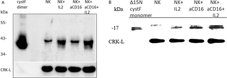
Figure 7. The levels of monomeric and dimeric cystatin F are increased following addition of anti-CD16mAb to the NK cells in the presence or absence of IL-2.

1 × 10⁶ NK cells/ml were treated with IL-2 (1000 units/ml), anti-CD16 antibody (3 μg/ml), or a combination of IL-2 (1000 units/ml) and anti-CD16 antibody (3 μg/ml) for 4 hours. Lysates from NK cells were separated by non-reducing A. and reducing B. SDS-PAGE and blots were probed with antibodies against cystatin F. Reactivity with intact dimeric cystatin F (A) and truncated Δ15 monomeric cystatin F (B) is also shown. CRK-L staining was used to show equivalent protein loading. One of three representative experiments is shown.
