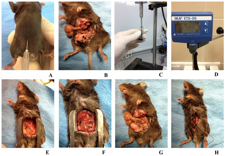
Figure 1. Animal model of peritoneal carcinomatosis and chemotherapy procedures.

A. To induce peritoneal carcinomatosis, the 1 × 10⁶ B16 cells were injected into the right lower abdomen quadrant of all animals from each group. B. Intraoperative picture of a mouse from the K3 group, one week after the B16 cells had been injected, the PC process at an advanced stage and exposed following preparation of the peritoneal and visceral tissue. C. Nanovehicles (DDS) were sonicated in 0.7 ml of sterile PBS for three minutes to obtain a homogenous solution. D. Continuous checking ensured that a consistent temperature was maintained across all study groups. E. A mouse from the A0-o-C1-chem-CD133 group just before the modified HIPEC procedure was performed. F. The same mouse as in E immediately after the application of liquid chemotherapy. G. The same mouse as in B just after the surgical cytoreduction of tumors. H. The same mouse as in E/F having sutures applied immediately after the experimental HIPEC procedure, a process consistently applied across all study and control groups.
