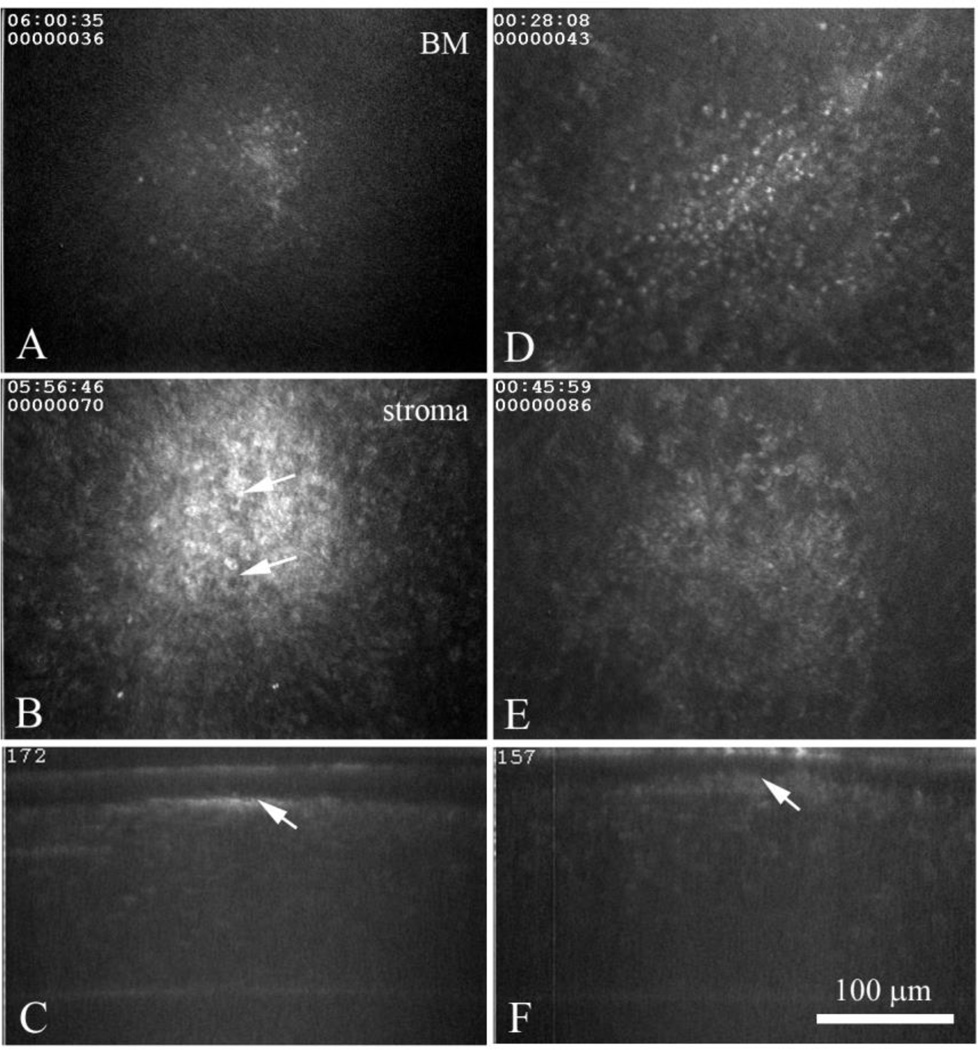
Figure 2.
In vivo confocal microscopic images from a wild type, latently infected rabbit eye at 35 Days (A-C) and 60 Days (D-F) post infection. Images through the basement membrane region (A and D) detected brightly reflecting, small cell infiltrates of variable size that overlaid granular deposits (B and E) that appeared to be composed of immune cell infiltrates (B, arrows). In the XZ projections (C and F) note the small region of light scattering (arrow) located at the basement membrane anterior stroma.

