Abstract
The present article deals with the rare documentation of elephant louse Haematomyzus elephantis from ceremonial elephants of Vrindavan, Mathura. The reports of this particular louse are very rare in literature as per the Indian context and before this, there is only one recent report of this parasite from India since the last decade. Interestingly, the louse found in a semi arid area, away from the natural habitat of elephants, where its existence in nature is very much a matter of debate. Finally, the morphology of the parasite, its feeding habit and its pathobiology are being described here in.
Keywords: Elephant, Haematomyzus elephantis, Lice
Introduction
Lice are perhaps one of the most adaptive creatures that thrive well in the habitat suitable for its host throughout the various altogether different ecological regimes across the globe (O’Toole 1986). The elephant louse, Haematomyzus elephantis, though rare in printable literature have been, sporadically documented from the Indian subcontinent (Godara et al. 2009) and the African grasslands (O’Toole 1986). Besides being a natural part of protected grasslands and forests, elephants are found in zoos and also as captive creatures with people for ceremonial purposes and elephant ride. The present communication deals with the rare presence of H. elephantis from ceremonial elephants of Vrindavan, Mathura area.
Materials and methods
Two elephants were found to be suffering from skin lesions on the axilla, groin region, ears and the adjacent areas on head and neck region during a routine visit. The elephants were being reared by a family of the landless but financially middle class owners, for entertaining uphill elephant ride and for ceremonial purposes during festival and marriage season. The skin lesions were carefully examined and visible ectoparasites were collected and brought to the divisional laboratory for further studies and permanent mounting. Micrometric measurements of different body parts of the parasite were taken and the louse was identified by morphological characteristics documented elsewhere in the literature (Lapage 1956; Bowman et al. 2003; Godara et al. 2009).
Results and discussion
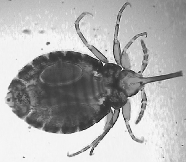
The lice infestation were observed more on hairy regions, especially folds of soft skin, in axilla, groin region, ears and the adjacent areas on head and neck and at the base of tail. The golden yellow adult louse was elongated to oval in shape and measured 2.0 ± 0.31 mm in length and 1.18 ± 0.17 mm in width at the fourth abdominal segment. The distinctly visible triangular head was much longer than thorax. In the pre antennal region, the head was armed with long and rigid snout called rostrum, bearing a pair of sharp mandible rasps, facilitating deep piercing into the thick skin of elephant, feeding blood and skin debris on surface. The morphology of the lice was very much in agreement with that described in the literature (Godara et al. 2009) and it was identified as H. elephantis (Fig. 1). The lice belong to suborder Mallophaga and feeds on epithelial debris on skin surface, its own eggs and skin scrapings including hair (Klots and Klots 1979; Arnest 1985). The lice is significant as it has no preferred site of predilection and is neither known to transmit pathogens nor is zoonotic in nature (Klots and Klots 1979). Strangely, the louse is believed to cause a massive drop of released eggs in the ovaries of breedable female elephants (Grzimek 1972). Though the lice is very much adaptive so as to survive the extreme geo-climatic fluctuations (Grzimek 1972; O’Toole 1986), yet it is very much difficult to explain how the lice gained entry onto the captive elephants in the instant case and how it survived in nature in this particularly elephant free area.
Fig. 1.

Adult louse of Haematomyzus elephantis
The detailed morphological account and veterinary significance of H. elephantis is lacking in the literature (Grzimek 1972; O’Toole 1986) and baring a single report (Godara et al. 2009), authors did not find any other substantial contribution from the Indian context in particular for the sake of discussion and/or criticism. Nevertheless, considering the finding of these lice in semi arid area coupled with the fact that elephant population is very much scare in the studied area, it is very much a matter of debate that how these lice survived in nature in absence of abundant hosts and/or from where these two elephants picked the infection. This seems to be the only second report on occurrence of the H. elephantis from captive elephants. Alongside it is significant because of its occurrence in such an area which is having comparatively much lower rainfall, but higher humidity and ambient temperature, in comparison with other natural habitats of the elephants in India.
Acknowledgments
The authors express their deep sense of gratitude and sincere thanks to the Hon’ble Vice Chancellor and the Dean, DUVASU for the facilities provided.
References
- Arnest RH. American insects: a handbook of the insects of America North of Mexico. New York: Van Nostrand Reinhold Company; 1985. [Google Scholar]
- Bowman DD, Lynn RC, Eberhard ML, Alcaraz A. Georgi’s parasitology for veterinarians. 8. St. Louis: Saunders-an imprint of Elsevier; 2003. pp. 1–82. [Google Scholar]
- Godara R, Singh S, Dev R, Sharma RL. Occurrence of Haematomyzus elephantis in semi-arid Rajasthan. J Vet Parasitol. 2009;23(1):69–71. [Google Scholar]
- Grzimek BKM. Grhizmek’s animal encyclopedia: volume 2 insects. New York: Van Nostrand Reinhold Company; 1972. [Google Scholar]
- Klots A, Klots A. Living insects of the wind. Garden City: Doubleday & Company Inc; 1979. [Google Scholar]
- Lapage G. Veterinary parasitology. 1. London: Oliver and Boyd LTD; 1956. pp. 364–588. [Google Scholar]
- O’Toole C. The encyclopedia of insects. New York: Faction File Publications; 1986. [Google Scholar]


