Abstract
The mechanisms of cellular and humoral immune responses against P. vivax parasite remain poorly understood. Several malaria immunological studies have been conducted in endemic regions where both P. falciparum and P. vivax parasites co-exist. In this study, a comparative analysis of immunity to Plasmodium vivax antigens in different geography and incidence of Plasmodium spp. infection was performed. We characterised antibodies against two P. vivax antigens, PvMSP-1 and PvAMA-1, and the cross-reactivity between these antigens using plasma from acute malaria infected patients living in the central region of China and in the western border of Thailand. P. vivax endemicity is found in central China whereas both P. vivax and P. falciparum are endemic in Thailand. There was an increased level of anti-PvMSP-1/anti-PvAMA-1 in both populations. An elevated level of antibodies to total P. vivax proteins and low level of antibodies to total P. falciparum proteins was found in acute P. vivax infected Chinese, suggesting antibody cross-reactivity between the two species. P. vivax infected Thai patients had both anti-P. vivax and anti-P. falciparum antibodies as expected since both species are present in Thailand. More information on humoral and cell mediated immunity during acute P. vivax-infection in the area where only single P. vivax species existed is of great interest in the relation of building up anti-disease severity caused by P. falciparum. This knowledge will support vaccine development in the future.
Keywords: malaria immunity, Plasmodium vivax, Plasmodium falciparum, antibody to malaria
Introduction
Malaria remains one of the most devastating infectious diseases due to its influence on more than 225 million individuals and approximately 700 thousand deaths each year. The majority of malaria infections are caused by Plasmodium falciparum in African countries (Sachs and Malaney, 2002[20]). However, as the most widely distributed human malaria parasite, Plasmodium vivax can also cause life-threatening symptoms even though it was previously considered benign. In contrast to P. falciparum, studies on P. vivax have lagged behind, largely due to the fact that this parasite cannot be cultured continuously in vitro. The mechanisms of cellular and humoral immune responses against P. vivax parasite also remain poorly understood. Other reasons for the neglect of “benign” P. vivax malaria could be the difficulty in accessing P. vivax-infected cases. Several studies have been conducted in endemic regions where both parasites co-exist (Snounou and White, 2004[23]; Rodrigues et al., 2005[18]; Oliveira et al., 2006[15]; Wickramarachchi et al., 2007[29]; Michon et al., 2007[13]; Chuangchaiya et al., 2010[3]; Kochar et al., 2010[9]; Lin et al., 2010[10]; Douglas et al., 2011[6]; Lin et al., 2011[11]). The strain-specific and serological cross-reactive immunity between P. falciparum and P. vivax blood stage antigens has been documented (Diggs and Sadun, 1965[4]; Woodberry et al., 2008[31]; Doolan et al., 2009[5]). However, very few studies were conducted in areas where only P. vivax causes infection (Jangpatarapongsa et al., 2012[8]). Therefore, a comparative study of immunity to P. vivax antigens in different endemic settings will contribute to a better understanding on the development and dynamics of host immunity to P. vivax infections.
Strong humoral immune responses to Plasmodium can be induced in residents of malaria endemic areas (Wipasa et al., 2002[30]) The level of total antimalarial antibodies increases with age and depends on the length and intensity of exposure to malaria. Antibody-mediated inhibition of parasites is more efficient in blood stage than in liver stage infections (Troye-Blomberg and Perlmann, 1988[27]). Antibodies also mediate antibody-dependent cellular cytotoxicity and phagocytosis involving polymorphonuclear cells, neutrophils or platelets (Bolad and Berzins, 2000[2]).
To understand the natural immune response during P. vivax infection in central China where only P. vivax is present and western Thailand with P. vivax and P. falciparum were almost equally prevalent (WHO, 2013[28]), we determined antibodies in the patients' sera against proteins extracted from P. vivax parasites and recombinant proteins PvMSP1(19) and PvAMA-1 produced in Escherichia coli (Soares et al., 1997[25]; 1999[24][26]; Rodrigues et al., 2003[17]). Our study aimed to characterize the level of IgG antibodies following P. vivax infection comparing two malaria endemic areas having different geography and incidence of Plasmodium spp. infection.
Materials and Methods
Study population
Plasma samples were collected from 76 patients with acute P. vivax infections (AC) at Wuhe County Hospital, Guzhen County Hospital, The First City Hospital, Bengbu city, Anhui Province, China. The patients were enrolled sequentially during June and July of 2009 and 2010. All patients enrolled in this study are inhabitants of Wuhe County, Guzhen County or the Bengbu City suburbs. Malaria transmission in this region is non-stable but can lead to malaria endemic in China. In the 1960s and 1970s, there were two malaria epidemics which were primarily caused by the P. vivax parasite. P. falciparum and P. vivax parasites were found together in this region until the end of the 1980s, but P. falciparum has not been found since the early 1990s. During the first decade of this century (from 2000 to 2010), malaria in this and other regions of China was mainly caused by the P. vivax parasite.
In Thailand plasma samples were collected from 52 patients from malaria clinics at Mae Sot and Mae Kasa, Tak Province, who were enrolled sequentially during 2009 and 2010. The diagnosis of P. vivax malaria infection was based on the examination of Giemsa-stained thick and thin blood films. Polymerase chain reaction (PCR) with species-specific primers was performed on DNA isolated from the blood samples to further verify P. vivax infections (Snounou et al., 1993[22]).
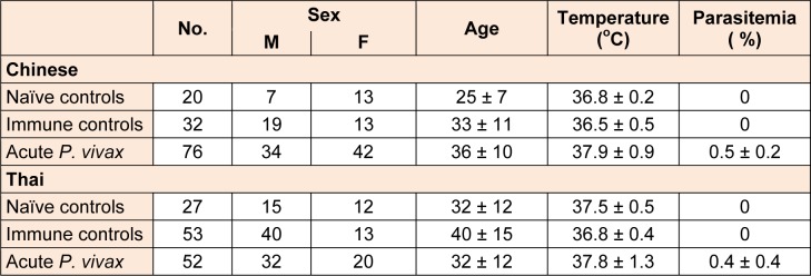
Blood samples were collected from 32 Chinese and 53 Thai people who did not suffer from P. vivax at the time of blood collection determined by both microscopy and PCR residing in the same P. vivax-endemic area as “immune controls” (IC). Another 20 healthy Chinese adults living in Wuxi city, China and 27 healthy Thai adults living in Bangkok, Thailand without previous malaria exposure were recruited to serve as “naïve controls” (NC). The clinical characteristics of the subjects are listed in Table 1(Tab. 1). This study was approved by Ethical Approval Committee of Biomedical Institute of Anhui Medical University and Committee on Human Rights Related to Human Experimentation, Mahidol University. Informed consents were obtained from each individual before a blood sample was taken.
Table 1. Information and clinical data of P. vivax patients, immune and naïve controls.
Parasite culture and antigen preparation
P. vivax-infected red blood cells (iRBC) purified from infected blood were used as crude antigens for coating. Briefly, P. vivax infected bloods were depleted of white blood cells by filtering through a sterile column of CF11 cellulose (Whatman®, Maidstone, UK) and the red blood cells were washed with RPMI-1640 by centrifugation at 1190 g for 5 minutes. The parasites were cultured for 24 - 30 hours at 5 % hematocrit in McCoy's 5A medium (GIBCO, Carlsbad, USA) supplemented with 25 % human AB serum. P. vivax parasites were maintained in an incubator containing 5 % CO2, 5 % O2 and 90 % N2 until matured to schizont stage ( 6 nuclei). P. faciparum culture was performed as described previously (Jangpatarapongsa et al., 2006[7]). The late stage iRBC were enriched by centrifugation using 60 % Percoll (GE Healthcare, Uppsala, Sweden) at 1190 g for 10 minutes. The enriched iRBC pellets were sonicated for 40 seconds at 150 watts and the protein concentration was determined by Bradford assay (Bio-Rad, Hercules, USA). The proteins were then aliquoted and stored at -70 °C until use. Uninfected RBCs were processed similarly as above and equivalent amount of protein concentration to the malaria antigens was stored at -70 °C to be used as a negative control.
Protein expression and purification
A blood filter paper from P. vivax-infected patients from Thailand was used to extract genomic DNA. PCR was used to obtain the MSP-1(19) fragment by using primers in Table 2(Tab. 2). Sequences were cut with BamH I and Xho I and cloned into pET28a vector. Protein was expressed in E. coli BL21. IPTG was used for protein induction and cell pellet was collected and sonicated. Protein was purified by using native condition as described by manufacturer's protocol (Qiagen, USA). Supernatant was collected and protein was purified by Ni-NTA beads. Finally, eluted protein was checked for expression by SDS-PAGE and kept at -20 °C.
Table 2. PCR primers and sequences.
For PvAMA-1, PCR was performed to amplify PvAMA-1 gene from a Thai P. vivax sample by using the designed primers in Table 2(Tab. 2). Sequences were cut using Nde I and Xho I and cloned into pET22b vector. Protein was expressed in E. coli BL21, purified and checked for expression in similar processes to PvMSP-1(19) as mentioned above.
ELISA analysis and IgG antibody level in plasma
There were three groups of plasma samples analyzed: AC group (n = 76), IC group (n = 32), and NC group (n = 20). There were five soluble antigens used: NRBC (10 μg/ ml), PV (10 μg/ml), PF (10 μg/ml), PvMSP-1 (0.25 μg/ml), and PvAMA-1 (0.1 μg/ml). 50 μl of antigens were pipetted to each ELISA well and plates were stored in wetted box at 4 °C overnight. Then the plates were washed and blocked with 5 % skimmed milk for 2 h at 37 °C. Plasma samples were diluted (1: 100) and added to each well (50 μl). The plates were enclosed in wetted box and incubated for 2 h at 37 °C. After washing 3 times using TBS-T, 50 μl of HRP-conjugated goat anti human IgG (H&L chain) antibody was added into each well, then plates were enclosed in wetted box again, and incubated for another 2 h at 37 °C. TBS-T was used to wash the plate then 50 μl of ABST substrate were added to each well. The plate was stored in the dark for 1 h at room temperature. All absorption was determined at A405 and the final OD values of PV and PF antigen were obtained by subtracting the OD values from NRBC group.
Results
A comparison of anti-P. vivax antibody after being intected with P. vivax parasite between Thai and Chinese patients
Anti-crude PV
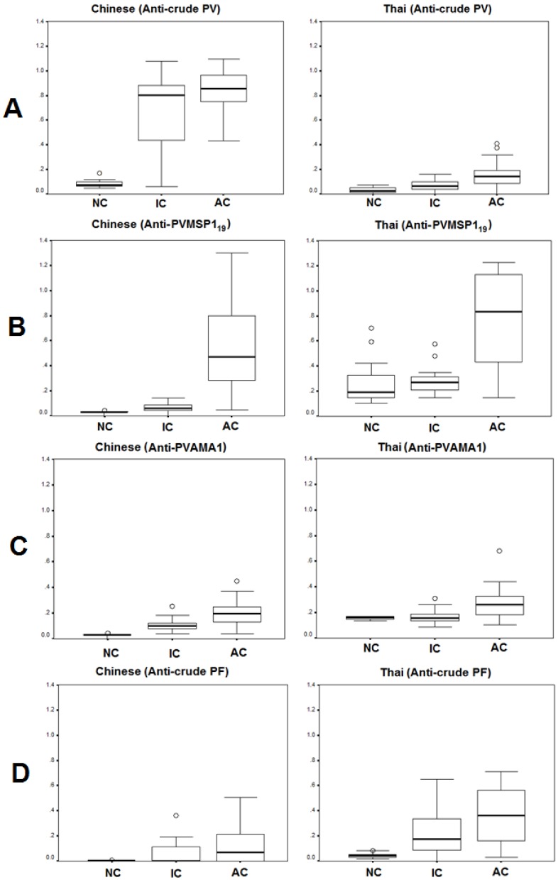
There was significant difference of base line antibody level among naïve controls against P. vivax-extracted antigens between Chinese (OD = 0.07) and Thai (OD = 0.03) (MD = 0.07, 95 % CI = 0.04-0.1, P < 0.001) (Figure 1A(Fig. 1)). After infection by P. vivax, significant elevation of antibodies were found in both patient groups Chinese (0.85) and Thai (0.14). However, the level of antibody in P. vivax infected Chinese patients was higher than that of the Thai patients. Similar to naïve controls, the immune Chinese controls (0.8) had higher levels of antibodies against the P. vivax antigens than that of the Thai controls (0.07).
Figure 1. Absorbance (405 nm) value of IgG antibody reacting with (A) crude P. vivax antigens, (B) recombinant P. vivax MSP-119 protein, (C) recombinant P. vivax AMA-1 protein, and (D) crude P. falciparum antigen respectively, in the naïve controls (NC), immune controls (IC), acute P. vivax infection (AC) comparing between Chinese and Thai patients. Data are shown in median, interquartile ranges (box plots), maximum and minimum (upper-lower lines).
Anti-P. vivax MSP-1(19)
Baseline values of mean level of antibodies to PvMSP-1(19) among healthy Thai controls and Chinese population living outside (naïve controls) and inside (immune controls) endemic areas were compared (Figure 1B(Fig. 1)). We found that the mean level of total antibodies in Thai naïve controls (OD = 0.3) was higher than that of Chinese naïve controls (OD = 0.03) (P < 0.001). Moreover, the level of antibodies in people living in P. vivax endemic area was significantly different between Thai (0.31) and Chinese populations (0.06) (P < 0.001). Two samples of Thai healthy controls had antibodies to PvMSP-1(19) as high as P. vivax infected patients as shown in above figure(Fig. 1), and were excluded from the baseline value. Among both population groups, the mean level of total antibodies to PvMSP-1(19) was significantly increased during acute P. vivax infection in both Thai (0.8) (P < 0.001) and Chinese (0.5) (P < 0.001) patients. Interestingly, the significantly higher levels of antibodies among P. vivax infected Thai compared to Chinese patients (P = 0.02) were observed.
Anti-P. vivax AMA-1
The mean level of antibodies against PvAMA-1 among healthy controls living outside the P. vivax endemic area was very low both in Thai (OD = 0.1) and in Chinese populations (OD = 0.03) (Figure 1C(Fig. 1)). However, the level was significantly higher among the Thai population compared to the Chinese population (P < 0.000). There was a higher level among Thai (0.2) than Chinese immune controls (0.1) (P < 0.001). There was no significant difference in the level of antibodies against PvAMA-1 in the healthy Thai donors living inside and outside endemic areas (P > 0.05) in contrast to that found in the Chinese donors (P < 0.001). In acute P. vivax infection, the level was significantly higher compared to naïve controls (Thai P =< 0.001, Chinese P < 0.0.001). However, among P. vivax patients, we did not find significant difference between Thai (0.3) and Chinese (0.2) populations (P > 0.05). We also found a sample of P. vivax infected patient that had obviously high level of antibodies to PvAMA-1, so this was excluded from the baseline value.
Cross reactivity between P. vivax and P. falciparum antigens
To examine the immune cross-reactivity between P. vivax and P. falciparum antigens, the P. vivax and P. falciparum extracted antigens were used as coated antigens on the ELISA plates. The median percentage of baseline level antibodies in naïve controls among Chinese was very low (OD = 0.003) and was significantly lower than that of Thai (OD = 0.04) donors (MD = 0.07, 95 % CI = 0.04-0.1, P < 0.001) (Figure 1D(Fig. 1)). Moreover, significantly higher levels of antibodies against P. falciparum antigens were observed in immune controls between Thai (0.18) and Chinese (0.01) (MD = 0.6, 95 %CI = 0.45-0.75). After infection with P. vivax, patients had higher levels of antibodies against P. falciparum in Thai patients (OD = 0.36) than in Chinese patients (OD = 0.07).
Discussion
In this study, we provide evidence that Thai villagers having had P. vivax infection and living in the endemic area did not produce high level of anti-P. vivax-specific antibodies (Jangpatarapongsa et al., 2006[7]) as tested with total proteins extracted from the parasites. We confirmed the presence of antibodies to P. vivax antigens by testing with recombinant P. vivax proteins, i.e. PvMSP1(19) and PvAMA1, then compared the level of natural antibodies between two P. vivax endemic areas in China and in Thailand. The species specific antibodies could tell us the cross-reactivity between P. falciparum and P. vivax which led to the examination of epidemiological status in those areas.
Previously, a passive transfer of immune IgG to Gambian children was shown to provide protection (McGregor, 1964[12]). The immunity against blood stage of P. falciparum infection is associated with class and subclass of IgG antibody (Shi et al., 1999[21]). Similarly, IgG1 and IgG3 are predominant among P. vivax-infected patients with history of malaria (Pinto et al., 2001[16]). Recent study has shown anti-P. vivax merozoite surface protein 1 (MSP-1) IgG among subjects with distinct degrees of malaria exposure in endemic area. The IgG1 and IgG3 against P. vivax are higher among the subjects with one year-exposure period than the group with longer years of exposure (Morais et al., 2005[14]). PvMSP-1(19) is shown to induce immunity in non-human primates (Rosa et al., 2006[19]).
Our study showed no significant difference of base line level anti-P. vivax antibodies among Chinese and Thai patients. We found higher levels of anti-P. vivax antibodies in Chinese than in Thai immune controls. Moreover, this level was very high in similarity to that shown in the acute P. vivax infection. This could be that P. vivax endemicity in China was greater than in Thailand, although the two areas in both countries are hypo-endemic.
However, in contrast to what we found in anti-P. vivax antibodies, the base line level of anti-MSP-1 and anti-AMA-1 antibodies was significantly lower in naïve Chinese controls than in naïve Thai controls. A possible reason could be that some of these malaria naïve Thai donors might have had malaria experiences but were asymptomatic and, therefore, there were lower levels of antibodies that persisted in their circulation.
In our study, we found the level of anti-MSP-1 antibodies was significantly lower in immune controls and acute P. vivax infected Chinese. A possible reason is that the recombinant protein rMSP-1(19) was obtained from the P. vivax parasites infecting Thai patients. There may be difference in the gene sequence of P. vivax MSP-1(19) between those from Thailand and those from China (Birkenmeyer et al., 2010[1]).
Crude P. falciparum antigen was used in this study to determine the species specific antibody and the cross-reactivity. We found very low levels of base line anti-P. falciparum antibodies among naïve controls and immune controls of Chinese donors. After infection with P. vivax, Chinese patients had somewhat higher levels of anti-P. falciparum antibodies. These results were in contrast to those found in Thailand. Higher levels of base line anti-P. falciparum antibody was found among naïve and immune Thai controls. Moreover, development of anti-P. falciparum antibodies in P. vivax infected patients in Thailand were much higher than that in China. Taken together, this suggests that a cross-reactivity among epitopes/antigens between the two malaria species do exist. However, it does not over rule the fact that the anti-P.falciparum antibody may be maintained in the P. vivax-infected Thai patients since both parasites are common in the area. Our findings led to the hypothesis that a protection against severe malaria caused by P. falciparum might be obtained via vaccination with a common antigen(s) of a benign malaria parasite such as P. vivax.
Acknowledgements
We thank Dr. Hua Hai Yong for supporting healthy donors plasma, Drs. Chen Chong Xin, Dr. Zhu Mu Shan, Dr. Gao Lai, Dr. Luo Wen Wen for immune controls and malaria patients’ sera.
This work was supported by the Office of Higher Education Commission and Mahidol University under the National Research Universities. The Thailand Research Fund (BRG498009) to RU, and D43TW006571 to LC from The Fogarty International Center, NIH, USA.
Conflict of interest
The authors declare that they have no conflict of interest.
Notes
Dr. Rachanee Udomsangpetch (E-mail: rachanee.udo@mahidol.ac.th) and Dr. Baiqing Li (E-mail: bb_bqli@yahoo.com) contributed equally as corresponding authors.
Hui Xia and Qiang Fang contributed equally to this work.
References
- 1.Birkenmeyer L, Muerhoff AS, Dawson GJ, Desai SM. Isolation and characterization of the MSP1 genes from Plasmodium malariae and Plasmodium ovale. Am J Trop Med Hyg. 2010;6:996–1003. doi: 10.4269/ajtmh.2010.09-0022. [DOI] [PMC free article] [PubMed] [Google Scholar]
- 2.Bolad A, Berzins K. Antigenic diversity of Plasmodium falciparum and antibody-mediated parasite neutralization. Scand J Immunol. 2000;52:233–239. doi: 10.1046/j.1365-3083.2000.00787.x. [DOI] [PubMed] [Google Scholar]
- 3.Chuangchaiya S, Jangpatarapongsa K, Chootong P, Sirichaisinthop J, Sattabongkot J, Pattanapanyasat K, et al. Immune response to Plasmodium vivax has a potential to reduce malaria severity. Clin Exp Immunol. 2010;160:233–239. doi: 10.1111/j.1365-2249.2009.04075.x. [DOI] [PMC free article] [PubMed] [Google Scholar]
- 4.Diggs CL, Sadun EH. Serological cross reactivity between Plasmodium vivax and Plasmodium Falciparum as determined by a modified fluorescent antibody test. Exp Parasitol. 1965;16:217–223. doi: 10.1016/0014-4894(65)90046-9. [DOI] [PubMed] [Google Scholar]
- 5.Doolan DL, Dobano C, Baird JK. Acquired immunity to malaria. Clin Microbiol Rev. 2009;22:13–36. doi: 10.1128/CMR.00025-08. [DOI] [PMC free article] [PubMed] [Google Scholar]
- 6.Douglas NM, Nosten F, Ashley EA, Phaiphun L, van Vugt M, Singhasivanon P, et al. Plasmodium vivax recurrence following falciparum and mixed species malaria: risk factors and effect of antimalarial kinetics. Clin Infect Dis. 2011;52:612–620. doi: 10.1093/cid/ciq249. [DOI] [PMC free article] [PubMed] [Google Scholar]
- 7.Jangpatarapongsa K, Sirichaisinthop J, Sattabongkot J, Cui L, Montgomery SM, Looareesuwan S, et al. Memory T cells protect against Plasmodium vivax infection. Microbes Infect. 2006;8:680–686. doi: 10.1016/j.micinf.2005.09.003. [DOI] [PubMed] [Google Scholar]
- 8.Jangpatarapongsa K, Xia H, Fang Q, Hu K, Yuan Y, Peng M, et al. Immunity to malaria in Plasmodium vivax infection: a study in central China. PLoS One. 2012;7:e45971. doi: 10.1371/journal.pone.0045971. [DOI] [PMC free article] [PubMed] [Google Scholar]
- 9.Kochar DK, Tanwar GS, Khatri PC, Kochar SK, Sengar GS, Gupta A, et al. Clinical features of children hospitalized with malaria - a study from Bikaner, northwest India. Am J Trop Med Hyg. 2010;83:981–989. doi: 10.4269/ajtmh.2010.09-0633. [DOI] [PMC free article] [PubMed] [Google Scholar]
- 10.Lin E, Kiniboro B, Gray L, Dobbie S, Robinson L, Laumaea A, et al. Differential patterns of infection and disease with P. falciparum and P. vivax in young Papua New Guinean children. PLoS One. 2010;5:e9047. doi: 10.1371/journal.pone.0009047. [DOI] [PMC free article] [PubMed] [Google Scholar]
- 11.Lin JT, Bethell D, Tyner SD, Lon C, Shah NK, Saunders DL, et al. Plasmodium falciparum gametocyte carriage is associated with subsequent Plasmodium vivax relapse after treatment. PLoS One. 2011;6:e18716. doi: 10.1371/journal.pone.0018716. [DOI] [PMC free article] [PubMed] [Google Scholar]
- 12.McGregor IA. The passive transfer of human malarial immunity. Am J Trop Med Hyg. 1964;13(Suppl):237–239. doi: 10.4269/ajtmh.1964.13.237. [DOI] [PubMed] [Google Scholar]
- 13.Michon P, Cole-Tobian JL, Dabod E, Schoepflin S, Igu J, Susapu M, et al. The risk of malarial infections and disease in Papua New Guinean children. Am J Trop Med Hyg. 2007;76:997–1008. [PMC free article] [PubMed] [Google Scholar]
- 14.Morais CG, Soares IS, Carvalho LH, Fontes CJ, Krettli AU, Braga EM. IgG isotype to C-terminal 19 kDa of Plasmodium vivax merozoite surface protein 1 among subjects with different levels of exposure to malaria in Brazil. Parasitol Res. 2005;95:420–426. doi: 10.1007/s00436-005-1314-x. [DOI] [PubMed] [Google Scholar]
- 15.Oliveira TR, Fernandez-Becerra C, Jimenez MC, Del Portillo HA, Soares IS. Evaluation of the acquired immune responses to Plasmodium vivax VIR variant antigens in individuals living in malaria-endemic areas of Brazil. Malar J. 2006;5:83. doi: 10.1186/1475-2875-5-83. [DOI] [PMC free article] [PubMed] [Google Scholar]
- 16.Pinto AY, Ventura AM, Souza JM. IgG antibody response against Plasmodium vivax in children exposed to malaria, before and after specific treatment. J Pediatr. 2001;77:299–306. doi: 10.2223/jped.238. [DOI] [PubMed] [Google Scholar]
- 17.Rodrigues MH, Cunha MG, Machado RL, Ferreira OC, Rodrigues MM, Soares IS. Serological detection of Plasmodium vivax malaria using recombinant proteins corresponding to the 19-kDa C-terminal region of the merozoite surface protein-1. Malar J. 2003;2(1):39. doi: 10.1186/1475-2875-2-39. [DOI] [PMC free article] [PubMed] [Google Scholar]
- 18.Rodrigues MH, Rodrigues KM, Oliveira TR, Comodo AN, Rodrigues MM, Kocken CH, et al. Antibody response of naturally infected individuals to recombinant Plasmodium vivax apical membrane antigen-1. Int J Parasitol. 2005;35:185–192. doi: 10.1016/j.ijpara.2004.11.003. [DOI] [PubMed] [Google Scholar]
- 19.Rosa DS, Iwai LK, Tzelepis F, Bargieri DY, Medeiros MA, Soares IS, et al. Immunogenicity of a recombinant protein containing the Plasmodium vivax vaccine candidate MSP1(19) and two human CD4+ T-cell epitopes administered to non-human primates (Callithrix jacchus jacchus) Microbes Infect. 2006;8:2130–2137. doi: 10.1016/j.micinf.2006.03.012. [DOI] [PubMed] [Google Scholar]
- 20.Sachs J, Malaney P. The economic and social burden of malaria. Nature. 2002;415:680–685. doi: 10.1038/415680a. [DOI] [PubMed] [Google Scholar]
- 21.Shi YP, Udhayakumar V, Oloo AJ, Nahlen BL, Lal AA. Differential effect and interaction of monocytes, hyperimmune sera, and immunoglobulin G on the growth of asexual stage Plasmodium falciparum parasites. Am J Trop Med Hyg. 1999;60:135–141. doi: 10.4269/ajtmh.1999.60.135. [DOI] [PubMed] [Google Scholar]
- 22.Snounou G, Viriyakosol S, Jarra W, Thaithong S, Brown KN. Identification of the four human malaria parasite species in field samples by the polymerase chain reaction and detection of a high prevalence of mixed infections. Mol Biochem Parasitol. 1993;58:283–292. doi: 10.1016/0166-6851(93)90050-8. [DOI] [PubMed] [Google Scholar]
- 23.Snounou G, White NJ. The co-existence of Plasmodium: sidelights from falciparum and vivax malaria in Thailand. Trends Parasitol. 2004;20:333–339. doi: 10.1016/j.pt.2004.05.004. [DOI] [PubMed] [Google Scholar]
- 24.Soares IS, da Cunha MG, Silva MN, Souza JM, Del Portillo HA, Rodrigues MM. Longevity of naturally acquired antibody responses to the N- and C-terminal regions of Plasmodium vivax merozoite surface protein 1. Am J Trop Med Hyg. 1999a;60:357–363. doi: 10.4269/ajtmh.1999.60.357. [DOI] [PubMed] [Google Scholar]
- 25.Soares IS, Levitus G, Souza JM, Del Portillo HA, Rodrigues MM. Acquired immune responses to the N- and C-terminal regions of Plasmodium vivax merozoite surface protein 1 in individuals exposed to malaria. Infect Immun. 1997;65:1606–1614. doi: 10.1128/iai.65.5.1606-1614.1997. [DOI] [PMC free article] [PubMed] [Google Scholar]
- 26.Soares IS, Oliveira SG, Souza JM, Rodrigues MM. Antibody response to the N and C-terminal regions of the Plasmodium vivax Merozoite Surface Protein 1 in individuals living in an area of exclusive transmission of P. vivax malaria in the north of Brazil. Acta Trop. 1999b;72:13–24. doi: 10.1016/s0001-706x(98)00078-3. [DOI] [PubMed] [Google Scholar]
- 27.Troye-Blomberg M, Perlmann P. T cell functions in Plasmodium falciparum and other malarias. Progr Allergy. 1988;41:253–287. doi: 10.1159/000415226. [DOI] [PubMed] [Google Scholar]
- 28.WHO, World Health Organization. WHO Malaria report 2013. Geneva:WHO, 2013. [Google Scholar]
- 29.Wickramarachchi T, Illeperuma RJ, Perera L, Bandara S, Holm I, Longacre S, et al. Comparison of naturally acquired antibody responses against the C-terminal processing products of Plasmodium vivax Merozoite Surface Protein-1 under low transmission and unstable malaria conditions in Sri Lanka. Int J Parasitol. 2007;37:199–208. doi: 10.1016/j.ijpara.2006.09.002. [DOI] [PubMed] [Google Scholar]
- 30.Wipasa J, Elliott S, Xu H, Good MF. Immunity to asexual blood stage malaria and vaccine approaches. Immunol Cell Biol. 2002;80:401–414. doi: 10.1046/j.1440-1711.2002.01107.x. [DOI] [PubMed] [Google Scholar]
- 31.Woodberry T, Minigo G, Piera KA, Hanley JC, de Silva HD, Salwati E, et al. Antibodies to Plasmodium falciparum and Plasmodium vivax merozoite surface protein 5 in Indonesia: species-specific and cross-reactive responses. J Infect Dis. 2008;198:134–142. doi: 10.1086/588711. [DOI] [PMC free article] [PubMed] [Google Scholar]


