Abstract
Background: The specific recognition of oesophageal atresia (OA) with or without a tracheal fistula in a foetus is a diagnostic challenge for prenatal medicine. The aim of the present work is to analyse the value of the measurement of gastric size in the diagnosis of this significant malformation. Materials and Methods: Altogether, the examinations of 433 pregnancies between the 18.4 and 39.1 weeks of gestation were retrospectively analysed. 59 of these foetuses exhibited an OA. By means of a linear regression analysis with normal foetuses, significant parameters influencing gastric size were examined. Subsequently the gastric sizes were transformed into z values and a comparison was made between OA with and without fistulae with the help of t tests. Results: In the normal foetuses there was a significant association between the gastric circumference and the abdominal circumference (circumference = 6.809 + 0.179 × abdominal circumference, r = 0.686, p < 0.0001). In the normal group the average was 43.0 (standard deviation [SD] 13.7) mm and those in foetuses with and without fistuale were 33.8 (SD 22.7) and 0.9 (SD 3.7) mm. In 34 (57.6 %) foetuses with an OA, the gastric circumference was below the 5th percentile. In detail, there were 13 (34.2 %) foetuses with a fistula and 21 (100 %) without a fistula. The average z values in the normal group and in the groups of OA with fistula and without fistula amounted to 0.0 (SD 1.0), −1.3 (SD 2.2) and −4.5 (SD 1.0). Conclusion: Measurements of the gastric circumference below the 5th percentile should lead to further diagnostic measures, especially when associated with polyhydramnios. Although OA without a fistula is always conspicuous, only about one in three OAs with fistula are associated with a significantly smaller stomach.
Key words: gastric circumference, oesophageal atresia, tracheo-oesophageal fistula
Abstract
Zusammenfassung
Fragestellung: Die gezielte Erkennung einer fetalen Ösophagusatresie (ÖA) mit oder ohne tracheale Fisteln stellt eine diagnostische Herausforderung für die Pränatalmedizin dar. Ziel der vorliegenden Arbeit war es, den Stellenwert der Messung des Magenumfangs für die Diagnostik dieser bedeutsamen Fehlbildung zu analysieren. Material und Methodik: Insgesamt wurden die Untersuchungen von 433 Schwangerschaften zwischen der 18,4 bis 39,1 Schwangerschaftswoche retrospektiv ausgewertet. 59 dieser Feten wiesen eine ÖA auf. Mittels linearer Regressionsanalyse wurden bei normalen Feten signifikante Einflussparameter auf den Magenumfang untersucht. Anschließend erfolgte die Umwandlung der Magenumfänge in z-Werte und der Vergleich zwischen den ÖA mit und ohne Fistel mit der Normalpopulation mithilfe eines t-Tests. Ergebnisse: Bei den normalen Feten zeigte sich eine signifikante Assoziation zwischen dem Umfang des Magens und dem Abdomenumfang (Umfang = 6,809 + 0,179 × Abdomenumfang, r = 0,686, p < 0,0001). Der mittlere Magenumfang lag in der normalen Gruppe bei 43,0 (Standardabweichung [STW] 13,7) mm und bei Feten mit und ohne Fistel bei 33,8 (STW 22,7) und 0,9 (STW 3,7) mm. Bei 34 (57,6 %) Feten mit einer ÖA lag der Magenumfang < 5er-Perzentile. Im Einzelnen waren es 13 (34,2 %) Feten mit einer Fistel und 21 (100 %) ohne Fistel. Die mittleren z-Werte in der normalen Gruppe und in der Gruppe der ÖA mit Fistel und ohne Fistel lagen bei 0,0 (STW 1,0), −1,3 (STW 2,2) und −4,5 (STW 1,0). Schlussfolgerung: Messungen des Magenumfangs < 5er-Perzentile sollten eine weiterführende Diagnostik nach sich ziehen. Während ÖA ohne Fisteln hierbei stets auffällig werden, weist nur ungefähr jede 3. ÖA mit Fistel einen signifikant verkleinerten Magen auf.
Schlüsselwörter: Magenumfang, Ösophagusatresie, tracheoösophageale Fistel
Introduction
During the 5th to 6th week of embryonal development the oesophagus and trachea separate from a common anlage within the early intestinal tube. An incomplete separation leads to the congenital malformation of oesophageal atresia (OA). In the majority of the cases (72–90 %) the trachea is also involved in the form of a tracheo-oesophageal fistula between the lower section of the trachea and the oesophagus. The prevalence varies from region to region and is estimated to amount to an average of 2.43 to 2.86 cases per 10 000 births 1, 2, 3. A large proportion of the afflicted infants also exhibit further malformations or syndromes (such as, e.g., VACTERL association, trisomy 18, CHARGE syndrome). The most frequent associated malformation is congenital heart disease. In current reports of the EUROCAT Register only 44.7 % of the cases are isolated findings 1. Beside the associated malformations, the regular occurrence of preterm births represents a significant prognostic factor. Because of the frequent occurrence of polyhydramnios, premature delivery ensues in 38.5 % of the cases 1, 4, 5. Survival rates of almost 100 % can be expected in isolated cases with adequate prenatal and neonatal management 6, 7, 8. Later complications and morbidities during childhood consist of swallowing difficulties on account of narrow anastomoses or gastro-oesophageal reflux as well as recurring infections 9, 10, 11.
The current data situation is contradictory with regard to benefits for the afflicted babies resulting from prenatal diagnosis. Whereas, for example, Brantberg and co-workers demonstrated a significantly better survival (100 vs. 73 %) for cases identified prior to birth, other groups found a poorer prognosis for this collective 12, 13. The latter can be explained by the over-representation of OA without fistulas and associated malformations in the group of prenatally detected cases. However, current opinion is that prenatal diagnosis must be considered as a benefit in that it provides a possibility for prenatal counselling of the parents and a reduction of postpartum transfers 14.
The existing detection rates within Europe vary between less than 10 % to more than 50 % (on average 36.5 %) 1, 15. Sonographic visualisation of the oesophagus stretches even modern ultrasound systems to the limits of their resolution. In cases without a fistula the inability to visualise gastric filling leads to the diagnosis 16. Cases with fistulas are not so frequently detected prenatally. Although a small stomach has been described by many groups as a sign, there is mostly a lack of an objectifiable limiting value for gastric size 13. The aim of the present study is to examine the gastric size as a potential diagnostic criterium for foetuses with OA not only with but also without tracheo-oesophageal fistulas.
Material and Methods
The underlying investigations were carried out in the Department of Prenatal Medicine of the University Hospital Tübingen and findings were assessed retrospectively.
Measurement methods
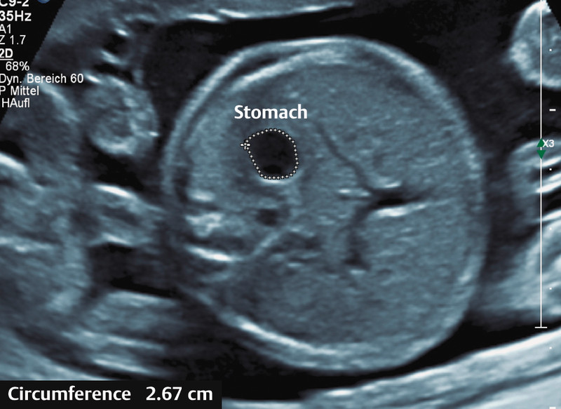
First of all the digitally saved images for gastric circumference measurements of routinely performed biometric examinations were examined. A prerequisite was the clear demonstration of the anatomic landmarks spinal column cross-section, parallel sections of rib pairs, the full stomach and the proximal part of the umbilical vein and its transition to the ductus venosus and to the right portal vein. The gastric filling was always measured in the biometry plane. For this the gastric circumference was determined manually on the digitally saved images (Fig. 1).
Fig. 1.

Illustration of the measurement of gastric circumference by manual tracing of the gastric circumference in the biometry plane.
Collective
To obtain an average value for the circumference, values from 374 normal singleton pregnancies were considered. The test collective consisted of 59 pregnant women in whom there was evidence for foetal OA.
In pregnancies with conspicuous foetuses the first examination undertaken in the Gynaecology Department of Tübingen University Hospital was considered in each case.
Registration of associated influencing factors
Beside the gastric circumference, we also recorded the head circumference, the abdominal circumference, the femur length as well as the gestational age at the time of the examination. Furthermore, the existance of polyhydramnios, defined as the largest vertical amniotic fluid depot over 8 cm, as well as performance of relief drainage measures were recorded.
Statistical Analysis
In normal foetuses significant factors influencing the gastric circumference were investigated by linear regression analysis. Subsequently the gastric circumference was transformed to a z value and comparisons between foetuses with OA with and without fistula and the normal population were made with the help of t tests. Differences with a p value < 0.05 were considered to be significant.
The results are given as median (range) or average (standard deviation SD) values.
Approval of ethical committee
Because of the retrospective anonymized character of our study, an approval of the local ethical committee was not necessary. However, the ethical committee was informed about the study (IEC number: 597/2015R).
Results
Characteristics of the enrolled pregnant women
Altogether 433 patients were included in the evaluation. The median body weight amounted to 62.3 kg (range 45.0–163.0 kg). The median gestational age was 23.3 weeks (range 18 + 2–39 + 1 weeks) whereby 261 (60.3 %) were seen before the 26 + 0 week and 172 (39.7 %) thereafter.
Distribution of foetuses with oesophageal atresia and gastric circumference
OA was present in 59 (13.7 %) of the 433 foetuses. Of these 38 (64.4 %) foetuses had OA with and 21 (35.6 %) OA without a tracheo-oesophageal fistula. Also 20 (33.9 %) of the foetuses had a chromosomal disorder, the proportions in the OA groups with and without a fistula were 23.7 and 52.4 %, respectively. 14 (36.8 %) of the foetuses with OA and a fistula also had a polyhydramnios. Among the group of foetuses with OA but without a fistula polyhydramnios was diagnosed in 12 (57.1 %) of them.
For 21 of the foetuses no stomach could be observed during the entire examination period. Of these 20 (95.2 %) had an OA without fistula. In these cases the gastric circumference was set at 0.1 mm. The average gastric circumference in the normal group amounted to 43.0 mm (SD 13.7 mm) and in foetuses with and without fistula to 33.8 mm (SD 22.7 mm) and 0.9 mm (SD 3.7 mm).
Association of gastric circumference with other factors
In the normal foetuses there was a significant association between gastric circumference and abdominal circumference (circumference = 6.809 + 0.179 × abdominal circumference, r = 0.686, p < 0.0001). In addition significant correlations with gestational age (r = 0.669, p < 0.0001), head circumference (r = 0.659, p < 0.0001) and femur length (r = 0.671, p < 0.001) were observed. The association with abdominal circumference was used for the further analyses.
Percentile and z values
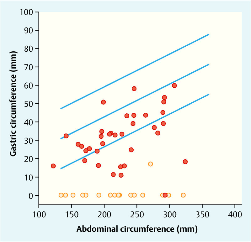
In 18 (4.8 %) of 374 normal patients the gastric circumference of the foetus was below the 5th percentile. In contrast this was the case with 34 (57.6 %) of the foetuses with OA. In detail, there were 13 (34.2 %) foetuses with a fistula and 21 (100 %) without a fistula (Fig. 2).
Fig. 2.

Graphic presentation of the normal distribution of gastric circumference in dependence on abdominal circumference including the 5th and 95th percentiles. The points show the gastric circumference values of foetuses with OA (red dots with fistula, orange dots without fistula).
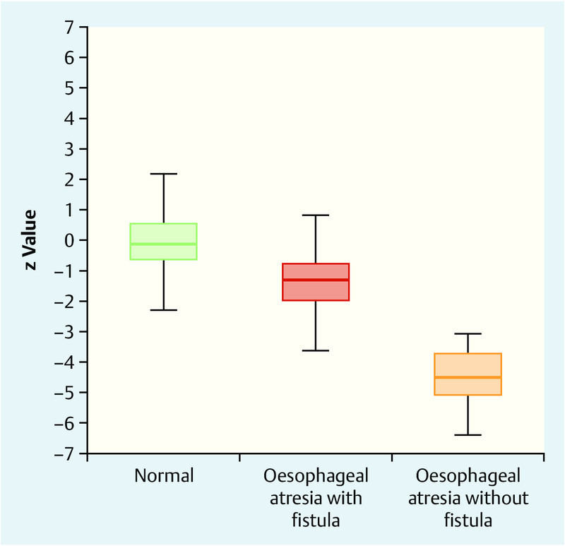
The average z values in the normal group and in the OA groups with and without fistula amounted to 0.0 (SD 1.0), −1.3 (SD 2.2) and −4.5 (SD 1,0) (t test: OA with fistula vs. normal p = 0.001; t test: OA without fistula vs. normal p < 0.001; t test: OA with fistula vs. OA without fistula p < 0.0001) (Fig. 3).
Fig. 3.

Boxplots of the z values for gastric circumference in the normal population, in foetuses with OA and fistulas and in foetuses with OA without fistulas.
Discussion
The present study has shown that the average gastric size, measured as gastric circumference at the level of the abdominal biometric plane is reduced not only in foetuses with OA and fistula but also in foetuses with OA but without fistula. In all cases without fistulas a completely or almost completely empty stomach was seen. The combination of a non-visualisable or very small stomach with polyhydramnios is a well known sign for the presence of an oesophageal obstruction 15, 17, 18. However, the appreciable false positive rates and the often subjective evaluation of gastric filling need to be taken into consideration 4, 19. Attempts to improve the prenatal detection rates concentrate on the prenatal visualisation of the oesophageal pouch sign that occurs through dilatation of the blind-ending proximal segment of the oesophagus 20, 21. But, in most cases, the OA results in this pathognomic blind sack being missed due to the fistula. In addition, the pouch sign can also be seen as a temporary transient finding in structurally inconspicuous foetuses, so that false positive findings are recorded 22.
Recognition of the majority of cases of OA with fistula represents the key problem. Impairments of foetal swallowing in such cases depend on the lumen of the tracheo-oesophageal fistula. In cases with large fistulas even the occurrence of polyhydramnios may be lacking. The sensitivity of polyhydramnios alone amounts to merely 73 % in some studies 13. In combination with the criterion of a small stomach at least a positive predictive value of up to 67 % can be expected 13. In our collective there was a comparatively low rate of polyhydramnios. Merely 36.8 % of the foetuses with OA and a fistula and 57.1 % of the foetuses with OA without a fistula exhibited polyhydramnios. The reason for this is to be seen in the high proportion of severe chromosomal disorders of 33.9 % that in most cases led to an early termination of the pregnancy and the stronger weighting in the 2nd trimester. Polyhydramnios develops in the course of the pregnancy to an ever increasing extent and is thus mostly to be found for the first time in an obvious degree in the 3rd trimester.
In many studies the assessment of gastric filling was purely subjective. An objective measurement by means of a standardised gastric circumference appears to be more reasonable in such cases. With the present study we have shown that the gastric size in at least 36.8 % of the afflicted foetuses with fistulas lies below the 5th percentile. Accordingly, the detection of a small stomach is a sign for further detailed diagnostic work-up.
The direct visualisation of the oesophagus either by means of high resolution 2D or 3D sonography or also by magnetic resonance imaging is extremely difficult and time-consuming 23, 24. In isolated case reports, the direct prenatal demonstration has been realised by concentration on the area between the aorta and the trachea 25. If detection of the pouch sign is successful its predictive value must be assessed as being high. In some studies the value not only for sonography but also for foetal MRI has been 100 %, while other studies refer to the possibility of false positive findings 19, 22, 26. This shows that even though a detailed sonographic examination of the oesophagus is technically possible, the method cannot be applied for unselected routine screening. Measurement of the gastric size can be helpful here since the presence of a small stomach can define a risk group for which a detailed examination of the oesophagus is required.
A further improvement in the diagnostics can be expected from the use of 3D sonography 27. However, use of the published normal values of three-dimensional biometry is at present limited due to the large discrepancies and the lack of methodological standardisation 28.
In very recent studies an approach involving biochemical analysis of the amniotic fluid has been described. Here, unchanged values with regard to L-leucine aminopeptidase (AMP) and elevated concentrations of alpha-foetoprotein (AFP) and gamma-glutamyl transpeptidase (GGT) are expected so that especially the quotient from both concentrations has exhibited very good sensitivities and specificities in the first studies (98 %/100 %) 29, 30. The currently available studies, however, are based on very small case numbers and require confirmation by larger trials. Furthermore, this is not suitable as a screening procedure due to its invasiveness and the thus associated interventional risks.
A limitation of the present study is its retrospective character and the thus associated lack of consideration of the dynamics of gastric filling. The latter are not constant and are subject to foetal swallowing cycles 31. However, this limitation holds for all included pregnancies and thus equally for the recorded gastric sizes of the afflicted foetuses as well as those of the normal population.
Practical Conclusions
The gastric size in foetuses with OA is reduced in cases with complete obstruction of the oesophagus. We have shown that reduced gastric filling below the 5th percentile is also found in 36.8 % of the foetuses with tracheo-oesophageal fistulas. Accordingly the measurement of gastric size can define a risk group requiring extensive further diagnostic work-up of the oesophagus.
Footnotes
Conflict of Interest None.
Supporting Information
References
- 1.Pedersen R N, Calzolari E, Husby S. et al. Oesophageal atresia: prevalence, prenatal diagnosis and associated anomalies in 23 European regions. Arch Dis Child. 2012;97:227–232. doi: 10.1136/archdischild-2011-300597. [DOI] [PubMed] [Google Scholar]
- 2.Depaepe A, Dolk H, Lechat M F. The epidemiology of tracheo-oesophageal fistula and oesophageal atresia in Europe. EUROCAT Working Group. Arch Dis Child. 1993;68:743–748. doi: 10.1136/adc.68.6.743. [DOI] [PMC free article] [PubMed] [Google Scholar]
- 3.Forrester M B, Merz R D. Epidemiology of oesophageal atresia and tracheo-oesophageal fistula in Hawaii, 1986–2000. Public Health. 2005;119:483–488. doi: 10.1016/j.puhe.2004.07.008. [DOI] [PubMed] [Google Scholar]
- 4.Abele H, Starz S, Hoopmann M. et al. Idiopathic polyhydramnios and postnatal abnormalities. Fetal Diagn Ther. 2012;32:251–255. doi: 10.1159/000338659. [DOI] [PubMed] [Google Scholar]
- 5.de Jong E M, de Haan M A, Gischler S J. et al. Pre- and postnatal diagnosis and outcome of fetuses and neonates with esophageal atresia and tracheoesophageal fistula. Prenat Diagn. 2010;30:274–279. doi: 10.1002/pd.2466. [DOI] [PubMed] [Google Scholar]
- 6.Deurloo J A, Ekkelkamp S, Schoorl M. et al. Esophageal atresia: Historical evolution of management and results in 371 patients. Ann Thorac Surg. 2002;73:267–272. doi: 10.1016/s0003-4975(01)03263-5. [DOI] [PubMed] [Google Scholar]
- 7.Konkin D E, Oʼhali W A, Webber E M. et al. Outcomes in esophageal atresia and tracheoesophageal fistula. J Pediatr Surg. 2003;38:1726–1729. doi: 10.1016/j.jpedsurg.2003.08.039. [DOI] [PubMed] [Google Scholar]
- 8.Lilja H E, Wester T. Outcome in neonates with esophageal atresia treated over the last 20 years. Pediatr Surg Int. 2008;24:531–536. doi: 10.1007/s00383-008-2122-z. [DOI] [PubMed] [Google Scholar]
- 9.Little D C, Rescorla F J, Grosfeld J L. et al. Long-term analysis of children with esophageal atresia and tracheoesophageal fistula. J Pediatr Surg. 2003;38:852–856. doi: 10.1016/s0022-3468(03)00110-6. [DOI] [PubMed] [Google Scholar]
- 10.Chetcuti P, Phelan P D. Gastrointestinal morbidity and growth after repair of oesophageal atresia and tracheo-oesophageal fistula. Arch Dis Child. 1993;68:163–166. doi: 10.1136/adc.68.2.163. [DOI] [PMC free article] [PubMed] [Google Scholar]
- 11.Chetcuti P, Phelan P D. Respiratory morbidity after repair of oesophageal atresia and tracheo-oesophageal fistula. Arch Dis Child. 1993;68:167–170. doi: 10.1136/adc.68.2.167. [DOI] [PMC free article] [PubMed] [Google Scholar]
- 12.Brantberg A, Blaas H G, Haugen S E. et al. Esophageal obstruction-prenatal detection rate and outcome. Ultrasound Obstet Gynecol. 2007;30:180–187. doi: 10.1002/uog.4056. [DOI] [PubMed] [Google Scholar]
- 13.Kunisaki S M, Bruch S W, Hirschl R B. et al. The diagnosis of fetal esophageal atresia and its implications on perinatal outcome. Pediatr Surg Int. 2014;30:971–977. doi: 10.1007/s00383-014-3562-2. [DOI] [PubMed] [Google Scholar]
- 14.Garabedian C, Sfeir R, Langlois C. et al. Does prenatal diagnosis modify neonatal treatment and early outcome of children with esophageal atresia? Am J Obstet Gynecol. 2015;212:3400–3.4E9. doi: 10.1016/j.ajog.2014.09.030. [DOI] [PubMed] [Google Scholar]
- 15.Houben C H, Curry J I. Current status of prenatal diagnosis, operative management and outcome of esophageal atresia/tracheo-esophageal fistula. Prenat Diagn. 2008;28:667–675. doi: 10.1002/pd.1938. [DOI] [PubMed] [Google Scholar]
- 16.Sase M, Asada H, Okuda M. et al. Fetal gastric size in normal and abnormal pregnancies. Ultrasound Obstet Gynecol. 2002;19:467–470. doi: 10.1046/j.1469-0705.2002.00695.x. [DOI] [PubMed] [Google Scholar]
- 17.Choudhry M, Boyd P A, Chamberlain P F. et al. Prenatal diagnosis of tracheo-oesophageal fistula and oesophageal atresia. Prenat Diagn. 2007;27:608–610. doi: 10.1002/pd.1745. [DOI] [PubMed] [Google Scholar]
- 18.Stringer M D, McKenna K M, Goldstein R B. et al. Prenatal diagnosis of esophageal atresia. J Pediatr Surg. 1995;30:1258–1263. doi: 10.1016/0022-3468(95)90480-8. [DOI] [PubMed] [Google Scholar]
- 19.Ethun C G, Fallon S C, Cassady C I. et al. Fetal MRI improves diagnostic accuracy in patients referred to a fetal center for suspected esophageal atresia. J Pediatr Surg. 2014;49:712–715. doi: 10.1016/j.jpedsurg.2014.02.053. [DOI] [PubMed] [Google Scholar]
- 20.Kalache K D, Wauer R, Mau H. et al. Prognostic significance of the pouch sign in fetuses with prenatally diagnosed esophageal atresia. Am J Obstet Gynecol. 2000;182:978–981. doi: 10.1016/s0002-9378(00)70357-7. [DOI] [PubMed] [Google Scholar]
- 21.Yagel S, Sonigo P, Rousseau V. et al. Esophageal atresia diagnosed with three-dimensional ultrasonography. Ultrasound Obstet Gynecol. 2005;26:307–308. doi: 10.1002/uog.1982. [DOI] [PubMed] [Google Scholar]
- 22.Solt I, Rotmensch S, Bronshtein M. The esophageal “pouch sign”: a benign transient finding. Prenat Diagn. 2010;30:845–848. doi: 10.1002/pd.2568. [DOI] [PubMed] [Google Scholar]
- 23.Brugger P C, Weber M, Prayer D. Magnetic resonance imaging of the normal fetal esophagus. Ultrasound Obstet Gynecol. 2011;38:568–574. doi: 10.1002/uog.9002. [DOI] [PubMed] [Google Scholar]
- 24.Malinger G, Levine A, Rotmensch S. The fetal esophagus: anatomical and physiological ultrasonographic characterization using a high-resolution linear transducer. Ultrasound Obstet Gynecol. 2004;24:500–505. doi: 10.1002/uog.1091. [DOI] [PubMed] [Google Scholar]
- 25.Develay-Morice J-E, Rathat G, Duyme M. et al. Ultrasonography of fetal esophagus: healthy appearance and prenatal diagnosis of a case of esophagus atresia with esotracheal fistula. Gynecol Obstet Fertil. 2007;35:249–257. doi: 10.1016/j.gyobfe.2007.01.016. [DOI] [PubMed] [Google Scholar]
- 26.Garabedian C, Verpillat P, Czerkiewicz I. et al. Does a combination of ultrasound, MRI, and biochemical amniotic fluid analysis improve prenatal diagnosis of esophageal atresia? Prenat Diagn. 2014;34:839–842. doi: 10.1002/pd.4376. [DOI] [PubMed] [Google Scholar]
- 27.Kusanovic J P, Nien J K, Gonçalves L F. et al. The use of inversion mode and 3D manual segmentation in volume measurement of fetal fluid-filled structures: comparison with Virtual Organ Computer-aided AnaLysis (VOCAL) Ultrasound Obstet Gynecol. 2008;31:177–186. doi: 10.1002/uog.5242. [DOI] [PMC free article] [PubMed] [Google Scholar]
- 28.Ioannou C, Sarris I, Salomon L J. et al. A review of fetal volumetry: the need for standardization and definitions in measurement methodology. Ultrasound Obstet Gynecol. 2011;38:613–619. doi: 10.1002/uog.9074. [DOI] [PubMed] [Google Scholar]
- 29.Czerkiewicz I, Dreux S, Beckmezian A. et al. Biochemical amniotic fluid pattern for prenatal diagnosis of esophageal atresia. Pediatr Res. 2011;70:199–202. doi: 10.1203/PDR.0b013e318220c08a. [DOI] [PubMed] [Google Scholar]
- 30.Muller C, Czerkiewicz I, Guimiot F. et al. Specific biochemical amniotic fluid pattern of fetal isolated esophageal atresia. Pediatr Res. 2013;74:601–605. doi: 10.1038/pr.2013.131. [DOI] [PubMed] [Google Scholar]
- 31.Sase M, Miwa I, Sumie M. et al. Gastric emptying cycles in the human fetus. Am J Obstet Gynecol. 2005;193:1000–1004. doi: 10.1016/j.ajog.2005.05.044. [DOI] [PubMed] [Google Scholar]
Associated Data
This section collects any data citations, data availability statements, or supplementary materials included in this article.


