Abstract
Accumulating evidence indicates that leukotriene B4 (LTB4) via its receptors BLT1 and/or BLT2 (BLTRs) could have an important role in regulating infection, tumour progression, inflammation, and autoimmune diseases. In the present study, we showed that LTB4 not only augments cytotoxicity by NK cells but also induces their migration. We found that approximately 30% of fresh NK cells express BLT1, 36% express BLT2, and 15% coexpress both receptors. The use of selective BLTR antagonists indicated that BLT1 was involved in both LTB4-induced migration and cytotoxicity, whereas BLT2 was involved exclusively in NK cell migration, but only in response to higher concentrations of LTB4. BLT1 and BLT2 expression increased after activation of NK cells with IL-2 and IL-15. These changes of BLTR expression by cytokines were reflected in enhanced NK cell responses to LTB4. Our findings suggest that BLT1 and BLT2 play differential roles in LTB4-induced modulation of NK cell activity.
1. Introduction
Human natural killer (NK) cells with the CD3− CD56+ phenotype comprise 10–15% of peripheral blood lymphocytes. They constitute a major component of the innate immune system especially in response to transformed and infected cells [1–3]. Even though priming is not necessary for NK cells to perform their cytolytic function, proinflammatory cytokines, such as IL-2 [4, 5] and IL-15 [6], can induce NK cell proliferation, cytotoxicity, or cytokine production. Chemokine-induced NK cell migration may explain the redistribution of NK cells from the bone marrow and lymph nodes to blood and other organs [7]. In addition to chemokines, NK cells respond to other chemoattractants such as N-formyl-methionyl-leucyl-phenylalanine (f-MLP), casein, and C5a [8].
Leukotriene B4 (LTB4) is a potent lipid mediator of allergic and inflammatory reactions, in addition to modulating immune responses [9, 10]. LTB4 is a major chemoattractant of granulocytes [11, 12] and can be responsible for T cell recruitment in asthma [13–15]. Two human LTB4 cell-surface receptors, BLTRs, high-affinity BLT1 and low-affinity BLT2, were cloned and identified in 1997 and 2000, respectively [16, 17]. It has been demonstrated that BLT1 expression is high in peripheral blood leukocytes and lower in other tissues, whereas BLT2 expression is ubiquitous in most human tissues with lower expression in peripheral blood leukocytes [18]. Studies using BLT1 −/− mice and specific BLT1 antagonists have demonstrated that BLT1 plays critical roles in both host defence and many inflammatory diseases by mediating multiple activities of LTB4, including inflammatory cell recruitment [19, 20], prolongation of inflammatory cell survival [21, 22], and activation of inflammatory cell functions [23, 24]. Recent studies with BLT2 −/− mice showed that BLT2 is involved in autoantibody-induced severe inflammatory arthritis [25] but is protective in DSS-induced colitis by enhancing epithelial cell barrier functions [26]. However, the functions and biological activity of BLT2 in lymphocytes are not completely known at this time.
It has been shown that LTB4 could augment the cytolytic function of human NK cells [27–29] and induce T lymphocyte recruitment to inflammatory sites [13–15]. These observations led us to examine whether LTB4 was chemotactic for NK cells and to define the contribution of BLT1 and/or BLT2 to NK cell migration and cytolysis in response to LTB4. We first determined BLT1 and BLT2 expression in NK cells, at both the mRNA and protein levels, and then studied the differential contribution of these receptors in LTB4-induced NK cell migration and cytotoxicity. We also evaluated the modulation of BLT1 and BLT2 expression after cytokine stimulation and the subsequent effect on NK cell responses to LTB4.
2. Materials and Methods
2.1. Antibodies and Reagents
Mouse anti-human CD56 and CD3 antibodies and 7AAD were purchased from BD Biosciences (Mississauga, ON, Canada). FITC-conjugated goat anti-rabbit IgG (GAR-FITC) and DTAF-conjugated streptavidin (SA-FITC) were from Jackson ImmunoResearch Laboratories (West Grove, PA, USA). Polyclonal rabbit anti-human BLT1R and BLT2R antibodies, LTB4, CAY10583, U75302, and LY255283 were from Cayman Chemical (Ann Arbor, MI, USA). Isotype control rabbit IgG was from InterSciences (Markham, ON, Canada). Biotinylated mouse anti-human BLTR antibody and isotype control were from AbD SeroTec (Raleigh, NC, USA). Human IL-2 and IL-15 were purchased from PeproTech (Dollard des Ormeaux, QC, Canada). MIP-1α was from Abcam (Cambridge, MA, USA). All other chemical agents were obtained from Sigma-Aldrich (Oakville, ON, Canada) unless otherwise mentioned.
2.2. Cell Culture
Peripheral blood mononuclear cells (PBMCs) and lymphocytes (PBLs) were isolated as described previously [30]. Briefly PBMCs were isolated from healthy volunteers' peripheral blood using density gradient centrifugation with Ficoll-Paque PLUS (GE healthcare) and PBLs were collected after monocyte depletion of PBMCs by adherence. Human NK cells were purified from fresh PBLs using Macs magnetic system (Miltenyi Biotec, Cambridge, MA, USA) with human NK cell enrichment kits (StemSep, Vancouver, BC, Canada), according to the manufacturer's directions. Enrichment routinely resulted in greater than 95% purity as determined by cytometric analysis with anti-CD56 antibodies. PBLs or NK cells (2 × 10⁶ cells/mL) were cultured in RPMI 1640 (Invitrogen, Burlington, ON, Canada) with 80 IU/mL penicillin G (Novopharm, Toronto, ON, Canada), and 100 μg/mL streptomycin and 5% FBS (PAA, Etobicoke, ON, Canada) in the absence or presence of IL-2 or IL-15, 10 ng/mL, in a humidified atmosphere with 5% carbon dioxide at 37°C. The antagonists, U75302 10 μM and LY255283 50 μM, were added 30 minutes prior to stimulation with LTB4.
2.3. Semiquantitative End Point or Real-Time PCR Analysis
After appropriate treatment, total cellular RNA was isolated using TRIzol reagent (Invitrogen). After treatment with RNasin (Promega, Madison, WI, USA) and DNase kit (Fermentas, Burlington, ON, Canada) to exclude genomic DNA contamination, 1 μg of RNA was converted to cDNA with oligo(dT) (Fermentas) and M-MLV reverse transcriptase (Promega) in a volume of 20 μL.
End point RT-PCR was performed in a final volume of 50 μL containing 2 μL cDNA, 1 μM primer, and the reaction buffer of Taq DNA polymerase kit (Feldan, Quebec, QC, Canada), using a Biometra thermocycler (Montreal Biotech, Montreal, QC, Canada) using an initial denaturation step at 95°C for 2 min, 24 cycles (for GAPDH) or 32 cycles (for BLT1/BLT2) of 30 s denaturation at 95°C/30 s annealing at 58°C/30 s extension at 72°C, and a final 8 min extension at 72°C. Positive controls of cloned human BLT1 or BLT2 cDNA were described previously [31]. Negative controls, in which the reverse transcription step was omitted, confirmed that the PCR products reflected mRNA levels rather than contaminating genomic DNA. PCR products (10 μL) were electrophoresed in a 1.2% (w/v) agarose gel and visualized with ethidium bromide. Densitometric quantification was done with NIH ImageJ software.
Real-time PCR was performed with Rotor-Gene 3000 system (Corbett Research, Concorde, NSW, Australia) using the SYBR Green I detection method. Each sample for the real-time PCR consisted of 1 μL of cDNA, 1 μM primer, 2.5 mM MgCl2, the reaction buffer of Taq DNA polymerase kit (Feldan), and 0.8 μL of SYBR Green I (1/1000 stock dilution; Molecular Probes, Invitrogen) in a reaction volume of 25 μL. The cycling program consisted of an initial denaturation at 95°C for 5 minutes, 45 cycles of amplification conditions as follows: 95°C (30 s), 58°C (30 s), and 72°C (30 s) and a final extension at 72°C for 6 min. Comparison of the expression of each gene between its control and stimulated states was determined with the delta-delta (ΔΔ)Ct, according to the following formula:
| (1) |
Results were then transformed into fold variation measurements: fold increase = 2ΔΔCt. Each experiment was performed in duplicate.
PCR primers (IDT, Coralville, IA, USA) were designed with Primer3, and their sequences are as follows: GAPDH (housekeeping gene, 246 bps) 5′-GAT GAC ATC AAG AAG GTG GTG AA-3′ (forward), 5′-GTC TTA CTC CTT GGA GGC CAT GT-3′ (reverse); hBLT1 (216 bps) 5′-GTT TTG GAC TGG CTG GTT GC-3′ (forward), 5′-GGT ACG CGA GGA CGG GTG TG-3′ (reverse); hBLT2 (183 bps) 5′-GAG ACT CTG ACC GCT TTC GT-3′ (forward), 5′-AAG GTT GAC TGA GTG GTA GG-3′ (reverse).
2.4. Flow Cytometry
2.4.1. Cell-Surface Staining
Freshly isolated cells (1 × 10⁶) were suspended in 5 μL PBS-2% BSA and labelled with 5 μL anti-BLTR-Biotin or isotype antibodies for 30 minutes on ice. After washing with PBS, cells were incubated with SA-FITC, anti-CD3-APC, and anti-CD56-PE antibodies for 30 minutes, then washed, and resuspended in 200 μL PBS.
2.4.2. Intracellular Staining
Freshly isolated cells (1 × 10⁶) were fixed with 2% paraformaldehyde and permeabilized with 0.1% saponin at room temperature. Cells were then incubated for 15 minutes with human IgG to block binding to Fc receptors, resuspended in 100 μL PBS-2% BSA, and labelled with polyclonal anti-BLT1 Ab (1 : 2000 dilution), polyclonal anti-BLT2 Ab (1 : 1000 dilution), or isotype control for 30 minutes at room temperature. After washing with PBS, cells were incubated with GAR-FITC, anti-CD3-APC, and anti-CD56-PE antibodies for 20 minutes at room temperature, then washed, and resuspended in 200 μL PBS.
2.4.3. Flow Cytometry Analysis
100,000 events/sample were recorded and analyzed with FACSCalibur (BD Biosciences) and Flowjo Software (Treestar, Ashland, OR, USA). Each experiment was performed in duplicate.
2.5. Chemokinesis and Chemotaxis Assays
NK cell chemotactic activity was evaluated using a modified Boyden chamber assay. A volume of 200 μL RPMI 1640-2% BSA with MIP-1α (1 ng/mL) and graded concentrations of LTB4 or control (medium or EtOH) alone was added to the lower chamber. A volume of cells (6 × 10⁵), which were prestained with anti-CD3-FITC/CD56-PE antibodies and preincubated with or without antagonists, in 200 μL RPMI 1640-2% BSA only or LTB4 (for the chemokinesis assay), was added to the upper chamber. The two chambers were separated by a 5 μm pore size polycarbonate filter (Neuroprobe, Gaithersburg, MD). After a 3-hour incubation, migrating cells were collected from the lower chamber and on the lower side of the filter for counting by flow cytometry with fixed time acquisition. Each experiment was performed in triplicate. The number of migrating NK cells was quantitated by counting CD3− CD56+ cells in the migrating population. The results were then converted to a migration index (MI): MI = mean number of cells migrating to chemoattractant/mean number of cells migrating to control (EtOH or medium).
2.6. Cytotoxicity Assay
Target cells, K562 (an erythroleukemia cell line, ATCC), were labelled with 0.1 μM CFSE for 5 minutes at room temperature, washed twice with PBS-2% FBS, and suspended at 5 × 10⁵ cells/mL in RPMI 1640-5% FBS. Effector cells, PBLs or enriched NK cells, were preincubated with or without antagonists. Effector and target cells were then coincubated at indicated effector : target ratios in a final volume of 200 μL. Graded concentrations of LTB4 or vehicle control were used during the 2-hour cytotoxicity assay. Target cells alone were incubated in medium to measure spontaneous cell death. After a 2 h incubation, 2 μL 7AAD was added to every sample and kept on ice for 15 min. Samples were immediately acquired by flow cytometry (BD FACSCalibur). The analysis was performed on gated cells that fell within the CFSE positive population (K562). Within this population of cells, we quantified the 7AAD labeling for each sample. Cytotoxicity was determined as
| (2) |
Each experiment was performed at least in duplicate.
2.7. Statistical Analysis
Data are presented as mean ± SEM. Statistical tests (Student's t-test, one-way ANOVA, or 2-way ANOVA, as appropriate) were performed using GraphPad Prism 5.0 (GraphPad Prism Software, San Diego, CA). P < 0.05 was considered significant.
3. Results
3.1. BLT1 and BLT2 Expression on NK Cells
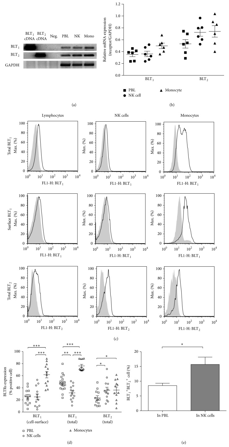
Initially it was believed that BLT1 was expressed only in phagocytes (granulocytes, eosinophils, and macrophages) [17, 32–34]. However, BLT1 mRNA was found in differentiated CD4+ TH cells and CD8+ TEFF cells and the LTB4-BLT1 pathway was found to be involved in inflammation-induced TH and TEFF cell recruitment [35, 36]. We, and others, have reported that LTB4 could augment NK cell cytotoxicity [27, 37–39]. Thus, we sought to determine the pattern of BLTR expression in NK cells. We assessed BLT1 and BLT2 mRNA expression by RT-PCR in each population of cells, PBLs, enriched NK cells, and monocytes (Figure 1(a)). Densitometric analysis of six donors' data, shown in Figure 1(b), indicated that BLT1 and BLT2 mRNA expression was similar in PBLs, NK cells, and monocytes, with a tendency for higher BLT2 mRNA expression in these cell populations.
Figure 1.
Expression of BLT1 and BLT2 in NK cells. RT-PCR of BLT1, BLT2, and GAPDH from PBLs, isolated NK cells, and monocytes were analyzed by agarose gel electrophoresis (a): lanes 1 and 2, human BLT1 and BLT2 cDNAs as positive controls; lane 3, polymerase-only, RT-negative samples for each oligonucleotide pair; lane 4, PBL mRNA; lane 5, NK cell mRNA; and lane 6, monocyte mRNA. (b) Expression levels of BLT1 and BLT2 mRNA were determined by densitometry and the ratio to GAPDH (the mean ± the SEM) from six donors is shown. (c) Whole cell and cell-surface expression of BLT1 (top and middle rows, resp.) and whole cell expression of BLT2 (bottom row) on PBLs, NK cells, and monocytes were measured by flow cytometry as described in Section 2. In the overlaid histogram graphs, grey background indicates control histograms, where cells were incubated with Ig isotype, and solid line indicates cells incubated with anti-BLTR antibodies. (d) Results represent the percentage of BLTR positive cells as population comparison analysis. Bars represent means ± SEM of ten to seventeen independent experiments. (e) Coexpression of BLT1 and BLT2 was measured as the percentage of events that were BLT1 and BLT2 double positive, gating on PBLs and NK cells (CD56+), respectively. Bar graphs represent means ± SEM of four independent experiments. ∗ P < 0.05; ∗∗ P < 0.01; ∗∗∗ P < 0.001, paired Student's t-test.
BLTR expression on fresh PBMCs was then evaluated by flow cytometry. Polyclonal anti-BLT1 and anti-BLT2 antibodies, which are directed toward an intracellular (C-terminus) domain of the receptor, were used to evaluate total (intracellular and extracellular) expression in permeabilized cells. In addition, a monoclonal anti-BLT1 antibody, which recognizes the N-terminal receptor epitope (extracellular), was used to evaluate cell-surface expression. Histogram graphs in Figure 1(c) illustrate the BLTR expression from a representative donor on lymphocytes, CD56+ NK cells, and monocytes, respectively. Figure 1(d) illustrates a compilation of individual donors and indicates that BLT1 receptors are expressed both intracellularly and on the cell-surface of all three cell populations. Around 25.19% ± 3.69% PBLs expressed BLT1 on the cell-surface, whereas almost 50% PBLs expressed BLT1 when intracellular receptors were taken into account (47.76% ± 3.23%). The discrepancy was smaller in NK cells and monocytes, as the expression of BLT1 on cell-surface (25.09% ± 4.75% and 61.76% ± 5.27%, resp.) was around 80% of total expression (31.64% ± 3.39% and 76.23% ± 2.56%). Although the antibodies used for cytometry were different, it appeared that the expression of BLT2 protein was lower than that of BLT1 in PBLs and monocytes, but it was similar in NK cells. Due to a lack of an antibody directed at the extracellular region of BLT2, we could not evaluate differences between its cell-surface and its total cellular expression. Interestingly, more NK cells (15.69% ± 2.49%) expressed both BLTRs than PBLs (8.53% ± 0.79%) (Figure 1(e)). Our findings of the heterogeneous expression of BLTR in NK cells led us to study the involvement of BLT1 and/or BLT2 in LTB4-mediated effects in these cells.
3.2. NK Cell Migration Response to LTB4
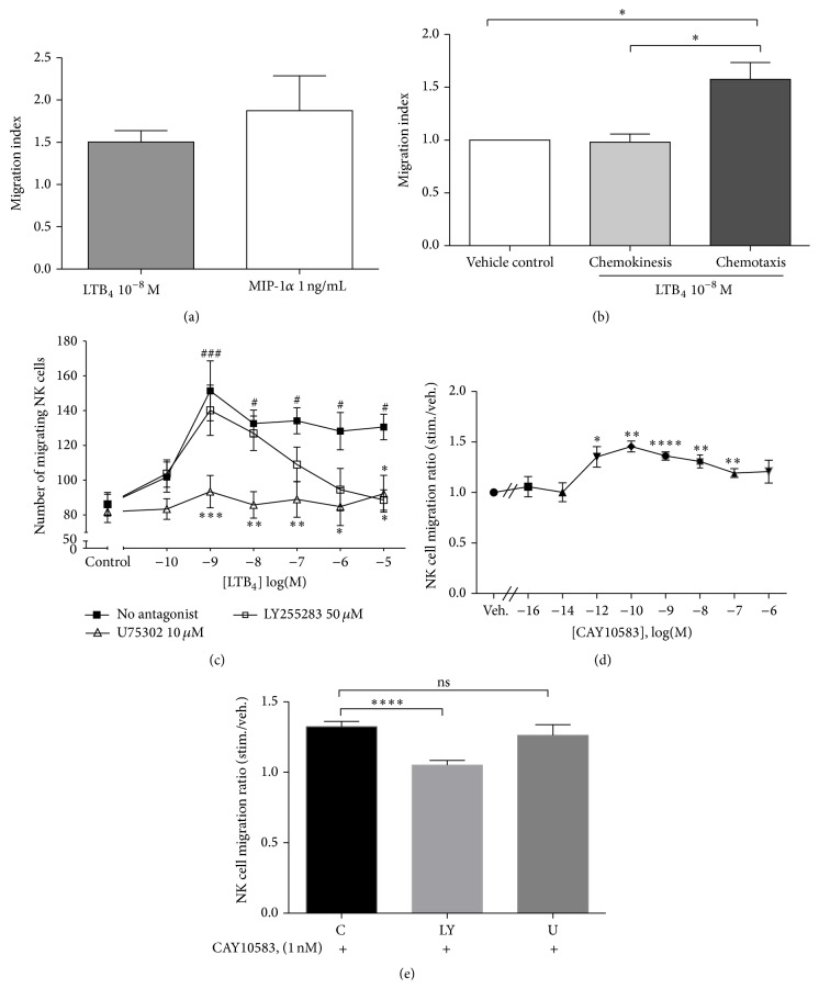
LTB4 is a potent neutrophil chemoattractant and recruits neutrophils to inflammatory sites in skin or lung by directing cell migration [40, 41]. Recent studies demonstrated that LTB4 can also induce migration of mast cells [42], dendritic cells [43], and T cells [35, 36, 44]. Thus, we investigated whether LTB4 could induce migration of human NK cells. As shown in Figure 2(a), LTB4 induced a 1.5-fold migration of NK cells at 10−8 M, and the magnitude of migration was similar to that induced by MIP-1α, a potent chemoattractant of NK cells [7]. This increase in chemotaxis was due to directional migration as LTB4 did not modulate NK cell chemokinesis (Figure 2(b)). NK cells migrated in response to a wide range of LTB4 concentrations, from 10−9 to 10−5 M, with a maximum at 10−9 M LTB4 (Figure 2(c)). In order to study the contribution of the two receptors to chemotaxis, we used selective antagonists. We found that LTB4-induced chemotaxis was abolished following preincubation with the BLT1 antagonist, U75302 at 10 μM, for 30 minutes. However, the BLT2 antagonist, LY255283, blocked LTB4-induced chemotaxis only at the highest concentrations of the ligand. Moreover, the selective BLT2 agonist CAY10583 was capable of inducing significant NK cell migration (Figure 2(d)) and this migration was only blocked by LY255283 (Figure 2(e)).
Figure 2.
NK cell migration in response to LTB4. (a) Bar graphs represent NK cell migration in response to MIPα, 1 ng/mL, and LTB4, 10−8 M. (MI: migration index, number of cells migrating in response to stimulus divided by number of cells migrating in response to control medium. Graph represents mean ± SEM of four independent experiments.) (b) Results illustrate the comparison of chemokinesis and chemotaxis to 10−8 M LTB4. Spontaneous migration in the presence of vehicle alone (ethanol 0.0033%) was normalized to 1. Bar graphs represent means ± SEM of four independent experiments, ∗ P < 0.05 paired Student's t-test. (c) PBLs were preincubated without antagonist (■), with U75302 10 μM (∆) or LY255283 50 μM (□) for 30 minutes at 37°C, before a chemotaxis assay with graded concentrations of LTB4 or vehicle control. The number of migrating cells was measured by FACS, gating on the NK cell population (CD3− CD56+). Each curve represents mean ± SEM of five independent experiments. # P < 0.05, and ### P < 0.001 by one-way ANOVA with Dunnett posttest to vehicle control. ∗ P < 0.05, ∗∗ P < 0.01, and ∗∗∗ P < 0.001 by two-way ANOVA with Bonferroni posttests to no-antagonist data. (d) NK cell migration in response to graded concentrations of CAY10583. Data are expressed as means ± SEM of ratios of migrating cells in response to CAY10583 versus vehicle (n = 8), ∗ P < 0.05, ∗∗ P < 0.01, ∗∗∗ P < 0.001, and ∗∗∗∗ P < 0.0001. (e) NK cell migration in response to 10−9 M CAY10583 in the absence or presence of LY255283 (LY) or U75302 (U). ∗∗∗∗ P < 0.0001, n = 5.
Our data suggest that the high-affinity BLT1 receptor mediates most of the LTB4-induced chemotactic activity in NK cells, with the lower affinity BLT2 receptor participating preferentially when LTB4 concentrations are very high.
3.3. BLT1 but Not BLT2 Mediates LTB4-Induced NK Cell Cytotoxicity
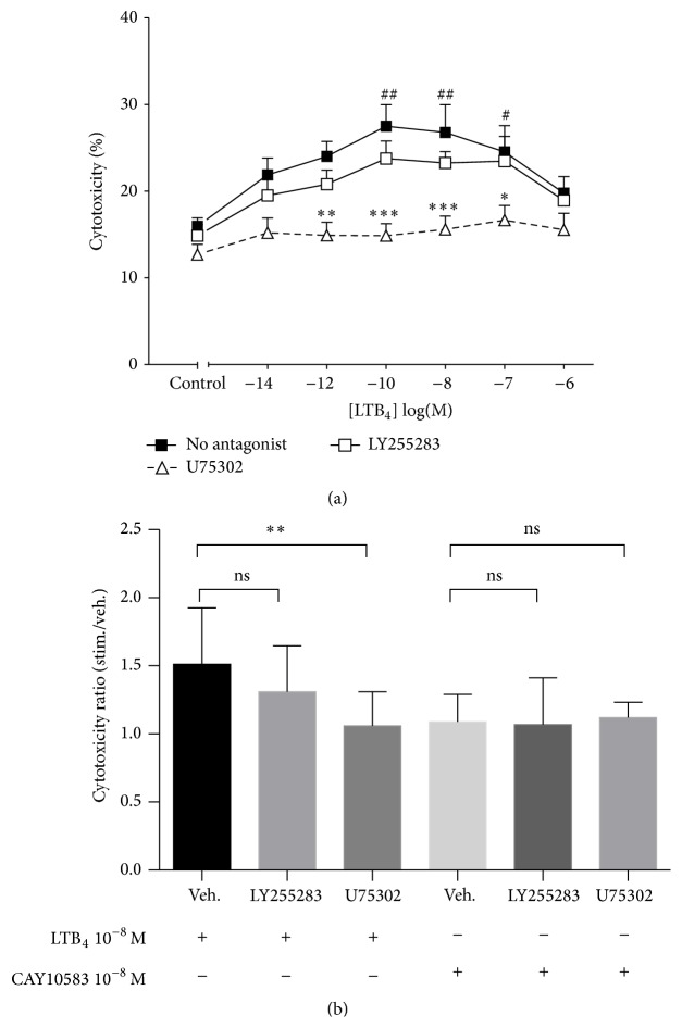
To determine which of the two receptors was necessary for the LTB4-induced effect on NK cell cytotoxic function, we again used the selective antagonists. LTB4 significantly enhanced NK cell cytolytic activity at 10−10 M to 10−7 M, with a maximal effect between 10−10 M and 10−8 M of LTB4 (Figure 3(a)). Preincubation with U75302 at 10 μM for 30 minutes abrogated the enhanced cytotoxicity. In contrast, LY255283 did not significantly block LTB4-induced cytotoxicity. Moreover, the selective BLT2 agonist Cay10583 failed to affect the level of NK cell activity (Figure 3(b)), suggesting that BLT2 was not involved in LTB4-induced cytotoxicity.
Figure 3.
(a) LTB4 enhances NK cell cytolytic function via the BLT1 receptor. PBLs (effector cells) were preincubated without antagonist (■) or with U75302 10 μM (∆) or LY255283 50 μM (□) for 30 minutes at 37°C and then were coincubated with CFSC-labelled K562 cells at E : T = 50 : 1 ratio in the presence of graded concentrations of LTB4. Cytotoxicity was measured using the percentage of 7AAD positive events gating on K562 cells. Each curve represents mean ± SEM of seven independent experiments. # P < 0.05, and ## P < 0.01 by one-way ANOVA with Dunnett posttest to controls. ∗ P < 0.05, ∗∗ P < 0.01, and ∗∗∗ P < 0.001 by two-way ANOVA with Bonferroni posttests to no-antagonist data. (b) Effects of LTB4 or CAY10583 on NK cell cytotoxicity in the absence or presence of LY255283 or U75302. Data are expressed as means ± SEM of cytotoxicity ratios. ∗∗ P < 0.01 using Student's paired t-test; n = 9.
3.4. Modulation of LTB4 Receptor Expression by IL-2 and IL-15
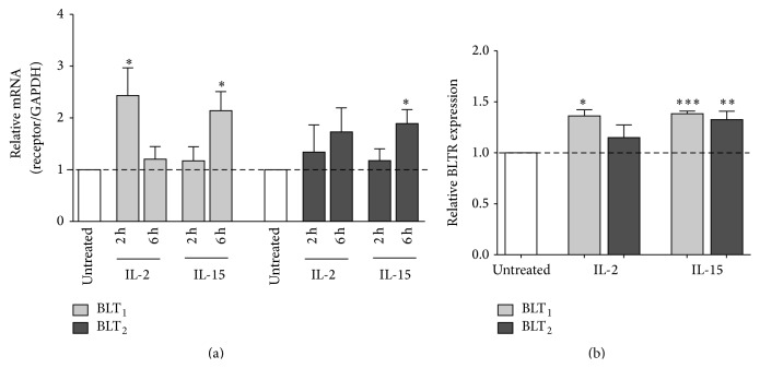
Pro- or anti-inflammatory cytokines can modulate BLT1 expression in human monocytes, with decreased expression induced by IFN-γ and TNFα, and increased expression stimulated by IL-10 and dexamethasone [45]. TNFα and dexamethasone also regulate BLT1 expression in neutrophils [21, 46]. Therefore, we sought to investigate whether IL-2 and IL-15, two NK-activating cytokines, could regulate BLTR expression. Real-time quantitative PCR was used to examine BLT1 and BLT2 mRNA expression in purified NK cells, incubated with IL-2 or IL-15 for 2 or 6 hours. IL-2 was found to induce a 2.4-fold increase of BLT1 expression after a 2 h stimulation but did not significantly increase BLT2 expression. IL-15 induced a 2.1-fold increase of BLT1 and a 1.9-fold increase of BLT2 expression after a 6 h incubation (Figure 4(a)). BLTR protein expression was then examined in NK cells incubated with IL-2 or IL-15 for 18 h. IL-2 only increased expression of BLT1, whereas IL-15-activated NK cells showed approximately 1.4- and 1.3-fold higher expression of both BLT1 and BLT2, respectively (Figure 4(b)).
Figure 4.
Modulation of BLTR expression on human NK cells by IL-2 and IL-15. (a) NK cells were incubated for 2 or 6 h in the absence or presence of IL-2 (50 ng/mL) or IL-15 (10 ng/mL). Total RNA was then extracted and analyzed by q-PCR. Bar graphs represent mean ± SEM of five independent experiments. ∗ P < 0.05, by one-way ANOVA with Dunnett posttest to untreated cells (control). (b) PBLs were incubated for 18 h in the absence or presence of IL-2 or IL-15. Total expression of BLT1 (light grey) and BLT2 (dark grey) on NK cells was measured by FACS and expressed as fold induction relative to untreated cells. Bar graphs represent mean ± SEM of seven independent experiments. ∗ P < 0.05, ∗∗ P < 0.01, and ∗∗∗ P < 0.001 by paired Student's t-test, treated versus untreated cells.
3.5. Regulation of LTB4-Induced NK Cell Migration by IL-2 and IL-15
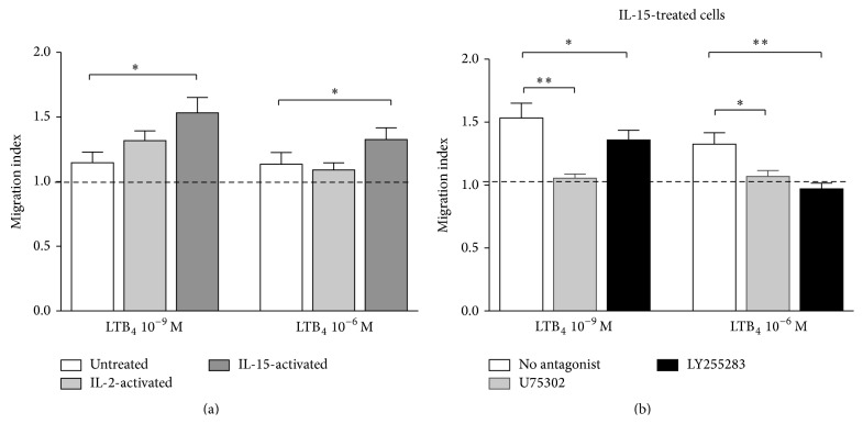
Dexamethasone-treated human neutrophils show a higher response to LTB4 in chemotaxis, as a result of enhanced BLT1 expression [21]. For this reason, we hypothesized that IL-2 and IL-15 may increase the chemotactic response of NK cells to LTB4. We treated PBLs with IL-2, IL-15, or medium for 18 h and NK cell migration in response to 10−9 M and 10−6 M LTB4 was examined (Figure 5(a)). IL-15 induced a significant augmentation of NK cell migration to both concentrations of LTB4. However, IL-2 did not significantly increase NK cell chemotactic response to LTB4 at either concentration. As illustrated in Figure 5(b), U75302 abolished the migration of IL-15-activated NK cells in response to both concentrations of LTB4; meanwhile, LY255283 totally abrogated the migration of IL-15-activated NK cells to higher concentration LTB4 (10−6 M) but only partially blocked the migration to lower concentration (10−9 M). Our data suggest that IL-15-enhanced chemotactic response of NK cells to LTB4 is also predominantly dependent on BLT1 signaling.
Figure 5.
Effect of IL-2 or IL-15 pretreatment on LTB4-induced NK cell migration. (a) PBLs were incubated for 18 h in the absence or presence of IL-2 (50 ng/mL) or IL-15 (10 ng/mL). Cells were stained with anti-CD3 and anti-CD56 antibodies and then placed in a chemotaxis assay in response to LTB4 10−9 M or 10−6 M. Migrating CD3− CD56+ cells were counted by FACS, and results were expressed as migration index (LTB4/vehicle). Bar graphs represent mean ± SEM of seven independent experiments. ∗ P < 0.05 by paired Student's t-test, treated versus untreated cells. (b) IL-15-activated PBLs were preincubated with or without U75302 or LY255283 before chemotaxis assays. Migrating CD3− CD56+ cells were counted by FACS, and results were expressed as migration index (LTB4/vehicle). Bar graphs represent mean ± SEM of seven independent experiments. ∗ P < 0.05 by paired Student's t-test.
3.6. Higher Response of IL-2- and IL-15-Activated NK Cells to LTB4 in Cytotoxicity
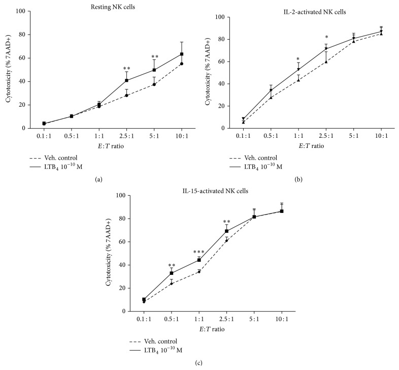
We next tested whether IL-2- and IL-15-activated NK cells could also increase their cytotoxic responses to LTB4. We treated purified NK cells with IL-2, IL-15, or medium for 18 h and then coincubated NK cells and K562 target cells at a graded effector : target cell ratio from 0.1 : 1 to 10 : 1. As shown in Figure 6, LTB4 induced a significant augmentation of cytotoxicity in resting NK cells at E : T ratios of 2.5 to 5 : 1. IL-2- and IL-15-activated NK cells showed an enhanced cytotoxicity in response to LTB4 at lower E : T ratios of 1 : 1 and 2.5 : 1 and 0.5 : 1 to 1 : 1, respectively. The BLT1 antagonist U75302, but not the BLT2 antagonist LY255283, blocked NK cell responsiveness to LTB4 (data not shown), suggesting that IL-2- and IL-15-induced upregulation of BLT1 expression may be involved in the augmented cytotoxic response to LTB4 by NK cells.
Figure 6.
Effect of IL-2 and IL-15 pretreatment on LTB4-induced NK cell cytotoxicity. Resting NK cells (a), IL-2-activated NK cells (b), and IL-15-activated NK cells (c) were coincubated with CFSE-labelled K562 cells at different E : T ratios for a 2 h cytotoxicity assay in the presence of LTB4 100 pM (solid lines) or vehicle control (dotted lines). The killing ability was measured using the percentage of 7AAD positive K562 cells. Each curve represents the means ± the SEM of three independent experiments. ∗ P < 0.05, ∗∗ P < 0.01, and ∗∗∗ P < 0.001 by two-way ANOVA with Bonferroni posttests to vehicle control.
4. Discussion
In the present study we investigated the differential involvement of BLT1 and BLT2 receptors in LTB4-induced chemotaxis and cytotoxic activity of NK cells.
Previously, we and others have shown that LTB4 increases NK cell cytotoxicity in several species [37, 39, 47]. However, BLTR expression in NK cells was still unclear. We found that the BLT1 receptor was expressed on human NK cells, which is in contrast to the findings of Pettersson et al. [48] who reported that no CD56+ cells were BLT1-positive, although they found low expression on some CD16+ cells. Interestingly, we found that the expression of BLT1 was quite variable from individual to individual. The highest BLT1 cell-surface expression on PBL and NK cells was 10 times higher than the lowest one. Moreover, we found expression of both mRNA and protein, which confirmed BLT1 and BLT2 expression in human NK cells. Meanwhile, we found that the expression of both receptors in monocytes and lymphocytes was similar to the findings of Islam et al. [13] and Yokomizo et al. [33]. The fact that protein expression of BLTR was different, especially that of BLT1, between PBLs, NK cells, and monocytes whereas mRNA expression was similar, suggests that there might be not only transcriptional regulation of BLT1 expression [49], but also posttranscriptional regulation of expression of these proteins. The higher expression of BLT2 mRNA in comparison to BLT1 mRNA might be due to the special gene structure, as the open reading frame of BLT2 is found in the promoter of BLT1 [49].
NK cells are rapidly distributed throughout the body in order to exercise their effector functions. However, the chemotactic signals responsible for their migration are not well known. We show here that LTB4 could induce a similar level of NK cell migration as MIP-1α, which has been shown as one of the most potent NK cell chemoattractants [7]. This suggests that LTB4 may induce early and rapid NK cell recruitment and trafficking to inflammatory sites, given that LTB4 is synthesized very rapidly [10] compared to chemokines. The peak of NK cell migration was in response to a low (1 nM) concentration of LTB4, which was lower than the optimal concentration inducing CD8+ T cell migration (10 nM) [36] or mast cell migration (100 nM) [42].
Similar to T cells [13–15] and neutrophils [11, 12], the BLT1 receptor was the principal signal transducer of LTB4-induced chemotaxis of human NK cells. However, unlike in the other cell types, BLT2 plays a partial role in the chemotactic response of NK cells to LTB4. This agrees with the observations of Yokomizo et al., who found that both BLT1 and BLT2 could mediate LTB4-induced cell migration in BLT1-BLT2 cotransfected CHO cells [33]. Since 12-HHT, a fatty acid derived from the COX pathway, may be the more effective agonist for BLT2 [50], it may also potentially be involved in NK cell chemotaxis.
On the other hand, our results show that BLT2 is probably not involved in LTB4-induced augmentation of NK cell cytotoxicity since the BLT1, but not the BLT2, antagonist could block all the effects of LTB4 and since the BLT2 agonist CAY10583 had no effect on NK cell cytotoxicity.
BLT1 and BLT2 belong to the seven transmembrane domains, G-protein-coupled receptor (GPCR) family. In some GPCRs, dimerization is associated with better receptor activation [51], and it has been shown that the BLT1:BLT1 homodimer does show higher affinity for LTB4 than the monomer [52]. However, BLT2 monomers have been shown to be more efficient at activating G proteins than the dimers [53]. In the present study, both BLT1 and BLT2 antagonists could block NK cell migration to higher concentrations of LTB4. This might suggest that, in NK cells, which coexpress the two receptors, BLT1:BLT2 heterodimers might exist, and although heterodimers of BLT receptors have not been investigated, it would not be surprising if one of the monomers influenced signal transduction [54].
We also observed that higher concentration of LTB4 seemed to induce a lower NK cell migration and cytotoxicity. It has been shown that rapid agonist-induced internalization and desensitization are important characteristics of BLT1 activation [55]. Thus, possibly, desensitization and internalization could reduce receptor responses at higher concentrations of LTB4.
It is now well established that pro- and anti-inflammatory cytokines as well as microbial products can modulate the expression of BLT1 in monocytes [45] and neutrophils [21]. In the present study, we found that cytokines which increase NK cell activation and cytotoxicity could also modulate BLT expression. We found that IL-15 upregulated both BLT1 and BLT2 expression, but IL-2 only increased BLT1 protein expression. These cytokines have also been shown to upregulate chemokine receptors in human NK cells [56, 57]. Although IL-2 and IL-15 belong to the same γc family of cytokines [6], IL-15R α-chain has a much broader tissue distribution than the IL2R α-chain, which is absent in NK cells [58]. IL-15 efficiently engages IL-2/15Rβγ with IL-15Rα for signal transduction, leading to the rapid upregulation of receptors for C-C chemokines, whereas IL-2 is unable to do so [57], in parallel with unaffected BLT2 expression.
Moreover, IL-2 and IL-15 augmented NK cell responses to LTB4 in terms of chemotaxis and cytolytic function. Preincubation with IL-2 did not increase NK cell migration to the same degree as treatment with IL-15, especially at higher concentration of LTB4, suggesting that BLT2 may be involved. The lower effect of IL-2 on BLT expression may be the consequence of the lack of IL-2Rα, which results in a receptor with lower affinity (IL-2Rβγ) for IL-2, and this in turn could result in a weaker modulation of BLT2 expression. However, IL-2- and IL-15-activated NK cells showed a similar augmentation in LTB4-induced cytotoxicity, which was mediated by BLT1, possibly due to the equivalent upregulation of BLT1 expression by both cytokines.
We previously showed that LTB4 augments IL-2Rβ expression in CD56+ NK cells and induces their responsiveness to 100-fold lower concentrations of IL-2 in terms of cytolytic activity [30]. Moreover, IL-15Rα mRNA expression was augmented in NK cells after a 30-minute stimulation with 100 nM LTB4 (data not shown). The evidence that cytokines and leukotrienes can increase each other's receptor expression suggests that IL-2, IL-15, and LTB4 may synergize to augment NK cell migration to tumour or infection sites and enhance cytotoxic responses.
In conclusion, functional BLT1 and BLT2 receptors are expressed in human NK cells and mediate, to a different degree, LTB4-induced chemotaxis and cytotoxicity. IL-2- and IL-15-dependent modulation of BLTR expression can upregulate the response of NK cells to LTB4.
Acknowledgment
This work was funded by an operating grant (MOP-102492) from the Canadian Institutes of Health Research to Jana Stankova and Marek Rola-Pleszczynski, who are members of the FRQS-funded Centre de recherche du Centre Hospitalier Universitaire de Sherbrooke.
Abbreviations
- 7AAD:
7-Aminoactinomycin D
- ADCC:
Antibody-dependent cellular cytotoxicity
- APC:
Allophycocyanin
- BLTR:
LTB4 receptor
- CFSE:
Carboxyfluorescein succinimidyl ester
- FITC:
Fluorescein isothiocyanate
- GAPDH:
Glyceraldehyde-3-phosphate dehydrogenase
- LTB4:
Leukotriene B4
- NK cell:
Natural killer cell
- PBLs:
Peripheral blood lymphocytes
- PE:
R-Phycoerythrin.
Conflict of Interests
The authors declare that there is no conflict of interests regarding the publication of this paper.
References
- 1.Paul W. E. Fundamental Immunology. Philadelphia, Pa, USA: Lippincott Williams & Wilkins; 2008. [Google Scholar]
- 2.Caligiuri M. A. Human natural killer cells. Blood. 2008;112(3):461–469. doi: 10.1182/blood-2007-09-077438. [DOI] [PMC free article] [PubMed] [Google Scholar]
- 3.Roitt I. M., Brostoff J., Male D. K. Immunology. Edinburgh, Scotland: Mosby; 2001. [Google Scholar]
- 4.Abo T., Miller C. A., Balch C. M., Cooper M. D. Interleukin 2 receptor expression by activated HNK-1+ granular lymphocytes: a requirement for their proliferation. The Journal of Immunology. 1983;131(4):1822–1826. [PubMed] [Google Scholar]
- 5.Wang K. S., Frank D. A., Ritz J. Interleukin-2 enhances the response of natural killer cells to interleukin-12 through up-regulation of the interleukin-12 receptor and STAT4. Blood. 2000;95(10):3183–3190. [PubMed] [Google Scholar]
- 6.Carson W. E., Giri J. G., Lindemann M. J., et al. Interleukin (IL) 15 is a novel cytokine that activates human natural killer cells via components of the IL-2 receptor. Journal of Experimental Medicine. 1994;180(4):1395–1403. doi: 10.1084/jem.180.4.1395. [DOI] [PMC free article] [PubMed] [Google Scholar]
- 7.Taub D. D., Sayers T. J., Carter C. R. D., Ortaldo J. R. α and β chemokines induce NK cell migration and enhance NK-mediated cytolysis. The Journal of Immunology. 1995;155(8):3877–3888. [PubMed] [Google Scholar]
- 8.Pohajdak B., Gomez J., Orr F. W., Khalil N., Talgoy M., Greenberg A. H. Chemotaxis of large granular lymphocytes. Journal of Immunology. 1986;136(1):278–284. [PubMed] [Google Scholar]
- 9.Borgeat P., Samuelsson B. Metabolism of arachidonic acid in polymorphonuclear leukocytes. Structural analysis of novel hydroxylated compounds. The Journal of Biological Chemistry. 1979;254(16):7865–7869. [PubMed] [Google Scholar]
- 10.Samuelsson B. Leukotrienes: mediators of immediate hypersensitivity reactions and inflammation. Science. 1983;220(4597):568–575. doi: 10.1126/science.6301011. [DOI] [PubMed] [Google Scholar]
- 11.Medoff B. D., Tager A. M., Jackobek R., Means T. K., Wang L., Luster A. D. Antibody-antigen interaction in the airway drives early granulocyte recruitment through BLT1. American Journal of Physiology—Lung Cellular and Molecular Physiology. 2006;290(1):L170–L178. doi: 10.1152/ajplung.00212.2005. [DOI] [PubMed] [Google Scholar]
- 12.Kim N. D., Chou R. C., Seung E., Tager A. M., Luster A. D. A unique requirement for the leukotriene B4 receptor BLT1 for neutrophil recruitment in inflammatory arthritis. The Journal of Experimental Medicine. 2006;203(4):829–835. doi: 10.1084/jem.20052349. [DOI] [PMC free article] [PubMed] [Google Scholar]
- 13.Islam S. A., Thomas S. Y., Hess C., et al. The leukotriene B4 lipid chemoattractant receptor BLT1 defines antigen-primed T cells in humans. Blood. 2006;107(2):444–453. doi: 10.1182/blood-2005-06-2362. [DOI] [PMC free article] [PubMed] [Google Scholar]
- 14.Miyahara N., Takeda K., Miyahara S., et al. Leukotriene B4 receptor-1 is essential for allergen-mediated recruitment of CD8+ T cells and airway hyperresponsiveness. The Journal of Immunology. 2005;174(8):4979–4984. doi: 10.4049/jimmunol.174.8.4979. [DOI] [PubMed] [Google Scholar]
- 15.Taube C., Miyahara N., Ott V., et al. The leukotriene B4 receptor (BLT1) is required for effector CD8+ T cell-mediated, mast cell-dependent airway hyperresponsiveness. Journal of Immunology. 2006;176(5):3157–3164. doi: 10.4049/jimmunol.176.5.3157. [DOI] [PubMed] [Google Scholar]
- 16.Yokomizo T., Izumi T., Chang K., Takuwa Y., Shimizu T. A G-protein-coupled receptor for leukotriene B4 that mediates chemotaxis. Nature. 1997;387(6633):620–624. doi: 10.1038/42506. [DOI] [PubMed] [Google Scholar]
- 17.Kamohara M., Takasaki J., Matsumoto M., et al. Molecular cloning and characterization of another leukotriene B4 receptor. The Journal of Biological Chemistry. 2000;275(35):27000–27004. doi: 10.1074/jbc.c000382200. [DOI] [PubMed] [Google Scholar]
- 18.Yokomizo T., Kato K., Terawaki K., Izumi T., Shimizu T. A second leukotriene B4 receptor, BLT2. A new therapeutic target in inflammation and immunological disorders. The Journal of Experimental Medicine. 2000;192(3):421–431. doi: 10.1084/jem.192.3.421. [DOI] [PMC free article] [PubMed] [Google Scholar]
- 19.Miyahara N., Takeda K., Miyahara S., et al. Requirement for leukotriene B4 receptor 1 in allergen-induced airway hyperresponsiveness. American Journal of Respiratory and Critical Care Medicine. 2005;172(2):161–167. doi: 10.1164/rccm.200502-205OC. [DOI] [PMC free article] [PubMed] [Google Scholar]
- 20.Terawaki K., Yokomizo T., Nagase T., et al. Absence of leukotriene B4 receptor 1 confers resistance to airway hyperresponsiveness and Th2-type immune responses. The Journal of Immunology. 2005;175(7):4217–4225. doi: 10.4049/jimmunol.175.7.4217. [DOI] [PubMed] [Google Scholar]
- 21.Stankova J., Turcotte S., Harris J., Rola-Pleszczynski M. Modulation of leukotriene B4 receptor-1 expression by dexamethasone: potential mechanism for enhanced neutrophil survival. Journal of Immunology. 2002;168(7):3570–3576. doi: 10.4049/jimmunol.168.7.3570. [DOI] [PubMed] [Google Scholar]
- 22.Murray J., Ward C., O'Flaherty J. T., et al. Role of leukotrienes in the regulation of human granulocyte behaviour: dissociation between agonist-induced activation and retardation of apoptosis. British Journal of Pharmacology. 2003;139(2):388–398. doi: 10.1038/sj.bjp.0705265. [DOI] [PMC free article] [PubMed] [Google Scholar]
- 23.Rola-Pleszczynski M., Staňková J. Leukotriene B4 enhances interleukin-6 (IL-6) production and IL-6 messenger RNA accumulation in human monocytes in vitro: transcriptional and posttranscriptional mechanisms. Blood. 1992;80(4):1004–1011. [PubMed] [Google Scholar]
- 24.Huang L., Zhao A., Wong F., et al. Leukotriene B4 strongly increases monocyte chemoattractant protein-1 in human monocytes. Arteriosclerosis, Thrombosis, and Vascular Biology. 2004;24(10):1783–1788. doi: 10.1161/01.atv.0000140063.06341.09. [DOI] [PubMed] [Google Scholar]
- 25.Mathis S. P., Jala V. R., Lee D. M., Haribabu B. Nonredundant roles for leukotriene B4 receptors BLT1 and BLT2 in inflammatory arthritis. The Journal of Immunology. 2010;185(5):3049–3056. doi: 10.4049/jimmunol.1001031. [DOI] [PubMed] [Google Scholar]
- 26.Iizuka Y., Okuno T., Saeki K., et al. Protective role of the leukotriene B4 receptor BLT2 in murine inflammatory colitis. The FASEB Journal. 2010;24(12):4678–4690. doi: 10.1096/fj.10-165050. [DOI] [PubMed] [Google Scholar]
- 27.Rola-Pleszczynski M., Gagnon L., Sirois P. Leukotriene B4 augments human natural cytotoxic cell activity. Biochemical and Biophysical Research Communications. 1983;113(2):531–537. doi: 10.1016/0006-291x(83)91758-8. [DOI] [PubMed] [Google Scholar]
- 28.Rola-Pleszczynski M., Gagnon L., Rudzinska M., Borgeat P., Sirois P. Human natural cytotoxic cell activity: enhancement by leukotrienes (LT) A4, B4 and D4, but not by stereoisomers of LTB4 or HETES. Prostaglandins, Leukotrienes and Medicine. 1984;13(1):113–117. doi: 10.1016/0262-1746(84)90111-2. [DOI] [PubMed] [Google Scholar]
- 29.Seaman W. E. Human natural killer cell activity is reversibly inhibited by antagonists of lipoxygenation. Journal of Immunology. 1983;131(6):2953–2957. [PubMed] [Google Scholar]
- 30.Stankova J., Gagnon N., Rola-Pleszczynski M. Leukotriene B4 augments interleukin-2 receptor-beta (IL-2R β) expression and IL-2R β-mediated cytotoxic response in human peripheral blood lymphocytes. Immunology. 1992;76(2):258–263. [PMC free article] [PubMed] [Google Scholar]
- 31.Gaudreau R., Le Gouill C., Métaoui S., Lemire S., Stankovà J., Rola-Pleszczynski M. Signalling through the leukotriene B4 receptor involves both alphai and alpha16, but not alphaq or alpha11 G-protein subunits. Biochemical Journal. 1998;335(part 1):15–18. doi: 10.1042/bj3350001. [DOI] [PMC free article] [PubMed] [Google Scholar]
- 32.Huang W.-W., Garcia-Zepeda E. A., Sauty A., Oettgen H. C., Rothenberg M. E., Luster A. D. Molecular and biological characterization of the murine leukotriene B4 receptor expressed on eosinophils. The Journal of Experimental Medicine. 1998;188(6):1063–1074. doi: 10.1084/jem.188.6.1063. [DOI] [PMC free article] [PubMed] [Google Scholar]
- 33.Yokomizo T., Izumi T., Shimizu T. Co-expression of two LTB4 receptors in human mononuclear cells. Life Sciences. 2001;68(19-20):2207–2212. doi: 10.1016/S0024-3205(01)01007-4. [DOI] [PubMed] [Google Scholar]
- 34.Yokomizo T., Izumi T., Shimizu T. Leukotriene B4: Metabolism and signal transduction. Archives of Biochemistry and Biophysics. 2001;385(2):231–241. doi: 10.1006/abbi.2000.2168. [DOI] [PubMed] [Google Scholar]
- 35.Tager A. M., Bromley S. K., Medoff B. D., et al. Leukotriene B4 receptor BLT1 mediates early effector T cell recruitment. Nature Immunology. 2003;4(10):982–990. doi: 10.1038/ni970. [DOI] [PubMed] [Google Scholar]
- 36.Goodarzi K., Goodarzi M., Tager A. M., Luster A. D., von Andrian U. H. Leukotriene B4 and BLT1 control cytotoxic effector T cell recruitment to inflamed tissues. Nature Immunology. 2003;4(10):965–973. doi: 10.1038/ni972. [DOI] [PubMed] [Google Scholar]
- 37.Thivierge M., Rola-Pleszczynski M. Enhancement of pulmonary natural killer cell activity by platelet activating factor: mechanisms of activation involving Ca2+, protein kinase C, and lipoxygenase products. American Review of Respiratory Disease. 1991;144(2):272–277. doi: 10.1164/ajrccm/144.2.272. [DOI] [PubMed] [Google Scholar]
- 38.Chang K.-J., Saito H., Tatsuno I., Tamura Y., Yoshida S. Role of 5-lipoxygenase products of arachidonic acid in cell-to-cell interaction between macrophages and natural killer cells in rat spleen. Journal of Leukocyte Biology. 1991;50(3):273–278. doi: 10.1002/jlb.50.3.273. [DOI] [PubMed] [Google Scholar]
- 39.Bray R. A., Brahmi Z. Role of lipoxygenation in human natural killer cell activation. The Journal of Immunology. 1986;136(5):1783–1790. [PubMed] [Google Scholar]
- 40.Martin T. R., Pistorese B. P., Chi E. Y., Goodman R. B., Matthay M. A. Effects of leukotriene B4 in the human lung. Recruitment of neutrophils into the alveolar spaces without a change in protein permeability. The Journal of Clinical Investigation. 1989;84(5):1609–1619. doi: 10.1172/jci114338. [DOI] [PMC free article] [PubMed] [Google Scholar]
- 41.Soter N. A., Lewis R. A., Corey E. J., Austen K. F. Local effects of synthetic leukotrienes (LTC4, LTD4, LTE4, and LTB4) in human skin. Journal of Investigative Dermatology. 1983;80(2):115–119. doi: 10.1111/1523-1747.ep12531738. [DOI] [PubMed] [Google Scholar]
- 42.Lundeen K. A., Sun B., Karlsson L., Fourie A. M. Leukotriene B4 receptors BLT1 and BLT2: expression and function in human and murine mast cells. The Journal of Immunology. 2006;177(5):3439–3447. doi: 10.4049/jimmunol.177.5.3439. [DOI] [PubMed] [Google Scholar]
- 43.Shin E. H., Lee H. Y., Bae Y.-S. Leukotriene B4 stimulates human monocyte-derived dendritic cell chemotaxis. Biochemical and Biophysical Research Communications. 2006;348(2):606–611. doi: 10.1016/j.bbrc.2006.07.084. [DOI] [PubMed] [Google Scholar]
- 44.de Souza Costa M. F., de Souza-Martins R., de Souza M. C., et al. Leukotriene B4 mediates γδ T lymphocyte migration in response to diverse stimuli. Journal of Leukocyte Biology. 2010;87(2):323–332. doi: 10.1189/jlb.0809563. [DOI] [PMC free article] [PubMed] [Google Scholar]
- 45.Pettersson A., Sabirsh A., Bristulf J., et al. Pro- and anti-inflammatory substances modulate expression of the leukotriene B4 receptor, BLT1, in human monocytes. Journal of Leukocyte Biology. 2005;77(6):1018–1025. doi: 10.1189/jlb.1204740. [DOI] [PubMed] [Google Scholar]
- 46.O'Flaherty J. T., Rossi A. G., Redman J. F., Jacobson D. P. Tumor necrosis factor-alpha regulates expression of receptors for formyl-methionyl-leucyl-phenylalanine, leukotriene B4, and platelet-activating factor. Dissociation from priming in human polymorphonuclear neutrophils. Journal of Immunology. 1991;147(11):3842–3847. [PubMed] [Google Scholar]
- 47.Vaillier D., Daculsi R., Gualde N., Bezian J. H. Effect of LTB4 on the inhibition of natural cytotoxic activity by PGE2. Cellular Immunology. 1992;139(1):248–258. doi: 10.1016/0008-8749(92)90117-8. [DOI] [PubMed] [Google Scholar]
- 48.Pettersson A., Richter J., Owman C. Flow cytometric mapping of the leukotriene B4 receptor, BLT1, in human bone marrow and peripheral blood using specific monoclonal antibodies. International Immunopharmacology. 2003;3(10-11):1467–1475. doi: 10.1016/s1567-5769(03)00145-0. [DOI] [PubMed] [Google Scholar]
- 49.Kato K., Yokomizo T., Izumi T., Shimizu T. Cell-specific transcriptional regulation of human leukotriene B(4) receptor gene. The Journal of Experimental Medicine. 2000;192(3):413–420. doi: 10.1084/jem.192.3.413. [DOI] [PMC free article] [PubMed] [Google Scholar]
- 50.Okuno T., Iizuka Y., Okazaki H., Yokomizo T., Taguchi R., Shimizu T. 12(S)-hydroxyheptadeca-5Z, 8E, 10E-trienoic acid is a natural ligand for leukotriene B4 receptor 2. The Journal of Experimental Medicine. 2008;205(4):759–766. doi: 10.1084/jem.20072329. [DOI] [PMC free article] [PubMed] [Google Scholar]
- 51.Bouvier M. Oligomerization of G-protein-coupled transmitter receptors. Nature Reviews Neuroscience. 2001;2(4):274–286. doi: 10.1038/35067575. [DOI] [PubMed] [Google Scholar]
- 52.Banères J.-L., Parello J. Structure-based analysis of GPCR function: evidence for a novel pentameric assembly between the dimeric leukotriene B4 receptor BLT1 and the G-protein. Journal of Molecular Biology. 2003;329(4):815–829. doi: 10.1016/s0022-2836(03)00439-x. [DOI] [PubMed] [Google Scholar]
- 53.Arcemisbéhère L., Sen T., Boudier L., et al. Leukotriene BLT2 receptor monomers activate the Gi2 GTP-binding protein more efficiently than dimers. Journal of Biological Chemistry. 2010;285(9):6337–6347. doi: 10.1074/jbc.m109.083477. [DOI] [PMC free article] [PubMed] [Google Scholar]
- 54.Mesnier D., Banères J.-L. Cooperative conformational changes in a G-protein-coupled receptor dimer, the leukotriene B4 receptor BLT1. The Journal of Biological Chemistry. 2004;279(48):49664–49670. doi: 10.1074/jbc.m404941200. [DOI] [PubMed] [Google Scholar]
- 55.O'Flaherty J. T., Redman J. F., Jacobson D. P. Cyclical binding, processing, and functional interactions of neutrophils with leukotriene B4 . Journal of Cellular Physiology. 1990;142(2):299–308. doi: 10.1002/jcp.1041420212. [DOI] [PubMed] [Google Scholar]
- 56.Inngjerdingen M., Damaj B., Maghazachi A. A. Expression and regulation of chemokine receptors in human natural killer cells. Blood. 2001;97(2):367–375. doi: 10.1182/blood.V97.2.367. [DOI] [PubMed] [Google Scholar]
- 57.Perera L. P., Goldman C. K., Waldmann T. A. IL-15 induces the expression of chemokines and their receptors in T lymphocytes. The Journal of Immunology. 1999;162(5):2606–2612. [PubMed] [Google Scholar]
- 58.Giri J. G., Kumaki S., Ahdieh M., et al. Identification and cloning of a novel IL-15 binding protein that is structurally related to the α chain of the IL-2 receptor. The EMBO Journal. 1995;14(15):3654–3663. doi: 10.1002/j.1460-2075.1995.tb00035.x. [DOI] [PMC free article] [PubMed] [Google Scholar]


