Abstract
The use of diced cartilage grafts in reconstructive surgery was first described by Peer in 1943 though it was not for rhinoplasty. A number of studies describing diced cartilage have followed since then, but the technique has never achieved widespread use. In recent years, however, an interest in using diced cartilage for augmentation rhinoplasty has resurfaced. As surgeons revisit this technique, it is important that this technique is subjected to critical evaluation in terms of materials, approaches, and indications of using using diced-cartilage augmentation. External rhinoplasty approach with diced cartilage as a graft was used to for augmenting the nasal dorsum in 32 patients. Cosmetic appearance improved in all cases both subjectively and objectively. Only one patient showed constriction of dorsum 09 months after surgery. None of the patient had any intra-operative complication, 02 had donor site complication in the form of aural haematoma in 01 patient and wound infection in 01 patient. Diced cartilage technique is an attractive option for use in rhinoplasties especially those requiring augmentation procedures.
Keywords: Rhinoplasty, Diced cartilage, Nose deformity
Introduction
Graft materials in rhinoplasty can be obtained from various sources namely autogenous grafts like cartilage and bone [1] or from alloplastic materials like Medpor, Goretex [2] or Homografts [3]. However, the ideal graft material is yet to be found with each biomaterial having its advantages and disadvantages. Out of these autogenous cartilage has the largest recorded literature and is currently the material of choice for nasal augmentation [4].
Diced cartilage grafts in rhinoplasty were in widespread use around 70 years ago [5, 6] but lost their popularity post world war II. In recent years however renewed interest in this technique has emerged. The use of diced cartilage rather than a solid piece of cartilage attract surgeons because of its greater flexibility, minimal risk of warping and, obviates requirement of single large graft. A critical issue for biomaterials is absorption or extrusion and same has been presented as a concern in diced cartilage rhinoplasty. As we are revisting this technique, it is eminent that it will be subjected to various criticisms as well as laudation. Diced cartilage has been used in different methods like diced cartilage, diced cartilage wrapped in fascia and diced cartilage covered in fascia [7]. We hereby present our experience with this technique at our tertiary care centre.
Methodology
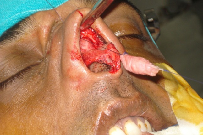
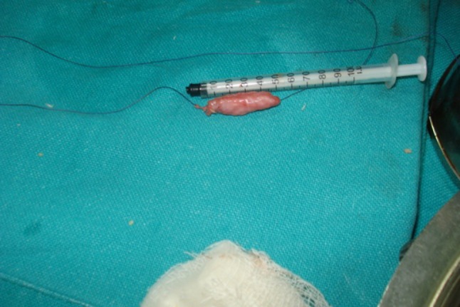
All rhinoplasties in our series were performed by one single surgical team headed by a senior ENT surgeon of our tertiary care centre with considerable experience in facial and aesthetic surgery. Open rhinoplasty technique was used in all cases. The common indications were to augment the nasal dorsum because of a supratip depression, bony saddle or combined bony and cartilaginous saddle. Preoperatively careful selection of patients was done by analysis of deformity in addition to expectations of the patient, psychological analysis and detailed counselling. Intra-operatively a trans-columellar inverted V shaped incision was connected to a bilateral rim incision and osteocartilaginous dorsum of nose was exposed (Fig. 1). Harvesting of cartilage was done from septal cartilage and associated deviation of nasal septum if any was corrected at the same time. If septal cartilage was found deficient or insufficient, conchal cartilage was harvested. Temporalis fascia was taken by supra-auricular incision. In cases where conchal cartilage was to be harvested, temporalis fascia as well conchal cartilage was harvested from post auricular incision. Obtained cartilage graft was diced into 0.5–1.0 mm pieces using no 11 blade. The diced cartilage is then filled in 1.0 cc tuberculin syringe. Sheet of temporalis fascia obtained was wrapped around cartilage filled syringe and closed with 4-0 vicryl. Tuberculin syringe was gradually withdrawn from the bag while simultaneously filling the bag with cartilage pieces (Fig. 2). Any extra cartilage was milked out. Due consideration was given to the size of dorsum graft which varied on case to case basis. This dorsum cartilage graft preparation is then placed in the defect and secured with suture passing beneath the flap and coming out through skin over radix for support and to avoid preoperative displacement. Any other procedures for the patient like osteotomies were obviously performed prior to placing the dorsal diced cartilage graft.
Fig. 1.

Exposure of nasal dorsum by external rhinoplasty/open approach
Fig. 2.

Diced cartilage wrapped in fascia made by filling diced pieces in tuberculin syringe
32 such procedures were performed in a period of 2 years from Aug 2011 and Dec 2013. Post operatively ant nasal packing was done which was removed after 24–48 h. Post-op analysis of correction was done after 3, 6 and 12 months.
Results
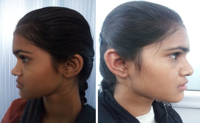
Out of the 32 cases performed, all were primary rhinoplasties. There were 22 female and 10 male patients. Common etiology was trauma in 20 cases, and 10 cases were of congenital deficiency and 02 following septal abscess. In 26 cases cartilage was harvested from septal cartilage and in 08 from both septum and concha. None of the patient had any intra-operative complication, 02 had donor site complication in the form of aural haematoma in 01 patient and wound infection in 01 patient. Only one patient showed constriction of dorsum 09 months after surgery. However patient was still quite satisfied with the outcome and did not want revision surgery. No case showed extrusion of graft. No warping was seen in any of the patient. Cosmetic appearance improved in all cases both subjectively and objectively (Fig. 3).
Fig. 3.

Comparison between pre and post operative nasal dorsum: A satisfactory one, both subjectively and objectively
Discussion
The goal of septorhinoplasty is reconstruction of the nasal skeleton to provide adequate structural support allowing for optimum functioning of the nasal airway while achieving an aesthetically pleasing result with the rest of the face. Autogenous grafts, particularly cartilage has been the gold standard because of its large acceptance rate, durability, virtual lack of an immunogenic response, low infection, and extrusion rates [8]. The use of diced cartilage grafts in reconstructive surgery was first described by Peer [9] in 1943. A number of additional reports describing diced cartilage have followed since then, but the technique has never achieved widespread use.
To overcome the disadvantages of its potential reported problems like palpability and visibility of diced grafts, surgeons have described the use of autogenous, synthetic, or alloplastic wraps to camouflage the cartilage construct. A great deal of controversy exists about the techniques that have been advocated, and the scaffold for delivering diced cartilage has yet to be determined.
In 2000, Erol [10] introduced the concept of a ‘‘Turkish Delight,’’ whereby diced cartilage wrapped in Surgicel was used as an adjunct to rhinoplasty. Most studies rejected this technique as it failed to correct the problem due to complete resorption by about 3 months and thereafter wrapping in temporalis fascia was started [11]. This technique being only a decade old has still been in great debate. Experimental studies have histologically proven diced cartilage wrapped in fascia to stay viable for more than 9 months. [12].
Adoption of any new grafting will require many questions to answer like what are its advantages and disadvantages over existing techniques and whether it is safe and likely to work on long term basis. We hereby discuss our experience of same. We found diced cartilage wrapped in fascia to be advantageous because it being an autograft rejection is never an issue. It is locally available and does not require large pieces of cartilage therefore ensuring better utilisation of available graft material. It is much easier to prepare and does not require careful carving of cartilage. The same preparation can be used to correct variety of deformities by digital manipulation. Minor postoperative residual deformities can be corrected by digital manipulation up to 10–15 days post operative which is just not a possibility with other techniques. Issues raised previously regarding resorption of diced cartilage in long term basis were not faced by us and there was no reduction in nasal dorsum post operatively after 1 year compared to 3 months evaluation. Disadvantage of this graft is that it does not provide any functional correction and does not give structural support. Another disadvantage is that it cannot be used where large amount of augmentation is required. In terms of long term viability we found that it undergoes minimal resorption which is not clinically significant.
Conclusion
Therefore we conclude that diced cartilage technique is an attractive option for use in rhinoplasties especially those requiring augmentation procedures. Its use should be encouraged till we find any substantial evidence against it. Adadpting to a new technique always has its opponents but only those who counter them emerge as winners.
References
- 1.Cheney ML. Reconstructive grafting by the open nasal approach. Facial Plast Surg Clinics of N Am. 1993;1:99–109. [Google Scholar]
- 2.Stucker FJ. Use of implantation in facial deformity. Laryngoscope. 1977;51:1523–1527. doi: 10.1288/00005537-197709000-00012. [DOI] [PubMed] [Google Scholar]
- 3.Kridel RWH, Konior RJ. Reconstructive grafting by the open nasal approach. Facial Plast Surg. 1993;3:141–144. [Google Scholar]
- 4.Stucker FJ, Gage White L. Survey of surgical implants. Facial Plast Surg. 1986;3:141–144. doi: 10.1055/s-2008-1064834. [DOI] [Google Scholar]
- 5.Burian F. The plastic surgery atlas. New York: Macmillan; 1968. [Google Scholar]
- 6.Denecke HJ, Meyer R. Plastic surgery of the head and neck. New York: Springer; 1967. p. 148. [Google Scholar]
- 7.Daniel RK. Diced cartilage grafts in rhinoplasty surgery: current techniques and applications. Plast Reconstr Surg. 2008;122:1883–1891. doi: 10.1097/PRS.0b013e31818d2104. [DOI] [PubMed] [Google Scholar]
- 8.Moshaver A, Gantous A. The use of autogenous costal cartilage graft in septorhinoplasty. Otolaryngol Head Neck Surg. 2007;137:862–867. doi: 10.1016/j.otohns.2007.06.740. [DOI] [PubMed] [Google Scholar]
- 9.Peer LA. Extended use of diced cartilage grafts. Plast Reconstr Surg. 1954;14:178–185. doi: 10.1097/00006534-195409000-00002. [DOI] [PubMed] [Google Scholar]
- 10.Erol O. The Turkish delight: a pliable graft for rhinoplasty. Plast Reconstr Surg. 2000;105:2229–2241. doi: 10.1097/00006534-200005000-00051. [DOI] [PubMed] [Google Scholar]
- 11.Daniel RK, Calvert JW. Diced cartilage grafts in rhinoplasty surgery. Plast Reconstr Surg. 2004;113:2156–2171. doi: 10.1097/01.PRS.0000122544.87086.B9. [DOI] [PubMed] [Google Scholar]
- 12.Calvert JW, Brenner KB, DaCosta-Iyer M, Evans GRD, Daniel RK. Histological analysis of human diced cartilage grafts. Plast Reconstr Surg. 2006;118:230. doi: 10.1097/01.prs.0000220463.73865.78. [DOI] [PubMed] [Google Scholar]


