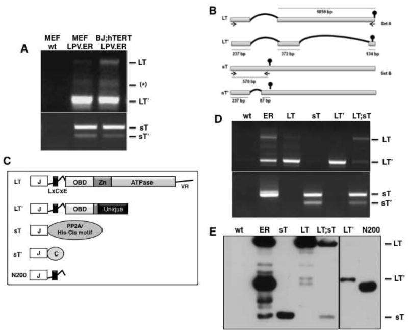
Figure 2. LPV.ER encodes four differentially spliced products.
A) RT-PCR analysis of MEFs and BJ;hTERT cells expressing LPV.ER. Three bands amplified with primer set A (materials and methods) are shown in the upper panel and correspond to LT (~2.1kb), LT′ (~750bs) and a PCR artifact (~1kb) confirmed by sequencing. Purified plasmids were used as controls (not shown). Two bands were amplified with primer set B (materials and methods) and are shown in the lower panel, corresponding to sT (~550b) and its spliced product sT′ (~300b). B) Schematic diagram of the splicing pattern generating LT, LT′, sT and sT′ from LPV.ER. The length of the segment in nts is indicated below each bar, and the vertical round head arrow shows the position of the stop codon for each transcript. Two sets of primers, A and B (materials and methods), were used to amplify the different components within LPV.ER and are indicated. C). Schematic representation of LPV proteins. Protein domains are inferred from sequence homology with SV40 LT. J indicates the J domain, OBD indicates the origin-binding-domain, Zn indicates the Zn binding domain, and ATPase indicates the ATPase domain. LPV LT contains similar domains and unstructured regions to SV40 LT, but lacks the C-terminal HR region and possess a longer linker region. LPV LT′ contains a J-domain, linker region and 4 amino acids of the OBD domain, and a unique C-terminus region due to a change in reading frame. LPV sT shares the J-domain with LT and LT′, but its C-terminus region contains putative PP2A interaction sites and a histidine -cysteine rich motif. sT′ contains a J domain and a unique C-terminus. N200 is a truncation mutant sharing the J domain and LxCxE motif with LT and LT′, but lacking the C-terminus unique region. D) RT-PCR analysis of MEFs expressing LPV.ER components independently or in combinations. The upper panel shows detection of LT and LT′ transcripts with primer set A; lower panel shows detection of sT and sT′ transcripts with primer set B. E). Western blot analysis of MEFs expressing LPV.ER products independently or in different combinations. Extracts were probed with Xt7 monoclonal antibody, which recognizes the J domain.

