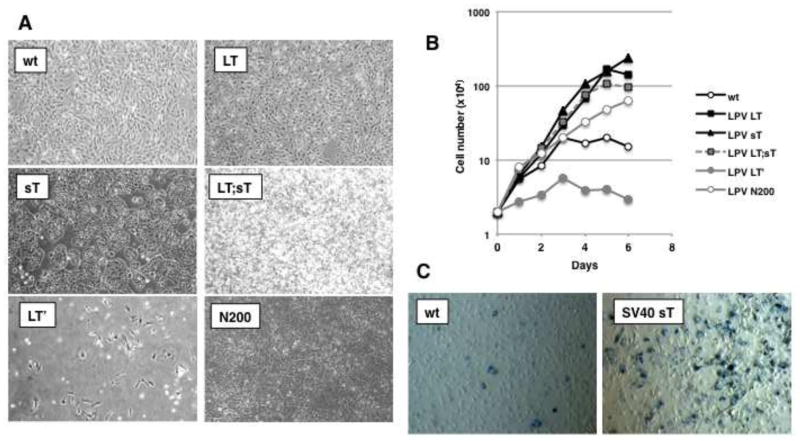
Figure 3. Different effects of LPV.ER components on cell proliferation.
A) Morphology of MEFs expressing different components of LPV.ER. Plating and manipulation of the cells was as described previously (Fig. 1A, materials and methods). B) Growth curve analysis of MEFs stably expressing LPV proteins. Pools of cells expressing different LPV components were allowed to proliferate in medium supplemented with 10% FBS, and the number of cells was monitored every two days (materials and methods). Each data point is an average of 2 separate experiments, performed using 2 biological replicates of each cell type. C) Beta-galactosidase staining to determine senescence in MEFs transduced with retroviruses containing empty vector (MEF) or SV40 sT cDNA (SV40 sT). The experiment was performed in triplicates and a representative image is shown. Blue-violet positive staining and enlarged morphology were observed only in SV40 sT expressing MEFs and not in control MEFs.

