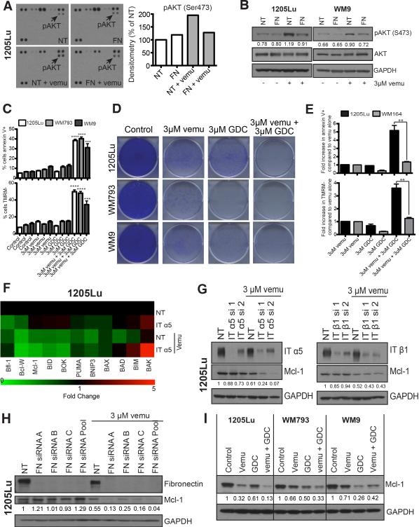
Figure 5. Treatment-associated integrin/FN signaling protects melanoma cells from vemurafenib-mediated cytotoxicity through increased PI3K/AKT signaling and control of Mcl-1 expression.
A: (left) Kinome array demonstrating that FN knockdown attenuates vemurafenib mediated AKT signaling (3 μM, 8 hrs.). (right) Quantification of pAKT densitometry on the kinome array B: Western blot confirmation of the kinome array from A demonstrating that FN knockdown prevents adaptive AKT signaling following BRAF inhibition (3 μM, 8 hrs.). pAKT was quantified using densitometry. C: Inhibition of PI3K/AKT signaling in combination with BRAF inhibition enhances cytotoxicity. Treatment with vemurafenib (3 μM) + GDC-0941 (3 μM) induces significantly more apoptosis compared to either inhibitor 25 alone. Cells were treated with the stated drug combinations for 48 hrs followed by Annexin-V/TMRM staining and flow cytometry. D: Combined BRAF/PI3K inhibition is associated with prolonged suppression of growth of BRAF6000E/PTEN- melanoma cell lines. Cells were treated with 3 μM vemurafenib and 3 μM GDC-0941 for 2 weeks. Colonies were visualized following staining with crystal violet. E: Inhibition of PI3K/AKT signaling in combination with BRAF inhibition does not enhance cytotoxicity in PTEN+ WM164 cells compared to PTEN- 1205Lu. Cells were treated with the stated drug combinations for 48 hrs. followed by Annexin-V/TMRM staining and flow cytometry. F: α5 integrin is required for maintenance of Mcl-1 expression following BRAF inhibition. LC-MRM experiment showing the relative fold changes in 11 BH3 only family proteins following siRNA knockdown of α5 integrin +/− 3 μM vemurafenib treatment in 1205Lu cell line, green indicating a decrease in expression and red indicating an increase in expression. G: Western blot showing siRNA knockdown of integrin α5 and β1 to be critical for the regulation of Mcl-1 expression in 1205Lu cell line. H: Induction of fibronectin regulates Mcl-1 expression. siRNA knockdown of FN is associated with decreased Mcl-1 expression following 3 μM vemurafenib treatment (72 hours) in 1205Lu cell line. I: Combined treatment with a BRAF and PI3K inhibitor downregulates Mcl-1 expression. Cells were treated with either 3 μM vemurafenib or 3 μM PI3K inhibitor (GDC-0941) for 72 hours followed by Western blotting for Mcl-1.

