Abstract
In infants, pleuropulmonary blastoma is a rare but aggressive tumor. The typical histopathological presentation includes the aggregation of malignant primitive small cells, usually observed in sheets. So as to provide proper and timely treatment, the differential diagnosis includes pulmonary blastoma, sarcomatoid mesothelioma, fetal rhabdomyoma, synovial sarcoma, and primitive neuroectodermal tumor. Herein, we will present one male pediatric patient with pleuropulmonary blastoma. The patient was a 4-month-old male infant, who had a prolonged cough and dyspnea for 4 months that was complicated by cyanosis for 3 days. A physical examination revealed a solid mass in the right lung that was sized 9.0 × 6.0 × 4.0 cm and had a grayish-white cross section. The boundary between the mass and lung tissue was clear; the mass already occupied a great portion of the lung. A microscopic examination suggested that the tumor was composed of round or orbicular-ovate primitive fetal cells. The cells were medium sized, having little cytoplasm, but had a clearly visualized nucleolus and active karyokinesis. The tumor mass was biphasic, namely, fasciculated sarcoma (composed of spindle-shaped cells and short spindle-shaped cells) and malignant fibrous histiocytoma containing well-differentiated cartilage islands or cartilaginous nodes. Immunohistochemistry was performed for further detection: vimentin (+), S-100 protein (+), CK (AE1/AE3), EMA and TTF-1 in residual epithelial components (+), NSE (focal +), SMA (mesenchymal cells, focal +), CD99 (weak +), Bcl-2 (weak +), desmin (-), myoglobin (-), calretinin (-), calponin (-), FLI (-), MyoD-1 (-), and CD34 (-). Pleuropulmonary blastoma is extremely rare but highly aggressive neoplasm in children. Its typical histopathological presentation is the aggregation of primitive malignant small cells. Combining imaging and histopathological examinations and clinical data should help in determining the diagnosis of pleural pulmonary blastoma.
Keywords: Pleuropulmonary blastoma, pathological diagnosis, differential diagnosis, immunohistochemistry
Background
Pleuropulmonary blastoma (PPB) is a rare, aggressive tumor that is most common in infants with a familial predisposition [1]. Although Manivel et al. first proposed PPB in 1988 [2], it was not until 1989 that PPB was considered as an independent entity in clinical pathology. The World Health Organization (WHO) classified PPB as a soft tissue tumor of the lung that is distinct from that of other pulmonary blastomas. Consisting of types I, II, and III, PPB presents with unique clinical and histopathological manifestations. We will report on one infant with PPB who was treated in our hospital. Combined with a literature review, the clinical pathology and immunohistochemistry features will be described. A special focus has been placed on the differential diagnosis of PPB.
Case presentation
The patient was a 4-month-old male infant, who mainly presented with a cough and dyspnea and occasional choking while drinking milk. During crying episodes, he experienced cyanosis for 3 days and was hospitalized for pneumonia. On February 21, 2011, a chest radiograph showed intact ribs and thorax. There were densities in the right upper lung field with unobvious mediastinal and tracheal displacement. The majority of the left lung field was obscured because it was blocked by the cardiac shadow. The mediastinum, hilar shadow, and cardiac border were obscured. Moreover, the diaphragmatic surface and bilateral costophrenic angles were obscured, but not significantly (Figure 1A). On February 23, 2011, a chest computed tomography (CT) scan showed an irregular soft tissue mass in the right upper lung that was sized 6.0 × 6.0 × 4.0 cm, with a CT value of approximately 18-40 Hu and a non-uniform density. Cystic-solid lesions were present. An aerial bronchogram could be locally observed; the inner and outer lateral borders were connected to the mediastinum, cardiac border, and chest wall; the mediastinum and cardiac shadow were considerably displaced to the left. The diagnosis of pulmonary blastoma was made preoperatively. During surgery, it was found that the mass was located in the right upper lobe and almost occupied the right thoracic cavity. The envelope was intact, and a cross section of the tumor was fish-like. The right middle and lower lobes were already compressed by the tumor. The tumor mass was resected, and a small portion of it was immediately frozen for pathological examination. The results showed that the tumor was composed of atypical spindle-shaped cells. An adenoid structure was sporadically observed, and the possibility of pulmonary blastoma could not be excluded. A right pneumonectomy was performed (Figure 1B). On March 2, 2011, a postoperative chest radiograph was performed that demonstrated the following: an intact thorax and ribs, with increased but obscured striates in bilateral lung fields; no mediastinal widening; a thickened, right hilar tumor shadow, with no right hilar enlargement; a cardiac shadow of normal size and morphology; a smooth diaphragmatic surface; sharpened bilateral costophrenic angles; a shadow cast by cannulation in the superior vena cava; and a drainage tube shadow in the right thorax (Figure 1C).
Figure 1.

A: Chest radiograph: intact thorax and ribs, with densities in the right upper lung field and unobvious mediastinal and tracheal displacement. The majority of the left lung field was obscured because it was blocked by the cardiac shadow; the mediastinum, hilar shadow, and cardiac border were obscured. Moreover, the diaphragmatic surface and bilateral costophrenic angles were obscured. B: Chest CT scan: an irregular soft tissue mass is shown in the right upper lung that was sized 6.0 × 6.0 × 4.0 cm. The CT value was approximately 18-40 Hu and had a non-uniform density. There were cystic-solid lesions. An aerial bronchogram could be locally observed; the inner and outer lateral borders were connected to the mediastinum, cardiac border, and chest wall; and the mediastinum and cardiac shadow were considerably displaced to the left. C: A postoperative chest radiograph demonstrated the following: intact thorax and ribs; increased but obscured striates in bilateral lung fields; no mediastinal widening; thickened right hilar tumor shadow; no right hilar enlargement; cardiac shadow with a normal size and normal morphology; smooth diaphragmatic surface; sharpened bilateral costophrenic angles; a shadow cast by cannulation in the superior vena cava; and a drainage tube shadow in the right thorax.
Histopathology
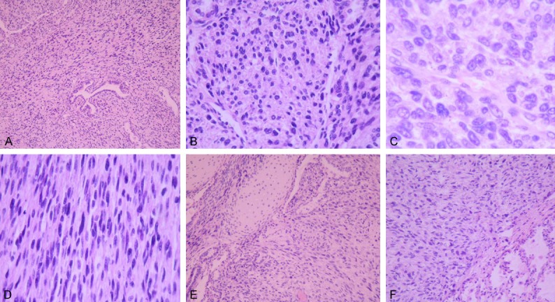
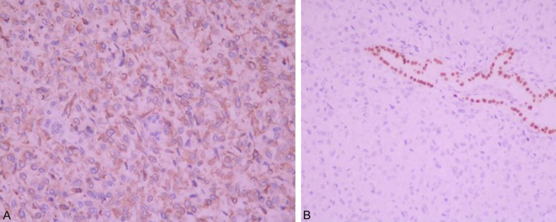
The tumor mass in the right lung was sized 9.0 × 6.0 × 4.0 cm, with a solid, soft-textured grayish-white cross section. The mass was clearly distinguishable from the lung, and a large part of the lung was occupied by the tumor. The specimen was fixed with 4% neutral formaldehyde and conventionally made into paraffin sections. HE staining was performed. The cells were detected using EnVison immunohistochemistry followed by DAB color development. The primary antibodies used for staining were Ki-67, AE1/AE3, EMA, CEA, TTF-1, vimentin, S100, NSE, bcl-2, CD99, Fli-1, CD34, SMA, desmin, MyoD-1, myoglobin, calponin, MC, and calretinin. All antibodies and EnVison kits were purchased from Beijing Zhong Shan Golden Bridge Biological Technology Co, Ltd. The tumor was mostly of a solid texture and locally nodular. Medium-sized primitive fetal cells of a round or orbicular-ovate shape were observed. The cytoplasm was eosinophilic and contained vacuoles; the nuclei were round or orbicular-ovate and deeply stained. The nucleolus was clearly visualized, and karyokinesis was active; the tumor mass had an aggregation of cells (Figure 2A-C). Some portion of the tumor was biphasic: fasciculated sarcoma (composed of spindle-shaped cells and short spindle-shaped cells) and malignant fibrous histiocytoma. The cells varied in density (Figure 2D), and well-differentiated cartilage islands or cartilaginous nodes (Figure 2E) were observed. In some regions, sunken or residual mature bronchioles or an alveolar epithelium was found. At the junction between the extensive solid tumor and lung tissues, a nodular structure was seen locally (Figure 2F). Immunohistochemistry revealed the following: vimentin (+) (Figure 3A), NSE (focal +), SMA (mesenchymal cells, focal +), CD99 (weak +), Bcl-2 (weak +), desmin (-), myoglobin (-), calretinin (-), calponin (-), FLI (-), MyoD-1 (-), and CD34 (-); in the residual epithelial components, CK (AE1/AE3), EM/A and TTF-1 (+) (Figure 3B), CEA (-), S-100 (cartilage, mesenchymal cells, focal +), and Ki-67 index (tumor cells accounting for about 45%; residual epithelial cells-). A pathologic diagnosis of type III (solid) pleuropulmonary blastoma was made.
Figure 2.

A: The tumor mass demonstrated an aggregation of primitive cells with a round or orbicular-ovate shape and with residual mature bronchioles. B: (medium magnification of a) Cells were medium sized and had little cytoplasm, and the nuclei were round or orbicular-ovate and deeply stained. The cells were highly malignant. C: (high magnification of a) The cytoplasm contained vacuoles; the nuclei were deeply stained; and the nucleolus was clearly visible. Karyokinesis was active. D: A fibrosarcoma-like lesion was interwoven with spindle-shaped cells and short spindle-shaped cells. E: A malignant fibrous histiocytoma contained well-differentiated cartilage. F: A junction between the tumor and lung tissue. The right comprises malignant tumor cells, while the left comprises normal lung tissue.
Figure 3.

A: Positive staining for vimentin; B: Positive for residual bronchial epithelia with TTF-1 staining.
Discussion
PPB usually occurs in infants and exhibits different clinical and epidemiological features from that of other pulmonary blastomas. About 93% of PPB patients are under 6 years of age [3,4], and adult cases are rare [5,6]. PPB does not demonstrate an obvious gender difference. There are three PPB types, namely, type I (multicystic), type II (multicystic tumor complicated by solid nodules), and type III (solid). The average age of onset for type I, type II, and type III is 10 months, 36 months, and 44 months, respectively [7]. Presenting with non-specific clinical manifestations, all three types are mainly associated with a prolonged cough, chest pain, difficulty breathing, and fever that are complicated by pleural effusion and atelectasis [8].
The gross specimen of the type I PPB in our study had a multicystic structure and thin cystic wall. The air-filled cysts could be easily distinguished from the surrounding tissues, and solid nodules or patches were not seen with the naked eye. The type II PPB specimen had a cystic-solid structure and thick cystic wall, combined with cystic type I PPB and solid type III PPB features. The solid nodules had grown into the cystic cavity in this case. The type III PPB mass was solid, with a soft, fish-like cross section, and contained bleeding or necrotic regions. Under a microscope, the type I PPB mass was lined with a multi-cystic structure of mature respiratory epithelial cells. There were primitive, malignant small cells aggregating under the epithelium. These cells were small and round and in a dense arrangement with a single-focal, multi-focal, or diffuse distribution. Rhabdomyoblastic cells of varying differentiation were also observed. The tumors mainly presented with discontinuous septa or an island-like structure, with or without cartilaginous nodes [9]. Type II PPB had a solid focal region based on type I PPB. The septal mesenchyma overgrew locally or completely. The mass was composed of sheets of insignificantly differentiated primitive malignant small cells, embryonal rhabdomyosarcoma, and bundle-like fasciculated sarcoma formed by patches or nodules. The solid region of the type III PPB exhibited mixed features including blastoma and sarcoma, with round or spindle-shaped cells. Histologically, the mass consisted of cartilage islands, cartilaginous nodes, rhabdomyoma, and anaplastic cells that were observed either separately or in a mixed condition. Bleeding, necrosis, and fibrosis of various extents were also occurred [10]. Because of progression or incomplete resection of PPB, type I PPB may develop into type II or III. With an early stage tumor, there is a transition between normal lung tissue and the tumor. The alveolar mesenchymal cells overproliferate, causing the alveolar septa to expand and thicken and to progress to type II. This point could be corroborated by the observation of residual cystic lesions in type II PPB [9]. The proliferating mesenchymal cells form solid nodes to replace the cystic region, and the tumor progresses to type III PPB. One salient feature of this development is the change in quantity of mesenchymal cells that only account for 6% in type I PPB. However, the proportion could be as high as 76.5% and 90.3% in type II and type III PPB, respectively. In our case, the solid region comprised the largest proportion, showing nodular edges, residual bronchioles, and locally expanded alveoli.
Currently, there are no specific molecular markers that could be used to diagnose PPB, especially if the marker expressions diverge in primitive small cells. The tumor cells could express vimentin, resulting in the corresponding immunophenotype with the differentiation of primitive mesenchymal cells. The commonly used muscle cell markers include desmin, Myo-D1, MSA, and SMA. The residual epithelium expresses CK, EMA, TTF-1, and other proteins. The results of the immunohistochemical staining showed that vimentin (spindle-shaped cells +) and muscle cell markers were not significantly expressed in our case. This suggested that the primitive mesenchymal cells did not undergo intensive myogenic differentiation. The expressions of CK (AE1/AE3) and EMA were only confined to the epithelium. The lack of expression of CEA and Ki-67 also indicated the existence of non-tumor respiratory epithelial cells growing into the tumor.
PPB may present as unilateral or bilateral local air-filled cysts upon an imaging examination. This could occur following difficulty breathing [11]. Alternately, PPB is found as a solid, mixed-density space-occupying lesion, or an irregular soft tissue mass with an obscure boundary and a non-uniform density. The septa are widened, and the cystic mass is visible. These imaging findings indicate that PPB is not a non-congenital adenomatoid malformation or congenital emphysema. However, Mut et al. [12] reviewed three PPB cases and concluded that PPB presented with diversified imaging findings. Some PPB may originate from a congenital pulmonary cyst.
In PPB, dyskaryosis is the amplification of chromosome 8, and a p53 mutation is implicated [13]. One recent research study showed that all included PPB cases with a familial predisposition carried a mutated DICER1 gene. In all cell types, one normal DICER1 gene and one abnormal DICER1 gene existed. It was suggested that PPB might derive from a mutation in DICER1 in one or several lung cells that is detrimental to cell function. Another research study showed that the occurrence of PPB was caused by an unknown mechanism for tumor induction. An abnormal DICER1 gene would downregulate signaling of the surrounding cells, inducing the transformation of normal cells to tumor cells. However, the cell carrying the abnormal DICER1 gene would not become a tumor cell.
Diagnosis or a relevant differential diagnosis of PPB is usually made based on a pathological examination (microscopically or with the naked eye) combined with clinical data and imaging findings (chest radiograph). However, a pathological examination is the only diagnostic tool that could be used to confirm the case. For type II PPB, both imaging findings and visual observation would reveal the presence of a simple cystic structure, which creates the need for differentiation with a histopathological examination. Type I PPB has to be pathologically differentiated from congenital pulmonary airway malformation (CPAM) in some cases. CPAM4 and type I PPB share common histological features, and the differentiation of clinical and imaging manifestations is difficult. The former is associated with a distal airway malformation and the presence of large cysts. Squamous epithelium or cubic epithelium overlaid the cystic wall, but no primitive malignant small cells were seen. In contrast, type I PPB contains primitive malignant small cells, which may be accompanied by fibroblast-like or adipose-like differentiation; the tumor cell nuclei usually show considerable atypia [9]. The immunohistochemical staining was positive for p53. Types II and III PPB need to be differentiated from the following tumors: (1) sarcomatoid mesothelioma. Sarcomatoid mesothelioma usually contains no primitive malignant small cells. Immunohistochemical staining is positive for actin and desmin. The diagnosis of sarcomatoid mesothelioma could be made if both the anti-calretinin antibody and mesothelial cell antibody were positive. Furthermore, the age of onset for sarcomatoid mesothelioma is much older. (2) Typical pulmonary blastoma: if PPB is associated with sunken epithelial components, it may be mistakenly diagnosed as typical pulmonary blastoma even though the epithelial components are non-cancerous [15]. However, typical pulmonary blastoma is more common in adults, and the microscopic manifestations include well-differentiated tubular glands and abundant mesenchymal components in cells. For either epithelial pulmonary blastoma or bidirectional pulmonary blastoma, the epithelial components are cancerous. The epithelium is primitive and malignant, as in fetal adenocarcinoma, which makes it easily distinguishable from the small quantity of normal respiratory epithelium in PPB. In addition, typical pulmonary blastoma contains no primitive malignant small cells. (3) Embryonal rhabdomyosarcoma: type II and III PPB may present with the differentiation of rhabdomyoblastic cells and could be confused with embryonal rhabdomyosarcoma. However, they differ in vulnerable organs: embryonal rhabdomyosarcoma is more common in the nasal cavity and vagina because they have a lumen, while PPB occurs mostly in the lung and rarely in the chest wall. Moreover, embryonal rhabdomyosarcoma is associated with unidirectional myogenic differentiation and is positive for myogenic markers [1]. (4) Synovial sarcoma: this tumor could be easily distinguished by age of onset and position of occurrence. Synovial sarcoma is most common in young people in deep soft tissues near the large joints in the four limbs. The spindle-shaped cells are morphologically different from those in PPB. In the former, the spindle-shaped cells are arranged near the vessels; in the latter, the cells are loosely arranged. Synovial sarcoma features bidirectional differentiation, but with no differentiation of the primitive mesenchymal cells and rhabdomyoblastic cells. Immunohistochemical staining may be simultaneously positive for CK, EMA, and vimentin. (5) Primitive neuroectodermal tumor (PNET): the prominent feature of PNET is the presence of Homer-Wright rosettes, ependymal tubules, and Flexner-Wintersteiner rosettes. Immunohistochemical staining indicates differentiation into neural cells. There are other non-cancerous lung lesions, from which PPB needs to be differentiated by analyzing both the clinical manifestations and pathological examinations.
Surgery is the first choice for PPB treatment and is usually combined with chemotherapy and radiotherapy, but the postoperative relapse rate is high. Autologous stem cell transplantation following large-dose chemotherapy is currently considered for clinical treatment [15]. In type II PPB that does not have any extrapulmonary involvement, simple tumor resection is sufficient; however, for type II and III PPB, tumor resection should be combined with chemotherapy and radiotherapy [16]. The administration of chemotherapy is to help relieve symptoms or for preoperative preparation and prevention of postoperative relapse. The frequently used chemotherapy scheme is vincristine (V), actinomycin D (A), and cyclophosphamide (C), or VAS. Douglas et al. [17] found that eight cases of 18 infants with type I PPB who received only surgical treatment had relapsed. In contrast, none of the 14 infants receiving postoperative chemotherapy relapsed. Chemotherapy seems to reduce PPB relapse considerably, although large-dose chemotherapy may induce complications and even death. Chemotherapy is primarily reserved for large tumors. However, one research study indicated that no obvious difference existed regarding the survival of patients receiving or not receiving chemotherapy; the two-year survival was 64% and 65%, respectively [18]. Like other tumors, PPB is also associated with metastatic loci: over 25% of type II and type III PPBs have metastatic loci. Moreover, PPB is more likely to result in metastasis to the central nervous system, as compared with other pediatric sarcomas. Therefore, the brain is the organ most vulnerable to metastasis. Other parts of the body that could be affected are the bones, lymph nodes, liver, spleen, kidney, and adrenal gland [7,19]. Type I PPB is associated with a better prognosis than type II and type III PPB [20]. Rubinas et al. [19] believed that the long-term survival of type I PPB was 83%, as opposed to only 42% for types II and III PPB [20].
Conclusion
PPB is a rare but highly aggressive neoplasm in children. Its typical histopathological presentation is the aggregation of primitive malignant small cells. Combining imaging and histopathological examinations and clinical data should help in determining the diagnosis of PPB.
Acknowledgements
Here we express our thanks to Professor Guang Liu (Department of Pathology, the General Hospital of Beijing Military Region, Beijing, China.) for his help in correcting this manuscript. Written informed consent was obtained from the patient for publication of this Case Report and accompanying images.
Disclosure of conflict of interest
None.
References
- 1.Fosdal MB. Pleuropulmonary blastoma. J Pediatr Oncol Nurs. 2008;25:295–302. doi: 10.1177/1043454208323292. [DOI] [PubMed] [Google Scholar]
- 2.Manivel JC, Priest JR, Watterson J, Steiner M, Woods WG, Wick MR, Dehner LP. Pleuropulmonary blastoma. The so-called pulmonary blastoma of childhood. Cancer. 1988;62:1516–1526. doi: 10.1002/1097-0142(19881015)62:8<1516::aid-cncr2820620812>3.0.co;2-3. [DOI] [PubMed] [Google Scholar]
- 3.Gupta K, Vankalakunti M, Das A, Marwaha RK. An autopsy report of a rare pediatric lung tumor: pleuropulmonary blastoma. Indian J Pathol Microbiol. 2008;51:225–227. doi: 10.4103/0377-4929.41663. [DOI] [PubMed] [Google Scholar]
- 4.Lucia-Casadonte C, Kulkarni S, Restrepo R, Gonzalez-Vallina R, Brathwaite C, Lee EY. An unusual case of pleuropulmonary blastoma in a child with jejunal hamartomas. Case Rep Pediatr. 2013;2013:140508. doi: 10.1155/2013/140508. [DOI] [PMC free article] [PubMed] [Google Scholar]
- 5.Smyth RJ, Fabre A, Dodd JD, Bartosik W, Gallagher CG, McKone EF. Pulmonary blastoma: a case report and review of the literature. BMC Res Notes. 2014;7:294. doi: 10.1186/1756-0500-7-294. [DOI] [PMC free article] [PubMed] [Google Scholar]
- 6.Van Loo S, Boeykens E, Stappaerts I, Rutsaert R. Classic biphasic pulmonary blastoma: a case report and review of the literature. Lung Cancer. 2011;73:127–132. doi: 10.1016/j.lungcan.2011.03.018. [DOI] [PubMed] [Google Scholar]
- 7.Priest JR, Magnuson J, Williams GM, Abromowitch M, Byrd R, Sprinz P, Finkelstein M, Moertel CL, Hill DA. Cerebral metastasis and other central nervous system complications of pleuropulmonary blastoma. Pediatr Blood Cancer. 2007;49:266–273. doi: 10.1002/pbc.20937. [DOI] [PubMed] [Google Scholar]
- 8.Aljehani YM, Elbaz AM, Moghazy KM, Alardi AA, Montaser AA, El-Ghoneimy YF. Pleuropulmonary blastoma. A rare childhood malignancy. Saudi Med J. 2007;28:1443–1445. [PubMed] [Google Scholar]
- 9.Hill DA, Jarzembowski JA, Priest JR, Williams G, Schoettler P, Dehner LP. Type I pleuropulmonary blastoma: pathology and biology study of 51 cases from the international pleuropulmonary blastoma registry. Am J Surg Pathol. 2008;32:282–295. doi: 10.1097/PAS.0b013e3181484165. [DOI] [PubMed] [Google Scholar]
- 10.Taube JM, Griffin CA, Yonescu R, Morsberger L, Argani P, Askin FB, Batista DA. Pleuropulmonary blastoma: cytogenetic and spectral karyotype analysis. Pediatr Dev Pathol. 2006;9:453–461. doi: 10.2350/06-02-0044.1. [DOI] [PubMed] [Google Scholar]
- 11.Reix P, Levrey H, Parret M, Louis D, Bellon G. Pulmonary cystic images as a presentation of a pleuropulmonary blastoma. Arch Pediatr. 2000;7:287–289. doi: 10.1016/S0929-693X(00)88747-3. [DOI] [PubMed] [Google Scholar]
- 12.Mut PR, Muro VM, Sanguesa NC, Ramirez LO. Pleuropulmonary blastoma in children: imaging findings and clinical patterns. Radiologia. 2008;50:489–494. doi: 10.1016/s0033-8338(08)76336-7. [DOI] [PubMed] [Google Scholar]
- 13.Vargas SO, Korpershoek E, Kozakewich HP, De Krijger RR, Fletcher JA, Perez-Atayde AR. Cytogenetic and p53 profiles in congenital cystic adenomatoid malformation: insights into its relationship with pleuropulmonary blastoma. Pediatr Dev Pathol. 2006;9:190–195. doi: 10.2350/06-01-0025.1. [DOI] [PubMed] [Google Scholar]
- 14.Hill DA, Ivanovich J, Priest JR, Gurnett CA, Dehner LP, Desruisseau D, Jarzembowski JA, Wikenheiser-Brokamp KA, Suarez BK, Whelan AJ, Williams G, Bracamontes D, Messinger Y, Goodfellow PJ. DICER1 mutations in familial pleuropulmonary blastoma. Science. 2009;325:965. doi: 10.1126/science.1174334. [DOI] [PMC free article] [PubMed] [Google Scholar]
- 15.Sciot R, Dal Cin P, Brock P, Moerman P, Van Damme B, De Wever I, Casteels-Van Daele M, Van den Berghe H, Desmet V. Pleuropulmonary blastoma (pulmonary blastoma of childhood): genetic link with other embryonal malignancies? Histopathology. 1994;24:559–563. doi: 10.1111/j.1365-2559.1994.tb00576.x. [DOI] [PubMed] [Google Scholar]
- 16.Indolfi P, Bisogno G, Casale F, Cecchetto G, De Salvo G, Ferrari A, Donfrancesco A, Donofrio V, Martone A, Di Martino M, Di Tullio MT. Prognostic factors in pleuro-pulmonary blastoma. Pediatr Blood Cancer. 2007;48:318–323. doi: 10.1002/pbc.20842. [DOI] [PubMed] [Google Scholar]
- 17.Miniati DN, Chintagumpala M, Langston C, Dishop MK, Olutoye OO, Nuchtern JG, Cass DL. Prenatal presentation and outcome of children with pleuropulmonary blastoma. J Pediatr Surg. 2006;41:66–71. doi: 10.1016/j.jpedsurg.2005.10.074. [DOI] [PubMed] [Google Scholar]
- 18.Priest JR, McDermott MB, Bhatia S, Watterson J, Manivel JC, Dehner LP. Pleuropulmonary blastoma: a clinicopathologic study of 50 cases. Cancer. 1997;80:147–161. [PubMed] [Google Scholar]
- 19.Rubinas TC, Manera R, Newman B, Picken MM. Pneumothorax and pulmonary cyst in a 2-year-old child. Pleuropulmonary blastoma. Arch Pathol Lab Med. 2006;130:e47–e49. doi: 10.5858/2006-130-e47-PAPCIA. [DOI] [PubMed] [Google Scholar]
- 20.Dehner LP. Pleuropulmonary blastoma is THE pulmonary blastoma of childhood. Semin Diagn Pathol. 1994;11:144–151. [PubMed] [Google Scholar]


