Abstract
There is a well-described association between infantile epilepsy and pervasive cognitive and behavioral deficits, including a high incidence of autism spectrum disorders. Despite the robustness of the relationship between early-life seizures and the development of autism, the pathophysiological mechanism by which this occurs has not been explored. As a result of increasing evidence that autism is a disorder of brain connectivity we hypothesized that early-life seizures would interrupt normal brain connectivity during brain maturation and result in an autistic phenotype. Normal rat pups underwent recurrent flurothyl-induced seizures from postnatal (P) day 5-14 and then tested, along with controls, for developmental alterations of development brain oscillatory activity from P18-25. Specifically we wished to understand how normal changes in rhythmicity in and between brain regions change as a function of age and if this rhythmicity is altered or interrupted by early life seizures.
In rat pups with early-life seizures, field recordings from dorsal and ventral hippocampus and prefrontal cortex demonstrated marked increase in coherence as well as a decrease in voltage correlation at all bandwidths compared to controls while there were minimal differences in total power and relative power spectral densities. Rats with early-life seizures had resulting impairment in the sociability and social novelty tests but demonstrated no evidence of increased activity or generalized anxiety as measured in the open field. In addition, rats with early-life seizures had lower seizure thresholds than controls, indicating long-standing alterations in the excitatory/inhibition balance. Bumetanide, a pharmacological agent that blocks the activity of NKCC1 and induces a significant shift of ECl toward more hyperpolarized values, administration at the time of the seizures precluded the subsequent abnormalities in coherence and voltage correlation and resulted in normal sociability and seizure threshold. Taken together these findings indicate that early-life seizures alter the development of oscillations and result in autistic-like behaviors. The altered communication between these brain regions could reflect the physiological underpinnings underlying social cognitive deficits seen in autism spectrum disorders.
Keywords: epilepsy, sociability, power spectrum, development, coherence, voltage correlations
Introduction
Autism is an early neurodevelopmental syndrome that often causes devastating impairments in social communication accompanied by restricted and repetitive behavior. It has been estimated that 1 in 68 children will be diagnosed with autism spectrum disorder (ASD) according to the estimates from CDC's Autism and Developmental Disabilities Monitoring (ADDM) Network (Autism and Developmental Disabilities Monitoring Network Surveillance Year 2010 Principal Investigators, 2014). The incidence of ASD appears to be rising, and this in combination with the limited effectiveness of treatments and the costs of care for these patients makes autism a growing public health problem (Autism and Developmental Disabilities Monitoring Network Surveillance Year 2010 Principal Investigators, 2014).
A singular pathophysiological mechanism responsible for the autistic phenotype is unlikely. There is substantial evidence that genetics plays an important role in ASD (Risch et al., 1999; Anney et al., 2010; Hallmayer et al., 2011; Pinto et al., 2014; Gaugler et al., 2014). There is also evidence that environmental and other non-genetic factors can be a factor in ASD (Hallmayer et al., 2011). An important clue to understanding the mechanism of autism is the observation that there is a strong relationship between early-life seizures (ELS), interictal epileptiform activity and ASD. Epilepsy and ASD coexist in up to 20% of children with either disorder (Tuchman and Cuccaro, 2011) and recent studies have suggested that ELS may place infants at a particular high risk for developing autism (Saemundsen et al., 2007; Saemundsen et al., 2008; Saemundsen et al., 2013). In addition to a high incidence of seizures in ASD, the incidence of epileptiform activity on the EEG is high in children with ASD with rates of epileptiform activity as high as 60% (Spence and Schneider, 2009). While the relationship between ASD and seizures and epileptiform EEG activity is clear, it is often difficult to determine whether the mechanism of ASD is due to the seizures or the underlying cause of the seizures. For example, ASD is common in children with tuberous sclerosis complex who have ELS (Bolton et al., 2002; Wiznitzer, 2004; Cohen et al., 2005). It is not known whether ASD is due to the consequences of an impaired mammalian target of rapamycin (mTOR) signaling pathway, the seizures themselves or both (Wiznitzer, 2004; Wang and Doering, 2013; Lugo et al., 2014a).
Given the difficulties in distinguishing the effects of seizures versus the etiology of seizures in the pathophysiology of ASD, we chose to study the role of frequent ELS in normal rats in the development of sociability deficits. Because altered brain connectivity appears to play a critical role in ASD (Stamou et al., 2013; Luckhardt et al., 2014), we used mulit-site electrophysiology in the ELS model of postnatal acquired epilepsy to study its resulting effects within and between brain regions that are critical for cognition, the prefrontal cortex (PFC), ventral (VH) and dorsal hippocampus (DH). We were particularly interested in evaluating coherence, as a measure of connectivity between brain regions. Coherence is a measure of “coupling” oscillations and therefore provides a dynamic link between brain areas required for the integration of distributed information (Varela et al., 2001; Thatcher, 2012). Alteration of coherences has been related to poor cognitive ability in rodents (Lee et al., 2014; O'Reilly et al., 2014) and autism in children (Coben et al., 2008; Leveille et al., 2010; Duffy and Als, 2012; Khan et al., 2013). In this study we also studied Pearson product correlation coefficients (PCC) as another measure of brain connectivity (Bonita et al., 2014). The PCC is a useful measure in that it measures the degree of association between EEG amplitude from two sources over a time interval, thus providing a distinctly different measure of connectivity from coherence measures.
Prior studies have demonstrated that bumetanide, a diuretic that inhibits the activity of the chloride importer NKCC1, and induces a significant shift of intracellular chloride concentration facilitating the transition of GABA from an excitatory to inhibitory neurotransmitter (Succol et al., 2012; Tyzio et al., 2014) has been used to treat ASD (Lemonnier et al., 2012) and can eliminate autistic-like behavior in the fragile X and valproate-induced models of autism through reduction of the excitatory:inhibitory balance (Tyzio et al., 2014). We further hypothesized that in a model early-life seizures that exhibit autism-like behavior (ALB), bumetanide treatment at the time of the seizures would normalize connectivity and improve sociability.
We show here that in normal rat pups, ELS during early development cause a significant increase in the coherence between the PFC and both the DH and VH, with a decrease in the PCC. Animals with this signature coherence and correlation deficits exhibited ALB. Both the ALB and abnormalities in connectivity were reversed by bumetanide treatment.
Methods
Overview of Experiments
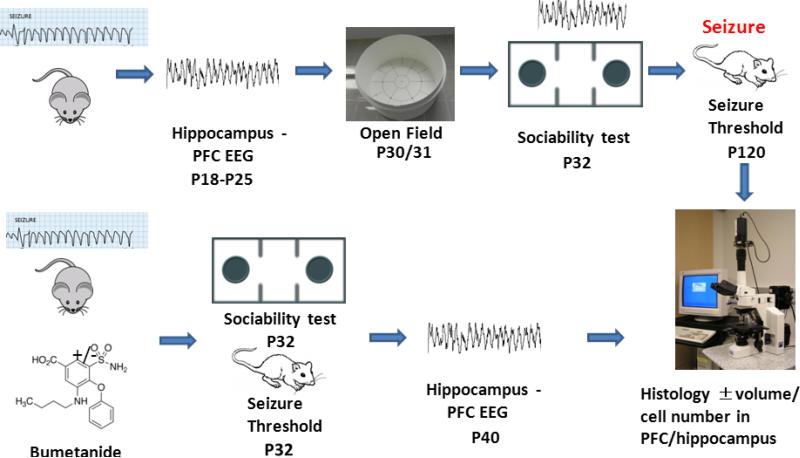
To assess seizure-induced alterations in brain oscillatory activity male rat pups were subjected to 70 flurothyl-induced seizures (7 seizures per day) from postnatal (P) day 5-14 (roughly corresponding to the first year of life in humans) (Avishai-Eliner et al., 2002). Intracranial EEG monitoring from the left prelimbic region of the prefrontal cortex (PFC), left ventral hippocampus (VH) and right dorsal hippocampus (DH) were initiated at P18 and continued to P25. To test for ALB the sociability and social novelty tests were assessed at P32 with a proportion of rats undergoing EEG monitoring concurrently with the task. Open field testing, to assess for generalized anxiety and activity level, and flurothyl seizure threshold test, to assess for seizure susceptibility, were performed in a subset of the rats. In a second cohort, animals were again subjected to 70 flurothyl-induced seizures from P5-P14 with a subset of animals receiving bumetanide treatment (i.p.) twice daily, once immediately before the first seizure induction and once following the cessation of seizure activity. This cohort of rats were studied in the sociability and social novelty tests and had EEGs and seizure threshold evaluated. A cartoon of the study design is shown in Figure 1.
Fig. 1.
Cartoon of the study design.
Animals
All experiments were performed in accordance with the guidelines provided by the National Institute of Heath and University of Vermont for the humane treatment of animals. The animal protocol was approved by the Institutional Animal Care and Use Committee of the University of Vermont.
Sample size/power calculations
The goal of the first study was to determine if early-life seizures in otherwise normal pups resulted in changes in the development of oscillations. Since we were particularly interested in coherence, a measure with a range from 0-1, where 1 = a phase angle between two waveforms of the same frequency which is stable and constant over time, i.e., phase locked, where 0 = indicates the phase angle between waveforms varies from moment-to-moment, we powered the study to determine whether there was a 0.1 difference in coherence between animals in the control and experimental group. Assuming a standard deviation of 0.1, we calculated that a total sample size of 44 (22 per group) would be necessary.
Sprague-Dawley rats were obtained from Charles River (Saint-Constant, QC). In the first series of experiments (Table 1), male Sprague-Dawley rats (n = 26) were subjected to 7 flurothyl-induced seizures daily from P5 to P14 for a total of 70 seizures using previously described methods (Karnam et al., 2009a; Lucas et al., 2011; Kleen et al., 2011b). A 10% flurothyl solution (Bis(2,2,2-trifluoroethyl) ether), an inhaled convulsive agent, was delivered to the pups, which were placed in a plastic container located in an airflow hood. Flurothyl (0.1 ml) was injected slowly onto filter paper placed on the inside of the container where it evaporated. Pups were removed from the flurothyl after approximately 2 min when tonic extension of both forelimbs and hindlimbs was observed. Littermate control pups (n = 30) were handled and removed from the dam during the time of the seizure to control for the effects of maternal separation stress.
Table 1.
Flowchart of studies done in the cohort of rats undergoing recurrent ELS.
| Exper. | Group | Rat# | Age of Sz | Age at EEG | Open Field | Sociability Test | Sociability Test with EEG | Seizure Threshold | Sacrificed |
|---|---|---|---|---|---|---|---|---|---|
| 1 | Cant. | 15 | P5-P14 | P18-P25 | >60 | ||||
| 1 | Sz | 14 | PE-P14 | P18-P25 | >60 | ||||
| 2 | Cant. | 10 | P5-P14 | P30/P31 (N=4) | P32 (N=8) | P120 | >130 | ||
| 2 | Sz | 10 | P5-P14 | P30/P31 (N=3) | P32 (N=5) | P120 | >130 | ||
| 3 | Cont. | 7 | P5-P14 | P32 | >60 | ||||
| 3 | Sz | 7 | PB-P14 | P32 | >60 |
Electrophysiology
In the first series of experiments P15-P18 rats underwent electrode implantation into the CA1 region of the DH, VH and prelimbic cortex of the PFC using a custom designed head stage (Suppl. Fig. 1A-C). Two 0.1 mm diameter, stainless steel electrodes with tips separated by ~0.5 mm were implanted at each location. The rats were anesthetized with inhaled isoflurane and placed in a stereotaxic frame. The skull was exposed and four screws inserted, two anterior to the left and right ends of bregma and two left and right over the cerebellum. The three pairs of electrodes were stereotaxically placed using coordinates from bregma, sagittal suture and skull surface using Sherwood and Timiras (Sherwood and Timiras, 1970): PFC: 2.5 mm anterior; 0.5 mm lateral, 3.3-3.5 mm below skull; DH: −2.3 mm posterior to bregma, 2.0 mm lateral, 2.4-2.6 mm below skull; VH: −3.8 mm posterior to bregma, 4.4 mm lateral, 6.0-6.3 mm below skull. Since this atlas did had coordinates for P10 and P21 rats, the coordinates for the rats implanted from P15-P18 were extrapolated with depth range based on age of the rat at the time of surgery. Five rats were sacrificed within three days of the electrode placement to verify correct location. A right cerebellar screw was used for grounding while two wires were placed in over the left cerebellum and used as reference electrodes. All implants were fixed to the skull via the skull screws and grip cement. The wound was sutured and a topical antibiotic was applied. The interval between surgery and the beginning of the cell screening process was a minimum of 48 hours. An example of EEGs recorded in each structure is shown in Supplementary Figure 1D.
The headstage was connected to a pre-amplifier with a cable connecting the rat to an amplifier and analog/digital board. EEG recordings were obtained from the rats from P18-P25 while the rat was in a cylindrical recording chamber. During testing in the open field and sociability test a light diode was attached to the headstage to allow tracking of movements with Any-maze software (Suppl. Fig. 1C)(Stoelting, Wood Dale, IL).
Field Recordings
All EEG analyses were performed using NeuroExplore 4 software (Nex Technologies, Madison, AL). Animals were studied between P18-P25. Recordings were performed for 15-30 minutes while the animal was in a cylindrical chamber. Video was digitally recorded for all animals during the EEG recordings. At each age, 5-8 rats in both the control and seizure groups were evaluated. Each individual rat was recorded from 2-4 days. Some rats started recordings earlier than others and no rats contributed data to more than four age points. Channels with excessive artifact were not used. Sixty sec artifact-free EEG epochs were used to calculate PSD, coherences and correlations.
The following oscillatory properties using local field potentials were calculated for each animal:
Coherence
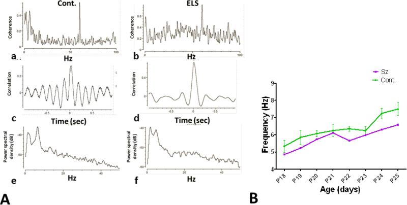
Coherence is defined as the normalized cross-power spectrum and phase delay as the “phase angle” and it is computed between two simultaneously recorded EEG signals from different brain region per frequency band. The Fast Fourier Transfrom (FFT) of the EEGs (after detrending and applying Hanning tapering) are calculated and then resampled to the specified frequency steps. Coherence values for delta, theta, alpha, beta, and gamma were calculated between the VH and DH and PFC was calculated. Figure 2a,b provide examples of coherence values from a control and ELS .
Fig. 2.
Electrophysiological recordings. A. Example of coherence (a,b), correlation (c,d) and PSD (e,f) from control (a,c,e) and seizure (b,d,f) rats. B. Peak theta frequency across ages in controls and ELS rats from the DH. No significant differences in groups were seen at any of the individual ages.
EEG Correlations
Cross-correlograms between EEGs in the VH with EEGs from the PFC and DH were obtained for 2 sec intervals and averaged over the 60 sec EEG recording (bin size = 3.4 × 10⁻⁴). The EEG voltages within the specified bin size were calculated and then the correlations between the reference EEG (VH) and other electrode computed. If x[i], i=1,...N is the reference voltage and y[i], i=1,...,N is another voltage, then c[t] = Pearson correlation coefficients (PCC) between vectors { x[1], x[2], ..., x[N-t] } and { y[t+1], y[t+2], ..., y[N] }. Mean correlation for all bins and peak correlation (mean of highest correlation using 0.1 sec intervals) was calculated. Figure 2c,d provide examples of correlations from cross-correlograms from a control and ELS rat.
Power spectral densities (PSD)
Frequencies from 0-100 Hz were analyzed with a FTT using the Hann function to reduce aliasing in Gaussian smoothing. Power spectral density is displayed on a log scale. Waveform frequencies were classified as follows: delta = 0-<4 Hz, theta 5-<10 Hz, alpha 10-<20 Hz, beta 20-<30 Hz, and gamma 30-100 Hz. Absolute and relative power (delta, theta, alpha, beta, and gamma divided by total power) were calculated for each region (DH, VH and PFC). Peak power theta frequency was determined using maximum peak power. Peak theta power was calculated only for animals in which a clear, distinguishing peak was present in the power spectrum. Figure 2e,f provide examples of PSD from a control and ELS rat.
Behavioral Studies
Open field
The open field test evaluates an animal's activity in a novel environment, as well as habituation, general anxiety, and exploration (Mestriner et al., 2013; Marques-Carneiro et al., 2014). A cylindrical chamber was used to assess activity level. The diameter of the open field was 76 cm. The chamber was divided into an inner circle (diameter = 54 cm) and outside circle (diameter = 76 cm). Animals were placed in the center of the chamber for 10 minutes and time spent, distance covered and average speed in both the center and peripheral portions of the field calculated using a video tracking system (ANY-Maze, SD Instruments, San Diego, CA).
Sociability and social novelty tests
The three chamber sociability test was used to test social behavior in the seizure and control rats (Nadler et al., 2004; Moy et al., 2004; Moy et al., 2007). The social test apparatus consists of a wooden box with removable partitions separating the box into three chambers (Suppl. Fig. 2). The size of the entire box was 122 × 41 cm (5002 cm2) with the middle chamber area of 1148 cm2 (28 × 41 cm) and two side chambers of 1927 cm2 (47 × 41 cm). The height of the walls was 43 cm. An aluminum metal cylinder (11 cm in height, bottom diameter 11 cm) with grating (1 cm apart) with a lid was placed in each of the end chambers. The test rat was placed in the middle chamber with the dividers closed to allow it to explore the middle chamber for five minutes. After this 5 min habituation period, an unfamiliar male rat was placed in one cylinder box while a marble block (object) was placed in the other cylinder. The doors were then opened and the animal was allowed 15 min to explore. Following this 15 min the test rat was removed from the box and placed in its home cage. The object was then substituted with a new (novel) rat. The test rat was then placed in the center of the maze and allowed another 15 min to explore. Measures were taken of the amount of time spent in each chamber and the number of entries into each chamber, by a human standing five feet from the apparatus. An entry was defined as all four paws in one chamber. Time spent in the chamber with the rat and object was calculated using ANY-Maze.
It is recognized that it is very difficult to extrapolate age in rats to age in humans since it is highly dependent on what measure is being evaluated. To estimate equivalent human age we relied on a model that integrates over 1000 empircally-derived neural events to translate neurodevelopmental time across mammalian species, including humans and rats (http://www.translatingtime.net)(Clancy et al., 2007; Workman et al., 2013). At P32, at the time of the sociability and social novelty tests, the equivalent human age would be a young child.
Flurothyl threshold
To test seizure threshold, time to generalized tonic seizures was assessed at P120 in 7 controls and 5 rats previously treated with flurothyl. Rats were placed in a clear plastic chamber (60 cm length, 30 cm width, 35 cm height; 6.3 × 104 cm3) with removable lid and two open ventilation holes on the side of the chamber. Flurothyl (0.2 ml) was injected every 30 seconds onto filter paper placed on the inside of the container where it evaporated. Time to a generalized tonic seizure was recorded for each rat. After the seizure onset the lid was removed and the animal allowed to recovery. The rat was then returned to the home cage and the chamber cleaned. All the threshold studies were done over 4 hours.
Bumetanide study
In a second set of experiments we determined whether treatment with bumetanide at the time of the seizures would alter coherence and sociability. Bumetanide, a chloride importer Na-K-Cl co-transporter antagonist, has been proposed as a treatment for neurological diseases, such as epilepsy or ischemic and traumatic brain injury that may involve deranged cellular chloride homeostasis. Bumetanide, which blocks the activity of NKCC1 and induces a significant shift of ECl toward more hyperpolarized values in vitro (Succol et al., 2012; Tyzio et al., 2014) has been used to treat ASD (Lemonnier et al., 2012). By shifting ECl toward more hyperpolarized values we anticipated that we would alter the excitatory:inhibitory ratio in the animals with ELS.
Sample size/power calculations
Based on results of the first set of experiments, that showed significant increases in coherence in the ELS group, we anticipated that a difference in coherence 0.2 between bumetanide versus non-bumetanide would be of behavioral significance. We used a standard deviation of 0.6 to calculate sample size. With a power of 90 and an alpha of 5 the sample size would be 4 per group. Because of concern that some animals would not provide evaluable data, we used 7 rats per group.
Bumetanide (0.5 mg/kg) was administered intraperitoneally prior to the first flurothyl-induced seizure and again after the last seizure each day in 7 rats (Table 2). Rats were weighed daily prior to the first bumetanide injection and the dosage calculated for that weight. Controls (n = 7) and ELS without bumetanide (n = 7) received equal volume injections of saline. Since it is known that bumetanide has anticonvulsant effects in developing animals (Mazarati et al., 2009; Sankar et al., 2010), all rats undergoing ELS were administered flurothyl until they displayed tonic seizures. The animals then underwent testing in the sociability and social novelty tests at P32 and then had electrodes stereotaxically placed at P37 for recording EEGs using coordinates from bregma, sagittal suture and skull surface using Sherwood and Timiras (1970): PFC: 3.8 mm anterior; 0.6 mm lateral, 4.0 mm below skull; DH: −3.6 mm posterior to bregma, 3.0 mm lateral, 3.0 mm below skull; VH: −4.2 mm posterior to bregma, 5.4 mm lateral, 5.6 mm below skull.
Table 2.
Flowchart of studies done in the cohort of rats undergoing recurrent ELS with and without bumetanide.
| Group | # Rat | Age of Sz | Dose of bumetanide | Sociability | Seizure Threshold | EEG |
|---|---|---|---|---|---|---|
| Controls -Saline | 7 | P5-P14 | P32 | P40 (N=4) | ||
| Sz + Bumetanide | 7 | P5-P14 | 0.5 mg/kg | P32 | P40 (N=4) | |
| Sz - Bumetanide | 7 | P5-P14 | P32 | P40 (N=4) | ||
| Controls - Saline | 10 | P5-P14 | P32 (N=10) | |||
| Sz + Bumetanide | 10 | P5-P14 | 0.5 mg/kg | P32 (N=5) | ||
| Sz - Bumetanide | 10 | P5-P14 | P32 (N=6) |
In a separate cohort of rats we tested seizure threshold at P32. Rats were placed in a clear plastic chamber (14 × 14 × 24 cm) with a removable lid. Flurothyl (0.1 ml) was injected every 60 seconds onto filter paper placed on the inside of the container where it evaporated. Time to a generalized tonic seizure was recorded for each rat. The threshold studies were done over 4 hours.
Histology
Following all behavioral and electrophysiological testing, rats were sacrificed. After deep anesthesia with isoflurane the rats were transcardially perfused with 0.1 M phosphate-buffered saline (PBS, pH 7.4) followed by 4% paraformaldehyde (PFA, Sigma). The brains were removed immediately, postfixed for 24 hours in 4% PFA and then immersed in 30% sucrose (w/v) at 4° C until the brains sank to the bottom of the chamber. Frozen coronal sections through the entire extent of the hippocampus were cut at 40 μm with a freezing microtome and then stored at −20° C. Every fourth section was stained with hematoxylin and eosin. Electrode placement in the prefrontal cortex, dorsal hippocampus and ventral hippocampus in animals undergoing electrophysiological evaluations was confirmed.
Cell counting was performed using the Nikon Eclipse E600 microscope with a digital video camera fitted with a 3D motorized stage. Unbiased stereological estimation of the number of neurons in the PFC and hippocampus were done using the Stereo Investigator (Version 11.03) software. Using the atlas of the rat brain from Paxinos (Paxinos and Watson, 1998) the PFC volume was calculated from sections between plates 7-9 and plates 33-35 for the CA1 and CA3 regions of the dorsal hippocampus. Volumetric analysis of the PFC was performed by dividing the region into three sections of equal area: Box 1 consisted of layers 1-3, Box 2 contained layers 4 & 5 and Box 3 contained layer 6. Computer generated random counting boxes of 50 μm × 50 μm were selected. The number of counting zones varied as a size of the zone of interest, ranging from 10-25 zones. A dissector height of 22 μm was used with guard zones of 2 μm at the top and bottom of the section. Cells were counted that were fully within the box and partially in the box if they crossed the top and right hand border of the box.
Statistical analysis
The t test was used to assess differences in controls and seizure animals in absolute PSD, relative PSD, coherence, cross-correlograms correlations, times spent in the various chambers in the sociability test and open field. Since the same rats were not used at all age point for the electrophysiological measurements, the repeated-measures ANOVA was not used. The one-way ANOVA was used to compare theta frequency as a function of age. The correlation data varied significantly from rat to rat with both positive and negative values and this data was normalized. When three groups were compared in the bumetanide experiments the ANOVA was used with individual groups compared with the Tukey's Multiple Comparison Test. Data is expressed throughout as the mean±sem.
Results
All of the rats had tonic seizures with administration of the flurothyl. Five rats died (4 during the period of time the flurothyl was administered and one after implantation of the electrodes). None of the controls died. The death rate for the entire seizure cohort was thus 12.5% (5 of 40). While none of the rats lost weight during the study, there was a significant reduction in weight in the seizure group. However, as adults there were no differences in weight between the controls and seizure groups (Cont. = 411.5±24.4 gms; ELS = 441.8±22.6 gms, p>0.05).
EEG Findings
Theta frequency
Peak spectral theta frequency increased in the DH from P18-P25 in both the controls (F(7,33) = 6.098, p < 0.001) and ELS rats (F(7,26) = 4.930, p = 0.001)(Fig. 2B). While at all the time points the mean theta frequency was higher in the controls than ELS rats, at no age points were these results significantly different Fig. 2B).
Coherences
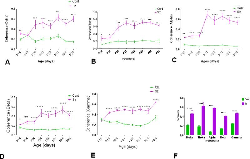
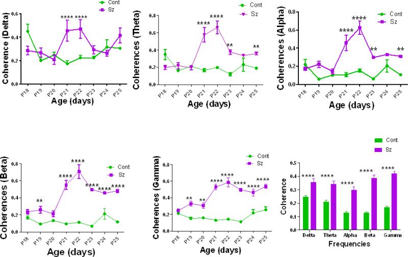
Marked increases in PFC/VH (Fig. 3) and DH/VH (Fig. 4) coherences were seen at all bandwidths across all ages in the ELS rats compared to the controls. While little change in age was seen in the controls, rats with recurrent seizures had increases in coherences with increasing age. When coherences were averaged over all age groups there were significantly increased coherences in all bands in both the PFC and DH when compared to the VH. Of interest, in the animals with ELS there were marked increases in coherence starting at approximately P21, at the time the animals were weaned. This was characteristic of both the DH and VH.
Fig. 3.
Comparison of coherence in control and ELS rats as a function of age in PFC compared to the VH. (A) delta, (B) theta, (C) alpha, (D) beta and (E) gamma frequencies. E. Mean coherences across all ages for bandwidths delta, theta, alpha, beta and gamma frequencies. * = p<0.05, ** = p<0.01, *** = p<0.001, **** = p<0.0001.
Fig. 4.
Comparison of coherence in control and ELS rats as a function across ages in DH compared to the VH. (A) delta, (B) theta, (C) alpha, (D) beta and (E) gamma frequencies. E. Mean coherences across all ages for bandwidths delta, theta, alpha, beta and gamma frequencies. * = p<0.05, ** = p<0.01, *** = p<0.001, **** = p<0.0001.
Correlations
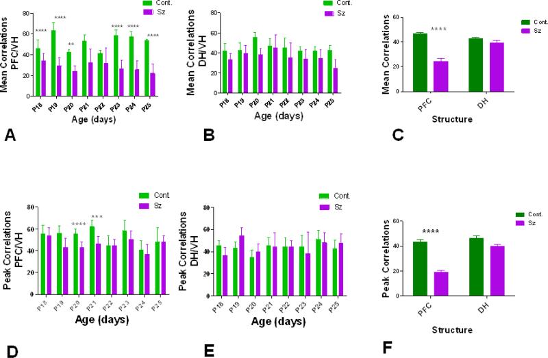
Mean and peak correlations of voltage over a 2 sec window were compared in the controls and ELS rats. Correlations were compared between the VH and PFC and VH and DH. Both mean and peak cross-correlogram for each age and group for PFC and DH (compared to the VH) are shown in Figure 5. Mean correlations were significantly higher for the controls than ELS rats at most ages in the PFC (Fig. 5A) but not the DH (Fig. 5B). Mean correlations averaged across all age groups showed the controls had higher mean correlations than the ELS rats in the PFC while there were no statistical differences in the DH (Fig. 5C). Peak correlations were also higher in the controls than the seizure animals at P20 and P21 (Fig. 5D) but no differences were seen in the DH (Fig. 5E). Peak correlations averaged across all age groups showed the controls had higher mean coherences than the controls in the PFC while there were no statistical differences in the DH (Fig. 5F).
Fig. 5.
Mean and peak PCC from PFC and DH. Mean correlations were significantly higher for the controls than seizure animals at most ages in the PFC (A) but not the DH (B). Mean correlations averaged across all age groups (P18-P25) showed the controls had higher mean correlations than the ELS rats in the PFC while there were no statistical differences in the DH (C). Peak correlations were also higher in the controls than the seizure animals at P20 and P21 (D) but no differences were seen in the DH (E) in the peak or mean correlations compared to VH. Peak correlations averaged across all age groups showed the controls had higher mean correlations than the controls in the PFC while there were no statistical differences in the DH (F). * = p<0.05, ** = p<0.01, *** = p<0.001, **** = p<0.0001.
PSD
PSD absolute and relative power was recorded for all groups from P18-P25 in the three brain locations for both the controls and ELS rats. Individual absolute power varied considerably across ages but no group differences for absolute power were seen (data not shown). Relative power varied little across age in both the control and ELS rats (Supp. Figs 4-7). While statistically significant differences were noted in the DH in the theta and gamma bandwidths (increases in relative power in theta and decreases in gamma power were seen in the ELS rats)(Supp. Fig. 4), no significant group differences were seen in the VH and PFC.
In summary, field recordings beginning shortly after the last seizure, between P18-P25 showed marked increases in coherence between the VH and PFC, and VH and DH, slower peak theta frequency and decreases in voltage correlations compared to control animals whereas there were few changes in relative power between groups. These electrophysiological studies indicate that shortly after ELS there are widespread alterations in oscillatory activity that clearly separate the developmental connectivity in the ELS rats from the controls.
Behavioral Testing
Sociability test
Rats with flurothyl-induced seizures spent significantly less time with the rat than the object (ANOVA of time spent with rat, object, or neither: F(2,27) = 22.17, p<0.0001, with more time spent with the object than the rat, p<0.05). The control rats behaved quite differently with the controls spending more time with the rat than the object (ANOVA of time spent with rat, object, or neither: F(2,27) = 86.84, p<0.0001, with more time spent with rat than the object, p<0.05). Rats with ELS spent less time with the rat t(18) = 5.981, p <0.0001) and more time with the object (t(18) = 6.484, p <0.0001) than the controls. Time spent with neither the rat or object did not differ significantly between groups (t(18) = 2.066, p = 0.054)(Fig. 6A).
Fig. 6.
Sociability test. A) Control rats spent significantly more time with the rat than the object. Rats with recurrent seizures spent similar amounts of time with the object and rat and less time with the rat than the controls. B) Control rats spent more time with the new rat than the rats with recurrent seizures. C) EEG coherence during the sociability test. The ELS rats had higher theta coherence when near both the object and rat than the control rat when near the object. While the controls had higher coherences when near the rat than the object the rats with prior seizures had higher coherences near the object. However, these differences were not statistically different; * = p<0.05, ** = p<0.01, *** = p<0.001, **** = p<0.0001.
Social Novelty Test
The control rats spent more time with the new (novel) rat than the object (ANOVA of time spent with rat, object or neither: F(2,27) = 14.81, p <0.0001, with more time spent with the novel rat than the old rat, P<0.05). In the rats with ELS no differences in time spent between the novel and old rat were noted (F(2,27) = 2.424, p = 0.107). When the ELS and controls were compared, the ELS rats spent less time with the novel rat than the controls (t(18) = 2.351, p = 0.030)(Fig. 6B).
This data demonstrates that rats with ELS have significant deficits in sociability as evidenced by decreased time spent with the conspecific (sociability test) and less time with the novel rat (social novelty test) than the controls. In addition, the rats with ELS spent more time with the object than the control, suggesting a behavioral disorder than involves more than social anxiety
A subset of rats (Cont. = 3, Sz = 4) underwent EEG recording during the sociability test. As shown in Fig. 6C, rats with a history of ELS showed higher coherences between the VH and PFC than the controls. Coherences were higher in the ELS rats than the controls whether near the object or rat. While the controls had higher coherences when near the rat than the object, the rats with prior seizures had higher coherences near the object. However, these differences were not statistically different. However, these differences were not statistically different. This data indicates that coherences are higher in the ELS rats than the control rats during the sociability test but that coherences did not vary significantly as to whether the rat was near another rat or object.
Open field
In order to rule out a component of general anxiety in the social anxiety paradigms shown herein, we conducted an open field task. In the open field test there were no differences in mean time or distance covered in either the inner or outer circle. Mean speed in the chamber did not differ between groups (data not shown).
Seizure threshold
All animals had generalized tonic seizures during the flurothyl inhalation test and survived the seizure. There was a significant difference in time (threshold) to the generalized tonic seizure with rats previously receiving flurothyl having a shorter latency (279±22 sec) than the controls (358±24 sec) with difference being statistically different (t(10) = 2.322, p = 0.042). To rule out a possible relationship between weight of the rat and flurothyl threshold a linear regression analysis was performed and no relationship between weight of the animals and flurothyl threshold was seen (F(1,10) = 2.852, p = 0.13).
Histology
Electrode placement in the PFC, DH and VH was confirmed in the majority of rats (Suppl. Fig. 6A-C). Because of the thickness of the brain slices in a few rats the entire track of the electrode, including the distal end, could not be seen. None of the rats with identifiable electrode tracks had incorrect placement.
Unbiased stereological estimation of the number of neurons in the PFC and hippocampus were done in the areas shown in Supplementary Fig. 6D-E. The PFC was divided into three sections of equal area for cell counts and volumetric studies of layers 1-3 (Box 1), 4-5 (Box 2) and 6 (Box 3)(Suppl. Fig. D). In the hippocampus (Suppl. Fig. E) the CA1 and CA3 areas shown above were used for volumetric measurements and cell counts. No differences were found between the controls and ELS rats in PFC, CA1 and CA3 volumes or cell counts (Table 3).
Table 3.
Cell counts and volumetric data from control and rats with ELS.
| Group | PFC–Cell Count (μm3) | PFC Volume (μm3) | CA1 – Cell Count (μm3) | CA1 Volume (μm) | CA3 – Cell Count (μm3 | CA3 Volume (μm3) |
|---|---|---|---|---|---|---|
| Controls | 9952±902 | 1.03(108)±1.10(107) | 4704±295 | 5.33(107)±3.26(106) | 2805±102 | 3.02(107)±2.06(106) |
| Fluirothyl Sz | 8717±731 | 8.82(107)±6.62(106) | 4677±296 | 5.37(107)±3.96(106) | 2582±79 | 2.95(107)±1.26(106) |
| p | 0.2941 | 0.2167 | 0.9497 | 0.9428 | 0.1022 | 0.8042 |
Treatment with bumetanide
EEG findings
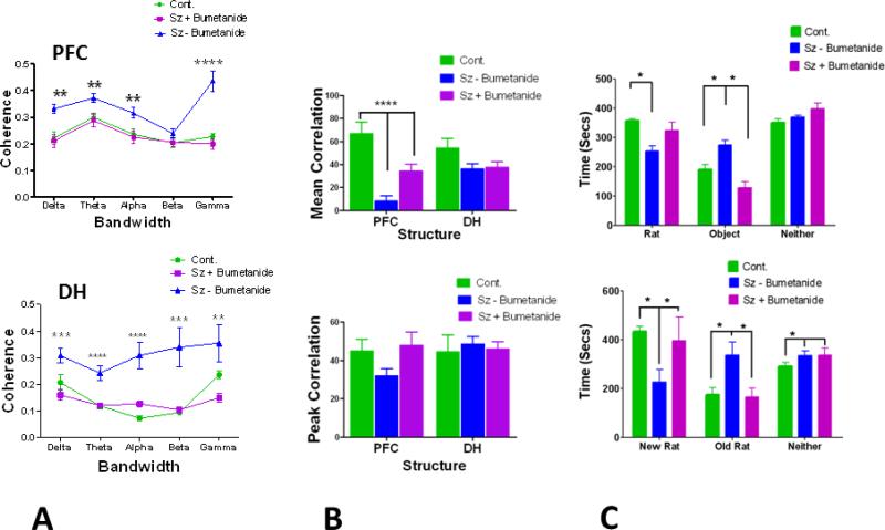
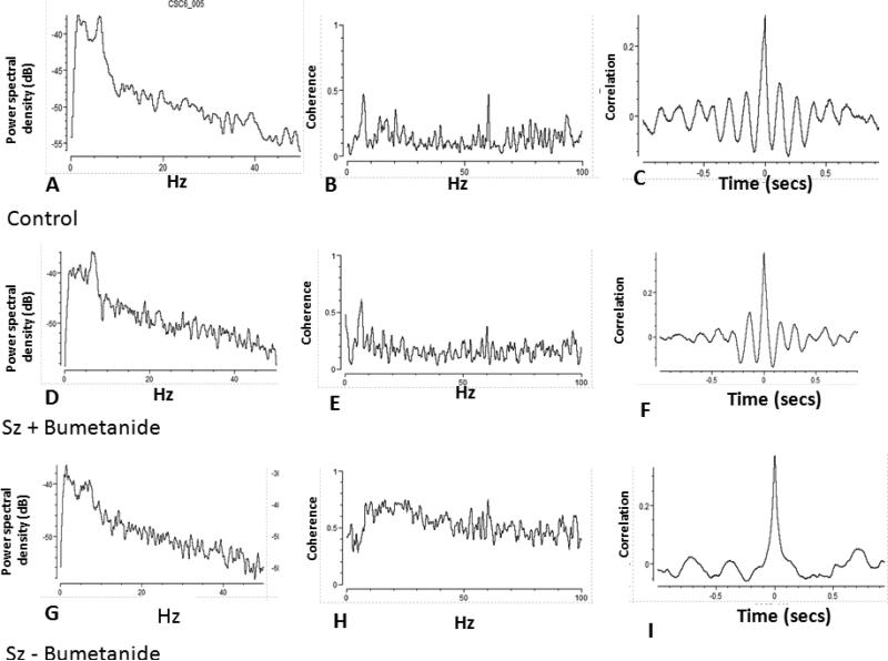
Rats treated with bumetanide during the seizures showed no differences in PSD than the controls or rats with seizures not treated with bumetanide (Suppl. Fig. 7). No differences in peak theta frequency were seen (Cont: 8.212±0.15 Hz; Sz with Bumetenide: 8.55±0.353 Hz; Sz without bumetanide Hz: 8.163±0.29, p>0.05) at P40. Coherences in rats with recurrent seizures treated with bumetanide did not differ significantly from controls, whereas the rats not receiving bumetanide had significantly higher coherences in both the PFC and DH (Fig. 7A). There were also significant group differences in voltage correlations between controls and rats with and without bumetanide treatment. ELS rats treated with bumetanide have higher correlations than the ELS rats not receiving bumetanide between both the VH and the PFC and the VH and DH (Fig. 7B). Examples of PSD, coherences and correlations in rats with recurrent seizures with and without bumetanide are shown in Fig. 8.
Fig. 7.
Effects of bumetanide treatment on coherences (A), correlations (B) and sociability and social novelty tests (C). A) Coherences. In both the PFC (top graph) and DH (bottom graph), rats treated with bumetanide did not differ in coherences from controls. Rats with ELS not treated with bumetanide had significantly higher coherences in both the PFC and DH than the controls and rats with ELS treated with bumetanide. B) Voltage correlation. Comparison of mean (top graph) and peak (bottom graph) correlations in controls, ELS rats without bumetanide treatment and ELS rats with bumetanide treatment. Correlations of the PFC and DH were compared to VH. C) Comparison of sociability test (top) and social novelty task (bottom) in control rats, rats with ELS seizures treated with bumetanide and rats with ELS seizures treated with bumetanide. * = p<0.05, ** =p<0.01, *** = p<0.0001.
Fig. 8.
Examples of PSD of VH (A,D,G), coherence between PFC and VH ( B,E,H) and Pearson correlations (C,F,I) in controls, ELS with bumetanide and ELS without bumetanide.
Sociability test
In the sociability test done at P35, group differences were noted for time spent with the rat (F(2,18) = 7.133, p = 0.005, with the rats not receiving bumetanide spending less time with the rat than the controls) and object (F(2,18) = 7.329, p = 0.005 with the rats not receiving bumetanide spending more time with the object than the controls and the treated rats, p<0.05). No differences were noted between time spent with neither the rat or object in the three groups (F(2,18) = 2.887, p = 0.083)(Fig. 7C, top graph).
Social Novelty test
Group differences were also noted for time spent with the new (novel) rat (F(2,18) = 8.390, p = 0.003, with the rats not receiving bumetanide spending less time with the novel rat than the controls and bumetanide-treated rats, p<0.05), old rat (F(2,18) = 5.683, p = 0.013, with the rats not receiving bumetanide spending more time with the old rat than the controls and bumetanide-treated rats, p<0.05) and time spent with neither the new or old rat (F(2,18) = 4.816, p = 0.021. with the ELS rats with or without bumetanide treatment spending less time with either the new or old rat than the controls, p<0.05)(Fig. 7C, bottom graph).
Seizure threshold
At the time of the seizure threshold test at P32 there were no group differences in weignt (Controls: 117.8±2.6 gms; ELS without bumentadie: 122.6±5.2 gms; ELS with bumetanide: 119.2±7,2, p>0.05). There was a significant difference in time (seizure threshold) to the generalized tonic seizure between groups (F(2,18) = 13.84, p = 0.0002) with ELS rats without bumetanide having a lower seizure threshold (52.7±5.2, secs, p <0.05) than the ELS rats with bumetanide (118.8±20.8 secs) and the controls (120.9 secs±6.1 secs). There was no difference in latency between the controls and ELS rats receiving bumetanide.
Histology
Electrode placement in the PFC, DH and VH in animals undergoing electrophysiological evaluations was confirmed.
This data demonstrates that bumetanide treatment at the time of the serial seizures prevents the post-seizure increase in coherence, decrease in voltage correlations, and corrects the sociability deficit.
Discussion
In this study, we show that ELS alters functional connectivity of the brain with the ELS rats showing widespread marked increases in coherence and decreased voltage correlations between VH and PFC and DH structures. These changes in connectivity began shortly after the seizures ended. Rats with ELS also had deficits in both the sociability and social novelty tests. Compared to control rats, rats with ELS spent more time with an object and less time with another rat in the sociability test. In the social novelty task the ELS rats spent less time with the new (novel) rat than the controls. These observations suggest that the behavioral changes following ELS extend beyond social anxiety. If social anxiety was the only behavioral abnormality in the ELS rats it would be expected that ELS rats would spend equal amounts of time with the rat and object which was not the case here. Previous studies with this model of ELS have shown deficits of spatial cognition (Karnam et al., 2009a; Kleen et al., 2011b; Lugo et al., 2014b), impaired auditory discrimination (Neill et al., 1996) and reduced behavioral flexibility (Kleen et al., 2011a). In addition, rats with ELS have persistent decreases in GABA currents in the hippocampus (Isaeva et al., 2006) and neocortex (Isaeva et al., 2010), enhanced excitation in the neocortex (Isaeva et al., 2010), impaired LTP (Zhou et al., 2007; Karnam et al., 2009a; Isaeva et al., 2013) and reduced place cell firing precision and stability (Karnam et al., 2009b). Thus, seizure-induced changes in the developing brain result in a complex cascade of physiological and behavioral changes that include far more than deficits in sociability.
While equating rat behavior to the complex symptoms of a child with ASD is difficult, the deficits in sociability and social novelty preference in rats has some similarities to the core features that are seen in children with ASD. Similar to the time spent with the conspecific and object by rats with ELS, children with ASD spend less time looking at people and longer times looking at objects compared to controls (Swettenham et al., 1998; Bhat et al., 2010). The findings show that ELS can result in ALB in immature rats, confirming the idea that ALB can be acquired following birth.
The most striking finding in our study were the changes in coherence and voltage correlations in the rats with ELS. On a frequency-by-frequency basis, EEG spectral coherence represents the consistency of the phase difference between two EEG signals when compared over time. Coherence is a measure of synchronization between two EEG signals and is based mainly on the conformity of phase differences between the EEG signals (Thatcher et al., 2008; Thatcher, 2012). High coherence values are taken as a measure of strong connectivity between the brain regions that produce the compared EEG signals. ELS results in markedly increased coherences, suggesting widespread functional “over-connectivity” in the developing brain.
Although increased coherences involved all three brain structures studied, of particular interest in this study was the high coherence between the VH and the PFC. The existence of a strong functional hippocampal-PFC interaction is supported by anatomical and electrophysiological data, demonstrating the existence of a monosynaptic pathway from the hippocampus to the medial PFC that can undergo activity-dependent modifications (Laroche et al., 1990; Jay et al., 1995; Takita et al., 1999; Laroche et al., 2000). A significant portion of neurons in the medial PFC of freely behaving rats are phase-locked to the hippocampal theta rhythm. This theta-entrained activity across cortico-hippocampal circuits is likely important for information flow and guiding the plastic changes that underlie the dynamic “back and forth” storage of information across these networks (Siapas et al., 2005; Fries, 2005).
Increased coherences were seen across all frequency bands. As with the current study, Duffy and Als (2012) found that in a study of coherences in children with ASD the ASD population coherence patterns tended to be remarkably stable across broad spectral ranges. It is interesting that the changes in coherences increased following the ELS. While some differences were present at the first day of recording (P18), the largest differences in coherence were seen beginning at P21. These findings indicate that the effect of ELS on oscillatory activity continues to occur for days following the last seizure.
It would be logical to assume that if coherence was impaired in autism it would be due to reduced coherences between highly integrated brain structures. Indeed, in rodent models of stress (Jacinto et al., 2013; Oliveira et al., 2013) and schizophrenia (Sigurdsson et al., 2010) coherences between VH and PFC are decreased. Likewise, the majority of studies in children with ASD have demonstrated reduced coherences (Coben et al., 2008; Khan et al., 2013). Neuronal synchrony in the brain is finely tuned and it is possible that functional “over connectivity” may be as detrimental as under connectivity as a network that is over-connected at baseline may not be able to adapt to increased cognitive demand. As shown in our laboratory previously, ELS can result in an imbalance of excitation/inhibition in the neocortex and hippocampus resulting in enhanced excitation that may predispose the developing brain to excessive connectivity (Isaeva et al., 2006; Isaeva et al., 2009; Isaeva et al., 2010). In this study we found that seizure threshold to flurothyl was reduced in rats with ELS compared to controls, suggesting that there are long-standing changes in brain excitability following ELS. Furthermore, early-life seizures and interictal spikes can result in increases in short-term plasticity in the PFC (Hernan et al., 2013; Hernan et al., 2014). High phase locking of neurons in mPFC and hippocampus likely results in neurons in both structures firing with excessive synchrony with a diminished ability to develop localized functional ensembles. We suggest that as with other electrophysiological processes there is an ideal “sweet spot” for coherence and that deviations in either a positive or negative direction can alter behavior. Our data further suggests that strategies for improving cognition in ELS or other models should be wary of interpreting increased synchrony as a positive measure of improved neural communication.
Another measure of brain connectivity, the voltage correlations was quite different between the controls and ELS rats. Amplitude or voltage correlation is a measure of the co-modulation of the amplitude envelopes, i.e. power, of oscillations in two brain areas (Bruns et al., 2000; Siegel et al., 2012). Amplitude correlation is also referred to as “power-to-power correlation” or “amplitude-amplitude coupling.” While coherences were increased in the rats with ELS, the voltage correlations were greater between the VH and PFC and DH in the controls than in the ELS rats. It is important to note that while both coherences and the voltage correlations are providing information about functional connectivity, they are providing distinctly different information. The PCC is a measure of the degree of association between amplitudes of the EEG across sites and does not calculate phase nor does it involve the measurement of the consistency of phase relationships seen with coherence. The mechanistic interpretation, and thus the functional significance, of amplitude coherence is less clear than the mechanistic interpretation of phase coherence. Amplitude correlations reflect state changes coupled across brain networks that are driven by neuromodulatory systems (Munk et al., 1996). Studies have shown that amplitude correlations provide a measure of large-scale interactions between cortical areas that are only indirectly connected via polysynaptic pathways (de Lange et al., 2008; Mazaheri et al., 2009; Donner et al., 2009; Mazaheri et al., 2010). For example, Mazaheri et al. (2010) found that reduced fronto-occipital interactions reflected attentional control deficits in children with attention deficit hyperactivity disorder. Amplitude correlation is thus an informative index of the large-scale cortical interactions that mediate cognition. The high voltage correlations between sites indicate that there is greater co-modulation of amplitude in the controls than the ELS rats and provides another possible reason for impaired neurological function following ELS.
The alterations in oscillatory patterns following ELS were confined primarily to coherence and voltage correlations across the VH and PFC and the VH and DH while power spectra showed no consistent differences between groups. While the peak theta frequency was slower in the ELS group than the controls, when the age groups were combined, significant differences were not reached at any specific age. One mechanism for increased coherence in autism is a failure of expected die-back of certain cortical-cortical connections with the aberrant over-connectivity interfering with normal cortical processing. Overgrowth of the frontal lobe has been reported in children with autism (Carper and Courchesne, 2005; Courchesne et al., 2011a; Courchesne et al., 2011b; Morgan et al., 2012). In addition, Kleen et al. (2011a) found increased thickness of the PFC in the same ELS model used in this study. However, we found no differences between groups in either PFC or hippocampal areas and volumes or PFC or hippocampal cell counts using unbiased stereological estimations of cell number. Since in clinical studies the overgrowth of frontal lobe occurs during early childhood, it is possible we would have seen group differences if the animals were sacrificed at an earlier age. It should be noted that Kleen et al. (2011a) also found no differences in PFC cell density in rats with ELS and controls. It is still possible that there is an increase in synaptic numbers and/or strength between these regions as a result of ELS. However, additional studies will be needed to directly address these issues.
It is not possible to say that the ALB seen following ELS is caused by the changes in oscillations that occur following the ELS. However, there are several lines of evidence showing that dynamic changes in coherence are related to performance, e.g. rats have increased theta phase coherence between the PFC and the hippocampus at the time they make a decision based on previous experience (Jones and Wilson, 2005; Benchenane et al., 2010). In addition, a study from our laboratory showed that rats with ELS were impaired with regard to learning a delayed-nonmatch-to-sample memory (DNMS) task and also had enhanced CA1-prefrontal theta and gamma coherence compared to non-seizure controls (Kleen et al., 2011b). However, rats with ELS had greater CA1-mPFC theta coherence in correct trials compared with incorrect trials when long delays were imposed, suggesting increased hippocampal-prefrontal cortex synchrony for the task in this group when memory demand was high. This was interpreted as an adaptive trait, and may well be for bridging temporal gaps in a DNMS task., The data presented herein suggest that this same trait could be maladaptive in terms of social development; however, a key difference in the two results is that the increases in coherence in Kleen et al. (2011b), were noted during a task of working memory. In the study herein, increased coherence was noted when the animal was not engaged in a cognitive task and therefore did not appear to be related to increased cognitive demand. This finding is suggestive of a circuit that is inflexible and unable to adapt to increased cognitive demand.
Support for the idea that ALB is related to altered connectivity and enhanced excitability comes from the bumetanide experiments. Rats with ELS treated with bumetanide had normal sociability skills and normal coherences and voltage correlations. In addition, rats treated with bumetanide have reduced excitability at the time that sociability was measured as indicated by high seizure thresholds compared to rats not receiving bumetanide. In our study bumetanide was given before and after the last seizure each day. We designed our study to eliminate the possibility that any effects of bumetanide would be due to the drug's anti-seizure properties. Animals receiving bumetanide had seizures identical in semiology and duration as the non-bumetanide treated group. While not directly studied here, our findings suggest that bumetanide through altering the ECl of immature neurons reduces excitability which drives connectivity. Similarly, Tyzio et al. (2014) found that maternal pretreatment with bumetanide eliminated ALB in the valproate and fragile × rodent models of autism. These rodent models of autism have elevated intracellular chloride levels, increased excitatory GABA, enhanced glutamatergic activity, and elevated gamma oscillations. Bumetanide corrected these electrophysiological changes. In our studies we do not know the mechanisms by which bumetanide altered coherences and deficits in social behavior. Of interest are recent observations that systemic injections of bumetanide result in very low brain concentrations of bumetanide (Li et al., 2011; Cleary et al., 2013; Tollner et al., 2014) raising fundamental questions of how bumetanide actually eliminates ALB.
Extrapolation of our findings to the clinical situation should be done cautiously. As described above, most studies examining coherence in children with ASD have found reduced, rather than increased coherences, particularly at short inter-electrode distances. In addition, the vast majority of children who have childhood epilepsy do not develop autism. We could find no correlations between coherence and voltage correlations with sociability skills. Finally, in the flurothyl seizure model, the behavior and cognitive consequences are complex and involve more than an isolated deficit in sociability. It is highly unlikely that bumetanide alone would prevent or cure ASD.
Taken as a whole, this study shows that ELS in otherwise normal rodents can result in acquired deficits in social behaviors. Our data suggests one possible pathway by which ELS can result in an autistic-like phenotype is through alterations in brain connectivity. The findings support the notion that ALB can occur as a result of enhanced coherences and reduced voltage correlations across a broad span of frequencies.
Supplementary Material
Bullet Points.
Early life seizures (ELS) result in autistic-like behaviors (ALB) in rats
ELS are impaired in sociability and social novelty tests
ELS cause high coherences and low voltage correlations between brain regions
Treatment with bumetanide reverses the ALB and aberrant functional connectivity
Acknowledgements
Supported by National Institute of Health grants NS074450, NS074450 and NS073083, the Emmory R. Shapses Research Fund and Michael J. Pietroniro Research Fund to GLH and NIH Grant Number P30 RR032135 from the COBRE Program of the National Center for Research Resources and P30 GM 103498 from the National Institute of General Medical Sciences.
Footnotes
Publisher's Disclaimer: This is a PDF file of an unedited manuscript that has been accepted for publication. As a service to our customers we are providing this early version of the manuscript. The manuscript will undergo copyediting, typesetting, and review of the resulting proof before it is published in its final citable form. Please note that during the production process errors may be discovered which could affect the content, and all legal disclaimers that apply to the journal pertain.
Reference List
- Anney R, et al. A genome-wide scan for common alleles affecting risk for autism. Hum Mol Genet. 2010;19:4072–4082. doi: 10.1093/hmg/ddq307. [DOI] [PMC free article] [PubMed] [Google Scholar]
- Autism and Developmental Disabilities Monitoring Network Surveillance Year 2010 Principal Investigators . Prevalence of Autism Spectrum Disorder Among Children Aged 8 Years - Autism and Developmental Disabilities Monitoring Network, 11 Sites, United States, 2010. Center for Surveillance, Epidemiology, and Laboratory Services, Centers for Disease Control and Prevention (CDC), U.S. Department of Health and Human Services; 2014. pp. 1–22. [Google Scholar]
- Avishai-Eliner S, Brunson KL, Sandman CA, Baram TZ. Stressed-out, or in (utero)? Trends Neurosci. 2002;25:518–524. doi: 10.1016/s0166-2236(02)02241-5. [DOI] [PMC free article] [PubMed] [Google Scholar]
- Benchenane K, Peyrache A, Khamassi M, Tierney PL, Gioanni Y, Battaglia FP, Wiener SI. Coherent theta oscillations and reorganization of spike timing in the hippocampal- prefrontal network upon learning. Neuron. 2010;66:921–936. doi: 10.1016/j.neuron.2010.05.013. [DOI] [PubMed] [Google Scholar]
- Bhat AN, Galloway JC, Landa RJ. Social and non-social visual attention patterns and associative learning in infants at risk for autism. J Child Psychol Psychiatry. 2010;51:989–997. doi: 10.1111/j.1469-7610.2010.02262.x. [DOI] [PMC free article] [PubMed] [Google Scholar]
- Bolton PF, Park RJ, Higgins JN, Griffiths PD, Pickles A. Neuro-epileptic determinants of autism spectrum disorders in tuberous sclerosis complex. Brain. 2002;125:1247–1255. doi: 10.1093/brain/awf124. [DOI] [PubMed] [Google Scholar]
- Bruns A, Eckhorn R, Jokeit H, Ebner A. Amplitude envelope correlation detects coupling among incoherent brain signals. NeuroReport. 2000;11:1509–1514. [PubMed] [Google Scholar]
- Carper RA, Courchesne E. Localized enlargement of the frontal cortex in early autism. Biol Psychiatry. 2005;57:126–133. doi: 10.1016/j.biopsych.2004.11.005. [DOI] [PubMed] [Google Scholar]
- Clancy B, Finlay BL, Darlington RB, Anand KJ. Extrapolating brain development from experimental species to humans. Neurotoxicology. 2007;28:931–937. doi: 10.1016/j.neuro.2007.01.014. [DOI] [PMC free article] [PubMed] [Google Scholar]
- Cleary RT, Sun H, Huynh T, Manning SM, Li Y, Rotenberg A, Talos DM, Kahle KT, Jackson M, Rakhade SN, Berry G, Jensen FE. Bumetanide enhances phenobarbital efficacy in a rat model of hypoxic neonatal seizures. PLoS ONE. 2013;8:e57148. doi: 10.1371/journal.pone.0057148. [DOI] [PMC free article] [PubMed] [Google Scholar]
- Coben R, Clarke AR, Hudspeth W, Barry RJ. EEG power and coherence in autistic spectrum disorder. Clin Neurophysiol. 2008;119:1002–1009. doi: 10.1016/j.clinph.2008.01.013. [DOI] [PubMed] [Google Scholar]
- Cohen D, Pichard N, Tordjman S, Baumann C, Burglen L, Excoffier E, Lazar G, Mazet P, Pinquier C, Verloes A, Heron D. Specific genetic disorders and autism: clinical contribution towards their identification. J Autism Dev Disord. 2005;35:103–116. doi: 10.1007/s10803-004-1038-2. [DOI] [PubMed] [Google Scholar]
- Courchesne E, Campbell K, Solso S. Brain growth across the life span in autism: age- specific changes in anatomical pathology. Brain Res. 2011a;1380:138–145. doi: 10.1016/j.brainres.2010.09.101. [DOI] [PMC free article] [PubMed] [Google Scholar]
- Courchesne E, Mouton PR, Calhoun ME, Semendeferi K, Ahrens-Barbeau C, Hallet MJ, Barnes CC, Pierce K. Neuron number and size in prefrontal cortex of children with autism. JAMA. 2011b;306:2001–2010. doi: 10.1001/jama.2011.1638. [DOI] [PubMed] [Google Scholar]
- de Lange FP, Jensen O, Bauer M, Toni I. Interactions between posterior gamma and frontal alpha/beta oscillations during imagined actions. Front Hum Neurosci. 2008;2:7. doi: 10.3389/neuro.09.007.2008. [DOI] [PMC free article] [PubMed] [Google Scholar]
- Donner TH, Siegel M, Fries P, Engel AK. Buildup of choice-predictive activity in human motor cortex during perceptual decision making. Curr Biol. 2009;19:1581–1585. doi: 10.1016/j.cub.2009.07.066. [DOI] [PubMed] [Google Scholar]
- Duffy FH, Als H. A stable pattern of EEG spectral coherence distinguishes children with autism from neuro-typical controls - a large case control study. BMC Med. 2012;10:64. doi: 10.1186/1741-7015-10-64. [DOI] [PMC free article] [PubMed] [Google Scholar]
- Fries P. A mechanism for cognitive dynamics: neuronal communication through neuronal coherence. Trends Cogn Sci. 2005;9:474–480. doi: 10.1016/j.tics.2005.08.011. [DOI] [PubMed] [Google Scholar]
- Gaugler T, Klei L, Sanders SJ, Bodea CA, Goldberg AP, Lee AB, Mahajan M, Manaa D, Pawitan Y, Reichert J, Ripke S, Sandin S, Sklar P, Svantesson O, Reichenberg A, Hultman CM, Devlin B, Roeder K, Buxbaum JD. Most genetic risk for autism resides with common variation. Nat Genet. 2014;46:881–885. doi: 10.1038/ng.3039. [DOI] [PMC free article] [PubMed] [Google Scholar]
- Hallmayer J, Cleveland S, Torres A, Phillips J, Cohen B, Torigoe T, Miller J, Fedele A, Collins J, Smith K, Lotspeich L, Croen LA, Ozonoff S, Lajonchere C, Grether JK, Risch N. Genetic heritability and shared environmental factors among twin pairs with autism. Arch Gen Psychiatry. 2011;68:1095–1102. doi: 10.1001/archgenpsychiatry.2011.76. [DOI] [PMC free article] [PubMed] [Google Scholar]
- Hernan AE, Alexander A, Jenks KR, Barry J, Lenck-Santini PP, Isaeva E, Holmes GL, Scott RC. Focal epileptiform activity in the prefrontal cortex is associated with long-term attention and sociability deficits. Neurobiol Dis. 2014;63:25–34. doi: 10.1016/j.nbd.2013.11.012. [DOI] [PMC free article] [PubMed] [Google Scholar]
- Hernan AE, Holmes GL, Isaev D, Scott RC, Isaeva E. Altered short-term plasticity in the prefrontal cortex after early life seizures. Neurobiol Dis. 2013;50:120–126. doi: 10.1016/j.nbd.2012.10.007. [DOI] [PMC free article] [PubMed] [Google Scholar]
- Isaeva E, Isaev D, Holmes GL. Alteration of synaptic plasticity by neonatal seizures in rat somatosensory cortex. Epilepsy Res. 2013 doi: 10.1016/j.eplepsyres.2013.03.011. [DOI] [PMC free article] [PubMed] [Google Scholar]
- Isaeva E, Isaev D, Khazipov R, Holmes GL. Selective impairment of GABAergic synaptic transmission in the flurothyl model of neonatal seizures. Eur J Neurosci. 2006;23:1559–1566. doi: 10.1111/j.1460-9568.2006.04693.x. [DOI] [PubMed] [Google Scholar]
- Isaeva E, Isaev D, Khazipov R, Holmes GL. Long-term suppression of GABAergic activity by neonatal seizures in rat somatosensory cortex. Epilepsy Res. 2009 doi: 10.1016/j.eplepsyres.2009.09.011. [DOI] [PMC free article] [PubMed] [Google Scholar]
- Isaeva E, Isaev D, Savrasova A, Khazipov R, Holmes GL. Recurrent neonatal seizures result in long-term increases in neuronal network excitability in the rat neocortex. Eur J Neurosci. 2010;31:1446–1455. doi: 10.1111/j.1460-9568.2010.07179.x. [DOI] [PMC free article] [PubMed] [Google Scholar]
- Jacinto LR, Reis JS, Dias NS, Cerqueira JJ, Correia JH, Sousa N. Stress affects theta activity in limbic networks and impairs novelty-induced exploration and familiarization. Front Behav Neurosci. 2013;7:127. doi: 10.3389/fnbeh.2013.00127. [DOI] [PMC free article] [PubMed] [Google Scholar]
- Jay TM, Burette F, Laroche S. NMDA receptor-dependent long-term potentiation in the hippocampal afferent fibre system to the prefrontal cortex in the rat. Eur J Neurosci. 1995;7:247–250. doi: 10.1111/j.1460-9568.1995.tb01060.x. [DOI] [PubMed] [Google Scholar]
- Jones MW, Wilson MA. Theta rhythms coordinate hippocampal-prefrontal interactions in a spatial memory task. PLoS Biol. 2005;3:e402. doi: 10.1371/journal.pbio.0030402. [DOI] [PMC free article] [PubMed] [Google Scholar]
- Karnam HB, Zhao Q, Shatskikh T, Holmes GL. Effect of age on cognitive sequelae following early life seizures in rats. Epilepsy Res. 2009a;85:221–230. doi: 10.1016/j.eplepsyres.2009.03.008. [DOI] [PMC free article] [PubMed] [Google Scholar]
- Karnam HB, Zhou JL, Huang LT, Zhao Q, Shatskikh T, Holmes GL. Early life seizures cause long-standing impairment of the hippocampal map. Exp Neurol. 2009b;217:378–387. doi: 10.1016/j.expneurol.2009.03.028. [DOI] [PMC free article] [PubMed] [Google Scholar]
- Khan S, Gramfort A, Shetty NR, Kitzbichler MG, Ganesan S, Moran JM, Lee SM, Gabrieli JD, Tager-Flusberg HB, Joseph RM, Herbert MR, Hamalainen MS, Kenet T. Local and long-range functional connectivity is reduced in concert in autism spectrum disorders. Proc Natl Acad Sci U S A. 2013;110:3107–3112. doi: 10.1073/pnas.1214533110. [DOI] [PMC free article] [PubMed] [Google Scholar]
- Kleen JK, Sesque A, Wu EX, Miller FA, Hernan AE, Holmes GL, Scott RC. Early-life seizures produce lasting alterations in the structure and function of the prefrontal cortex. Epilepsy Behav. 2011a;22:214–219. doi: 10.1016/j.yebeh.2011.07.022. [DOI] [PMC free article] [PubMed] [Google Scholar]
- Kleen JK, Wu EX, Holmes GL, Scott RC, Lenck-Santini PP. Enhanced oscillatory activity in the hippocampal-prefrontal network is related to short-term memory function after early-life seizures. J Neurosci. 2011b;31:15397–15406. doi: 10.1523/JNEUROSCI.2196-11.2011. [DOI] [PMC free article] [PubMed] [Google Scholar]
- Laroche S, Davis S, Jay TM. Plasticity at hippocampal to prefrontal cortex synapses: dual roles in working memory and consolidation. Hippocampus. 2000;10:438–446. doi: 10.1002/1098-1063(2000)10:4<438::AID-HIPO10>3.0.CO;2-3. [DOI] [PubMed] [Google Scholar]
- Laroche S, Jay TM, Thierry AM. Long-term potentiation in the prefrontal cortex following stimulation of the hippocampal CA1/subicular region. Neurosci Lett. 1990;114:184–190. doi: 10.1016/0304-3940(90)90069-l. [DOI] [PubMed] [Google Scholar]
- Lee H, Dvorak D, Fenton AA. Targeting Neural Synchrony Deficits is Sufficient to Improve Cognition in a Schizophrenia-Related Neurodevelopmental Model. Front Psychiatry. 2014;5:15. doi: 10.3389/fpsyt.2014.00015. [DOI] [PMC free article] [PubMed] [Google Scholar]
- Lemonnier E, Degrez C, Phelep M, Tyzio R, Josse F, Grandgeorge M, Hadjikhani N, Ben-Ari Y. A randomised controlled trial of bumetanide in the treatment of autism in children. Transl Psychiatry. 2012;2:e202. doi: 10.1038/tp.2012.124. [DOI] [PMC free article] [PubMed] [Google Scholar]
- Leveille C, Barbeau EB, Bolduc C, Limoges E, Berthiaume C, Chevrier E, Mottron L, Godbout R. Enhanced connectivity between visual cortex and other regions of the brain in autism: a REM sleep EEG coherence study. Autism Res. 2010;3:280–285. doi: 10.1002/aur.155. [DOI] [PubMed] [Google Scholar]
- Li Y, Cleary R, Kellogg M, Soul JS, Berry GT, Jensen FE. Sensitive isotope dilution liquid chromatography/tandem mass spectrometry method for quantitative analysis of bumetanide in serum and brain tissue. J Chromatogr B Analyt Technol Biomed Life Sci. 2011;879:998–1002. doi: 10.1016/j.jchromb.2011.02.018. [DOI] [PMC free article] [PubMed] [Google Scholar]
- Lucas MM, Lenck-Santini PP, Holmes GL, Scott RC. Impaired cognition in rats with cortical dysplasia: additional impact of early-life seizures. Brain. 2011;134:1684–1693. doi: 10.1093/brain/awr087. [DOI] [PMC free article] [PubMed] [Google Scholar]
- Luckhardt C, Jarczok TA, Bender S. Elucidating the neurophysiological underpinnings of autism spectrum disorder: new developments. J Neural Transm. 2014 doi: 10.1007/s00702-014-1265-4. [DOI] [PubMed] [Google Scholar]
- Lugo JN, Smith GD, Arbuckle EP, White J, Holley AJ, Floruta CM, Ahmed N, Gomez MC, Okonkwo O. Deletion of PTEN produces autism-like behavioral deficits and alterations in synaptic proteins. Front Mol Neurosci. 2014a;7:27. doi: 10.3389/fnmol.2014.00027. [DOI] [PMC free article] [PubMed] [Google Scholar]
- Lugo JN, Swann JW, Anderson AE. Early-life seizures result in deficits in social behavior and learning. Exp Neurol. 2014b;256:74–80. doi: 10.1016/j.expneurol.2014.03.014. [DOI] [PMC free article] [PubMed] [Google Scholar]
- Marques-Carneiro JE, Faure JB, Cosquer B, Koning E, Ferrandon A, de Vasconcelos AP, Cassel JC, Nehlig A. Anxiety and locomotion in Genetic Absence Epilepsy Rats from Strasbourg (GAERS): inclusion of Wistar rats as a second control. Epilepsia. 2014 doi: 10.1111/epi.12738. [DOI] [PubMed] [Google Scholar]
- Mazaheri A, Coffey-Corina S, Mangun GR, Bekker EM, Berry AS, Corbett BA. Functional disconnection of frontal cortex and visual cortex in attention-deficit/hyperactivity disorder. Biol Psychiatry. 2010;67:617–623. doi: 10.1016/j.biopsych.2009.11.022. [DOI] [PubMed] [Google Scholar]
- Mazaheri A, Nieuwenhuis IL, van DH, Jensen O. Prestimulus alpha and mu activity predicts failure to inhibit motor responses. Hum Brain Mapp. 2009;30:1791–1800. doi: 10.1002/hbm.20763. [DOI] [PMC free article] [PubMed] [Google Scholar]
- Mazarati A, Shin D, Sankar R. Bumetanide inhibits rapid kindling in neonatal rats. Epilepsia. 2009;50:2117–2122. doi: 10.1111/j.1528-1167.2009.02048.x. [DOI] [PMC free article] [PubMed] [Google Scholar]
- Mestriner RG, Miguel PM, Bagatini PB, Saur L, Boisserand LS, Baptista PP, Xavier LL, Netto CA. Behavior outcome after ischemic and hemorrhagic stroke, with similar brain damage, in rats. Behav Brain Res. 2013;244:82–89. doi: 10.1016/j.bbr.2013.02.001. [DOI] [PubMed] [Google Scholar]
- Morgan JT, Chana G, Abramson I, Semendeferi K, Courchesne E, Everall IP. Abnormal microglial-neuronal spatial organization in the dorsolateral prefrontal cortex in autism. Brain Res. 2012;1456:72–81. doi: 10.1016/j.brainres.2012.03.036. [DOI] [PubMed] [Google Scholar]
- Moy SS, Nadler JJ, Perez A, Barbaro RP, Johns JM, Magnuson TR, Piven J, Crawley JN. Sociability and preference for social novelty in five inbred strains: an approach to assess autistic-like behavior in mice. Genes Brain Behav. 2004;3:287–302. doi: 10.1111/j.1601-1848.2004.00076.x. [DOI] [PubMed] [Google Scholar]
- Moy SS, Nadler JJ, Young NB, Perez A, Holloway LP, Barbaro RP, Barbaro JR, Wilson LM, Threadgill DW, Lauder JM, Magnuson TR, Crawley JN. Mouse behavioral tasks relevant to autism: phenotypes of 10 inbred strains. Behav Brain Res. 2007;176:4–20. doi: 10.1016/j.bbr.2006.07.030. [DOI] [PMC free article] [PubMed] [Google Scholar]
- Munk MH, Roelfsema PR, Konig P, Engel AK, Singer W. Role of reticular activation in the modulation of intracortical synchronization. Science. 1996;272:271–274. doi: 10.1126/science.272.5259.271. [DOI] [PubMed] [Google Scholar]
- Nadler JJ, Moy SS, Dold G, Trang D, Simmons N, Perez A, Young NB, Barbaro RP, Piven J, Magnuson TR, Crawley JN. Automated apparatus for quantitation of social approach behaviors in mice. Genes Brain Behav. 2004;3:303–314. doi: 10.1111/j.1601-183X.2004.00071.x. [DOI] [PubMed] [Google Scholar]
- Neill JC, Liu Z, Sarkisian M, Tandon P, Yang Y, Stafstrom CE, Holmes GL. Recurrent seizures in immature rats: effect on auditory and visual discrimination. Brain Res Dev Brain Res. 1996;95:283–292. doi: 10.1016/0165-3806(96)00099-5. [DOI] [PubMed] [Google Scholar]
- O'Reilly KC, Kao HY, Lee H, Fenton AA. Converging on a core cognitive deficit: the impact of various neurodevelopmental insults on cognitive control. Front Neurosci. 2014;8:153. doi: 10.3389/fnins.2014.00153. [DOI] [PMC free article] [PubMed] [Google Scholar]
- Oliveira JF, Dias NS, Correia M, Gama-Pereira F, Sardinha VM, Lima A, Oliveira AF, Jacinto LR, Ferreira DS, Silva AM, Reis JS, Cerqueira JJ, Sousa N. Chronic stress disrupts neural coherence between cortico-limbic structures. Front Neural Circuits. 2013;7:10. doi: 10.3389/fncir.2013.00010. [DOI] [PMC free article] [PubMed] [Google Scholar]
- Paxinos G, Watson C. The Rat Brain in Stereotaxic Coordinates. Academic Press; San Diego: 1998. [Google Scholar]
- Pinto D, et al. Convergence of genes and cellular pathways dysregulated in autism spectrum disorders. Am J Hum Genet. 2014;94:677–694. doi: 10.1016/j.ajhg.2014.03.018. [DOI] [PMC free article] [PubMed] [Google Scholar]
- Risch N, et al. A genomic screen of autism: evidence for a multilocus etiology. Am J Hum Genet. 1999;65:493–507. doi: 10.1086/302497. [DOI] [PMC free article] [PubMed] [Google Scholar]
- Saemundsen E, Ludvigsson P, Hilmarsdottir I, Rafnsson V. Autism spectrum disorders in children with seizures in the first year of life - a population-based study. Epilepsia. 2007;48:1724–1730. doi: 10.1111/j.1528-1167.2007.01150.x. [DOI] [PubMed] [Google Scholar]
- Saemundsen E, Ludvigsson P, Rafnsson V. Risk of autism spectrum disorders after infantile spasms: a population-based study nested in a cohort with seizures in the first year of life. Epilepsia. 2008;49:1865–1870. doi: 10.1111/j.1528-1167.2008.01688.x. [DOI] [PubMed] [Google Scholar]
- Saemundsen E, Magnusson P, Georgsdottir I, Egilsson E, Rafnsson V. Prevalence of autism spectrum disorders in an Icelandic birth cohort. BMJ Open. 2013;3 doi: 10.1136/bmjopen-2013-002748. [DOI] [PMC free article] [PubMed] [Google Scholar]
- Sankar R, Auvin S, Kwon YS, Pineda E, Shin D, Mazarati A. Evaluation of development- specific targets for antiepileptogenic therapy using rapid kindling. Epilepsia. 2010;51(Suppl 3):39–42. doi: 10.1111/j.1528-1167.2010.02607.x. [DOI] [PMC free article] [PubMed] [Google Scholar]
- Sherwood NM, Timiras PS. A Stereotaxic Atlas of the Developing Rat Brain. University of California Press.; Berkely, California: 1970. [Google Scholar]
- Siapas AG, Lubenov EV, Wilson MA. Prefrontal phase locking to hippocampal theta oscillations. Neuron. 2005;46:141–151. doi: 10.1016/j.neuron.2005.02.028. [DOI] [PubMed] [Google Scholar]
- Siegel M, Donner TH, Engel AK. Spectral fingerprints of large-scale neuronal interactions. Nat Rev Neurosci. 2012;13:121–134. doi: 10.1038/nrn3137. [DOI] [PubMed] [Google Scholar]
- Sigurdsson T, Stark KL, Karayiorgou M, Gogos JA, Gordon JA. Impaired hippocampal prefrontal synchrony in a genetic mouse model of schizophrenia. Nature. 2010;464:763–767. doi: 10.1038/nature08855. [DOI] [PMC free article] [PubMed] [Google Scholar]
- Spence SJ, Schneider MT. The role of epilepsy and epileptiform EEGs in autism spectrum disorders. Pediatr Res. 2009;65:599–606. doi: 10.1203/01.pdr.0000352115.41382.65. [DOI] [PMC free article] [PubMed] [Google Scholar]
- Stamou M, Streifel KM, Goines PE, Lein PJ. Neuronal connectivity as a convergent target of gene x environment interactions that confer risk for Autism Spectrum Disorders. Neurotoxicol Teratol. 2013;36:3–16. doi: 10.1016/j.ntt.2012.12.001. [DOI] [PMC free article] [PubMed] [Google Scholar]
- Succol F, Fiumelli H, Benfenati F, Cancedda L, Barberis A. Intracellular chloride concentration influences the GABAA receptor subunit composition. Nat Commun. 2012;3:738. doi: 10.1038/ncomms1744. [DOI] [PMC free article] [PubMed] [Google Scholar]
- Swettenham J, Baron-Cohen S, Charman T, Cox A, Baird G, Drew A, Rees L, Wheelwright S. The frequency and distribution of spontaneous attention shifts between social and nonsocial stimuli in autistic, typically developing, and nonautistic developmentally delayed infants. J Child Psychol Psychiatry. 1998;39:747–753. [PubMed] [Google Scholar]
- Takita M, Izaki Y, Jay TM, Kaneko H, Suzuki SS. Induction of stable long-term depression in vivo in the hippocampal-prefrontal cortex pathway. Eur J Neurosci. 1999;11:4145–4148. doi: 10.1046/j.1460-9568.1999.00870.x. [DOI] [PubMed] [Google Scholar]
- Thatcher RW. Coherence, phase differences, phase shift, and phase lock in EEG/ERP analyses. Dev Neuropsychol. 2012;37:476–496. doi: 10.1080/87565641.2011.619241. [DOI] [PubMed] [Google Scholar]
- Thatcher RW, North DM, Biver CJ. Development of cortical connections as measured by EEG coherence and phase delays. Hum Brain Mapp. 2008;29:1400–1415. doi: 10.1002/hbm.20474. [DOI] [PMC free article] [PubMed] [Google Scholar]
- Tollner K, Brandt C, Topfer M, Brunhofer G, Erker T, Gabriel M, Feit PW, Lindfors J, Kaila K, Loscher W. A novel prodrug-based strategy to increase effects of bumetanide in epilepsy. Ann Neurol. 2014;75:550–562. doi: 10.1002/ana.24124. [DOI] [PubMed] [Google Scholar]
- Tuchman R, Cuccaro M. Epilepsy and autism: neurodevelopmental perspective. Curr Neurol Neurosci Rep. 2011;11:428–434. doi: 10.1007/s11910-011-0195-x. [DOI] [PubMed] [Google Scholar]
- Tyzio R, Nardou R, Ferrari DC, Tsintsadze T, Shahrokhi A, Eftekhari S, Khalilov I, Tsintsadze V, Brouchoud C, Chazal G, Lemonnier E, Lozovaya N, Burnashev N, Ben-Ari Y. Oxytocin-mediated GABA inhibition during delivery attenuates autism pathogenesis in rodent offspring. Science. 2014;343:675–679. doi: 10.1126/science.1247190. [DOI] [PubMed] [Google Scholar]
- Varela F, Lachaux JP, Rodriguez E, Martinerie J. The brainweb: phase synchronization and large-scale integration. Nat Rev Neurosci. 2001;2:229–239. doi: 10.1038/35067550. [DOI] [PubMed] [Google Scholar]
- Wang H, Doering LC. Reversing autism by targeting downstream mTOR signaling. Front Cell Neurosci. 2013;7:28. doi: 10.3389/fncel.2013.00028. [DOI] [PMC free article] [PubMed] [Google Scholar]
- Wiznitzer M. Autism and tuberous sclerosis. J Child Neurol. 2004;19:675–679. doi: 10.1177/08830738040190090701. [DOI] [PubMed] [Google Scholar]
- Workman AD, Charvet CJ, Clancy B, Darlington RB, Finlay BL. Modeling transformations of neurodevelopmental sequences across mammalian species. J Neurosci. 2013;33:7368–7383. doi: 10.1523/JNEUROSCI.5746-12.2013. [DOI] [PMC free article] [PubMed] [Google Scholar]
- Zhou JL, Shatskikh TN, Liu X, Holmes GL. Impaired single cell firing and long-term potentiation parallels memory impairment following recurrent seizures. Eur J Neurosci. 2007;25:3667–3677. doi: 10.1111/j.1460-9568.2007.05598.x. [DOI] [PubMed] [Google Scholar]
Associated Data
This section collects any data citations, data availability statements, or supplementary materials included in this article.


