Abstract
Overexpression of beta-human chorionic gonadotropin (β-hCG) is frequently associated with germ cell tumours, especially choriocarcinoma. Ectopic secretion of β-hCG by non-small cell lung cancer is exceptional. We present an exceedingly rare case of pulmonary adenocarcinoma that secretes β-hCG. Our patient is a 62-year-old postmenopausal woman, a nonsmoker, who presented with a six-month history of progressive dyspnoea, associated with decreased appetite and significant weight loss. Her serum β-hCG was very high (11211.9 mIU/ml), which prompted investigations to exclude germ cell tumour. Radiological imaging revealed a 10-cm right lung mass with adrenal metastasis. No other focal lesions were detected. Microscopy of the lung biopsy specimen showed replacement of normal lung tissue by sheets of malignant cells, forming vague glands in some areas. Immunohistochemically, the malignant cells showed focal immunopositivity for thyroid transcription factor 1 (TTF-1), napsin A, cytokeratin 7 (CK7) and β-hCG. A diagnosis of β-hCG-secreting pulmonary poorly differentiated adenocarcinoma was rendered. Serum β-hCG level decreased significantly to 168.6 mIU/ml after the first cycle of chemotherapy. In conclusion, β-hCG expression in lung cancer should be recognised to facilitate prompt diagnosis and initiation of appropriate intervention.
Keywords: HCG-beta, carcinoma; non-small cell lung; adenocarcinoma; germ cell tumour
Introduction
Human chorionic gonadotropin (hCG), a 38-kDa glycoprotein hormone, is physiologically produced by syncytiotrophoblast cells in the placenta during pregnancy. It comprises two subunits (α and β polypeptide chains) which are joined by noncovalent bonds (1). Since the α-subunit of hCG has a sequence homology similar to other pituitary hormones, including luteinising hormone (LH), thyroid-stimulating hormone (TSH) and follicle-stimulating hormone (FSH), the β-subunit of hCG is assayed.
Typically, serum β-hCG levels are markedly raised in germ cell tumours, particularly pure choriocarcinoma. Ectopic secretion of β-hCG by other non-germ cell neoplasms is rare. Paraneoplastic syndromes associated with ectopic β-hCG production have been previously described in oropharyngeal squamous cell carcinoma, urothelial carcinoma of bladder, clear cell renal cell carcinoma, leiomyosarcoma and malignant phyllodes tumour of the breast (2). To date, fewer than 100 cases of β-hCG secreting lung carcinomas have been reported in the English literature (2–5). We described a case of ectopic β-hCG-secreting poorly differentiated adenocarcinoma of the lung, which was initially thought to be choriocarcinoma clinically. In addition, we presented a review of the literature, as well as a discussion of the clinicopathological characteristics, prognosis and treatment options of these rare tumours.
Case Report
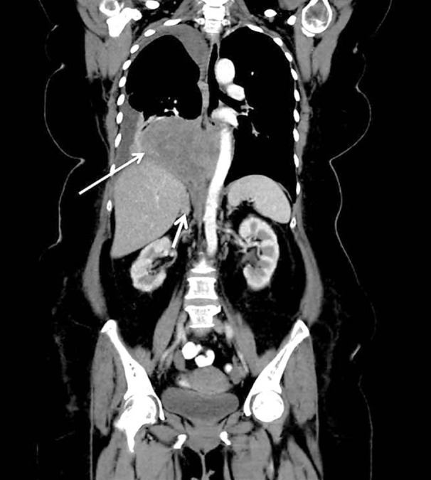
A 62-year-old postmenopausal female, a nonsmoker, presented with a six-month history of progressive dyspnoea, associated with decreased appetite and significant weight loss. Chest examination revealed dullness to percussion of the right lower zone with associated decreased breath sound. A computed tomography (CT) scan of the thorax, abdomen and pelvis following an abnormal chest radiograph disclosed a 10-cm right lower lobe lung mass with associated right adrenal metastasis (Figure 1). No other focal lesions were detected. Abdominal and pelvic ultrasonography yielded no abnormal findings. A series of serum tumour markers (alphafetoprotein, β-hCG, carcinoembryonic antigen, cancer antigen 125 (CA125), cancer antigen 19-9 (CA19-9) were examined. Incidentally, serum β-hCG levels were found to be high, measuring 11211.9 mIU/ml (normal upper limit 0.5 mIU/ml in nonpregnant woman), which prompted the initial suspicion of and further investigations to exclude a germ cell tumour. A CT-guided biopsy of the right lung mass was subsequently performed.
Figure 1:

Radiological features of β-hCG secreting lung adenocarcinoma. Coronal reconstruction contrast enhanced computed topography (CT) abdomen and thorax shows a heterogenously enhancing right lower lobe mass (long arrow) which displaces and abuts the descending thoracic aorta. The right adrenal is enlarged in keeping with adrenal metastasis (short arrow).
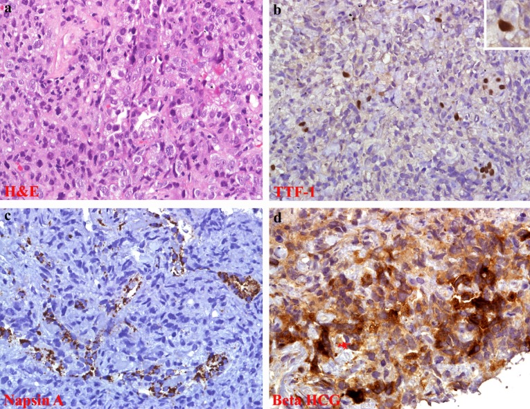
Histopathological examination revealed replacement of normal lung parenchyma by sheets of malignant cells. In areas, vague glandular formation was noted (Figure 2a). The malignant cells displayed moderate nuclear pleomorphism, vesicular nuclei with prominent nucleoli and abundant vacuolated to eosinophilic cytoplasm. Atypical mitosis was occasionally encountered (Figure 2a). There were no multinucleated giant cells, syncytiotrophoblasts and cytotrophoblasts to suggest choriocarcinoma.
Figure 2:
Histological and immunohistochemistry features of β-hCG secreting lung adenocarcinoma; (a) sheets of poorly differentiated malignant cells, in areas, vague glandular formation are seen; labelling for (b) TTF-1 and (c) Napsin A are focally expressed by malignant cells, indicating lung in origin; (d) some of the malignant cells display β-hCG immunoreactivity.
Immunohistochemically, the malignant cells were diffusely positive for cytokeratin AE1/AE3 (CKAE1/AE3), and exhibited focal (10%) immunoreactivity for TTF-1 (Figure 2b), napsin A (Figure 2c) and cytokeratin 7 (CK7). Other immunomarkers i.e. cytokeratin 20 (CK20), leucocyte common antigen (LCA), cytokeratin 5/6 (CK5/6), p63, neuroendocrine markers (chromogranin and synaptophysin) and S100 were negative. Some of the malignant cells also displayed immunopositivity for β-hCG (Figure 2d). A histological diagnosis of β-hCG-secreting poorly differentiated adenocarcinoma of the lung was rendered. No epidermal growth factor receptor (EGFR) mutation was detected.
The patient was started on chemotherapy with platinum-based agent (carboplatin) and etoposide. Biochemical response was observed after the first cycle of chemotherapy with significant reduction in serum ß-hCG levels to 168.6 mIU/ml. Further chemotherapy was withheld due to multiple complications. The patient developed febrile neutropenia complicated with urinary tract infection and catheter-related bacteraemia. Serial chest radiographs showed worsening of right lung opacity, which was attributed to parapneumonia-effusion. A CT scan three weeks post chemotherapy did not show any objective tumour response. A bone scan further revealed diffuse axial and appendicular skeletal metastasis. In view of progressive clinical and radiological deterioration, the patient and family opted for best supportive care.
Discussion
Ectopic hormone production (e.g. antidiuretic hormone, parathyroid hormone and adrenocorticotropic hormone) manifesting as paraneoplastic syndromes is not uncommon in pulmonary small-cell carcinomas and a smaller proportion of non-small cell lung carcinomas (NSCLC), particularly squamous cell type (6). Ectopic β-hCG expression by NSCLC is exceptional. To the best of our knowledge, about 60 cases of β-hCG-secreting NSCLC have been described in the English literature (2–5).
Expression of serum β-hCG is not associated with any specific histological subtypes, although some authors have found that this phenomenon is more characteristic of adenocarcinoma (3). In most reported cases of β-hCG-secreting lung adenocarcinoma, serum β-hCG levels were only slightly elevated (not exceeding 200 mIU/ml) (3). Intriguingly, our patient had unusually high levels of β-hCG compared to other published data.
The mean age at diagnosis for β-hCG-secreting NSCLC from published data is 59 years with a range of 31 to 73 years (2–5), much older than the usual age range for choriocarcinoma (younger than 20 years). The vast age difference between NSCLC and choriocarcinoma patients provides an invaluable clue for the correct diagnosis. The clinical manifestations pertaining to high β-hCG secretion include amenorrhoea mimicking pregnancy, dysfunctional uterine bleeding in women and gynaecomastia in men (2,4,5).
The precise mechanism of β-hCG secreted by nontrophoblastic tumours is poorly understood. Some authors have proposed the concept of a trophoblastic metaplasia within the carcinomatous tissue (1). To date, few published reports have attempted to address this issue by exploring ectopic β-hCG production at the molecular level. Earlier data showed that fetal lung contains trace to undetectable amounts of β-hCG mRNA transcripts and that its expression in the lung was developmentally regulated (7). In view of this data, the presence of β-hCG-specific mRNA in the primary lung cancer tissues was discovered (1), which suggested the possibility of reactivation of β-hCG gene transcription in the malignant transformed cells, leading to ectopic β-hCG protein expression.
Besides their important biological role in promoting progesterone production and vasculogenesis in the uterine vasculature during pregnancy, free β-subunit and hyperglycosylated hCG were recently found to play a major role in the tumourigenesis of nontrophoblastic tumours. They acted as an autocrine antiapoptotic and angiogenic growth factor, resulting in cancer cell growth (8). This may also explain the chemoresistance and aggressiveness of β-hCG-secreting lung cancer. Some authors have shown a high expression of membrane-associated complete β-hCG in metastatic lung cancer cell lines, which may attribute to the metastatic potential of such secreting tumours (9). The findings supported the potential use of serum β-hCG to predict patients who are at risk of developing metastatic disease.
At present, no information about the therapeutic approaches for such rare tumours is available. A chemotherapy regimen (platinum and etoposide) that fitted both lung adenocarcinoma and germ cell tumours was commenced for our patient. Interestingly, biochemical response with significant reduction in serum β-hCG level was observed after the first cycle of chemotherapy. A similar observation was reported in two cases of β-hCG-secreting gastric adenocarcinoma administered with a similar chemotherapy regimen (10).
Conclusion
We presented a rare case of β-hCG-secreting lung adenocarcinoma, which was initially confused with choriocarcinoma clinically. This unusual ectopic hormone production by lung cancer should be recognised to facilitate prompt diagnosis and initiation of appropriate intervention.
Acknowledgments
None.
Footnotes
Conflict of interest
None.
Funds
None.
Authors’ Contributions
Analysis and interpretation of the data: YPW, SA, SP
Drafting of the article: YPW, SP
Critical revision of the article for important intellectual content: GCT, SA, FI
Final approval of the article: GCT, FI
Collection and assembly of data: YPW, SP
References
- 1.Marcillac I, Troalen F, Bidart JM, Ghillani P, Ribrag V, Escudier B, et al. Free human chorionic gonadotropin beta subunit in gonadal and nongonadal neoplasms. Cancer Res. 1992;52:3901–3907. [PubMed] [Google Scholar]
- 2.Vicier C, Tabouret E, Tallet A, Goncalves A, Chetaille B, Viens P, et al. BetaHCG secretion by a pulmonary adenocarcinoma. World J Surg Oncol. 2013;11:228. doi: 10.1186/1477-7819-11-228. doi: 10.1186/1477-7819-11-228 . [DOI] [PMC free article] [PubMed] [Google Scholar]
- 3.Szturmowicz M, Slodkowska J, Zych J, Rudzinski P, Sakowicz A, Rowinska-Zakrzewska E. Frequency and clinical significance of beta-subunit human chorionic gonadotropin expression in non-small cell lung cancer patients. Tumour Biol. 1999;20:99–104. doi: 10.1159/000030052. doi: 10.1159/000030052 . [DOI] [PubMed] [Google Scholar]
- 4.Yoshida J, Nagai K, Nishimura M, Takahashi K, Kakinuma R, Nishiwaki Y, et al. Secretion of hCG/beta-hCG by squamous cell carcinoma of the lung in a 31-year-old female smoker. Jpn J Clin Oncol. 2000;30(3):163–166. doi: 10.1093/jjco/hyd028. doi: 10.1093/jjco/hyd028 . [DOI] [PubMed] [Google Scholar]
- 5.Khattri S, Vivekanandarajah A, Varma S, Kong F. Secretion of beta-human chorionic gonadotropin by non-small cell lung cancer: A case report. J Med Case Reports. 2011;5:19. doi: 10.1186/1752-1947-5-19. doi: 10.1186/1752-1947-5-19 . [DOI] [PMC free article] [PubMed] [Google Scholar]
- 6.Heinemann S, Zabel P, Hauber HP. Paraneoplastic syndromes in lung cancer. Cancer Ther. 2008;6:687–698. [Google Scholar]
- 7.Rothman PA, Chao VA, Taylor MR, Kuhn RW, Jaffe RB, Taylor RN. Extraplacental human fetal tissues express mRNA transcripts encoding the human chorionic gonadotropin-beta subunit protein. Mol Reprod Dev. 1992;33(1):1–6. doi: 10.1002/mrd.1080330102. [DOI] [PubMed] [Google Scholar]
- 8.Cole LA. HCG variants, the growth factors which drive human malignancies. Am J Cancer Res. 2012;2(1):22–35. [PMC free article] [PubMed] [Google Scholar]
- 9.Acevedo HF, Hartsock RJ. Metastatic phenotype correlates with high expression of membrane-associated complete beta-human chorionic gonadotropin in vivo. Cancer. 1996;78(11):2388–2399. doi: 10.1002/(sici)1097-0142(19961201)78:11<2388::aid-cncr18>3.0.co;2-x. doi: 10.1002/(SICI)1097-0142(19961201)78:11 . [DOI] [PubMed] [Google Scholar]
- 10.Germann N, Gross-Goupil M, Wasserman E, Emile JF, Misset JL, Reynes M, et al. The chemotherapy of metastatic gastric adenocarcinomas with hypersecretion of alpha-fetoprotein or beta-human chorionic gonadotrophin: Report of two cases. Ann Oncol. 2002;13(4):632–636. doi: 10.1093/annonc/mdf026. doi: 10.1093/annonc/mdf026 . [DOI] [PubMed] [Google Scholar]


