Abstract
Background
Ensuring genetic stability in pluripotent stem cell (PSC) cultures is essential for the development of successful cell therapies. Although most instances lead to failed experiments and go unreported in the literature, many laboratories have found the emergence of genetic abnormalities in PSCs when cultured in vitro for prolonged amounts of time. These cells are primarily cultured in non-physiological stiff substrates like tissue culture polystyrene (TCPS) which raises the possibility that the cause of these abnormalities may be influenced by substrate mechanics.
Findings
In order to investigate this, human PSCs were grown on substrates of varying stiffness such as a range of polyacrylamide formulations, TCPS, and borosilicate glass coverslips. These substrates allowed for the testing of a stiffness range from 5kPa to 64GPa. Two human induced PSC (iPSC) lines were analyzed in this study: 19-9-11 iPSCs and 19.7 clone F iPSCs. Centrosome and DNA staining revealed that 19-9-11 iPSCs range from 1–8.5 % abnormal mitoses under the different culture conditions. A range of 4.4–8.1 % abnormal mitoses was found for 19.7 clone F iPSCs.
Conclusions
Abnormal cell division was not biased towards one particular substrate. It was confirmed by Analysis of Variance (ANOVA) and Tukey’s Honest Significant Difference test that there was no statistically significant difference between passage numbers, cell lines, or substrates.
Electronic supplementary material
The online version of this article (doi:10.1186/s12952-015-0042-8) contains supplementary material, which is available to authorized users.
Keywords: Induced pluripotent stem cells, Chromosome missegregation, Mitosis, Substrate stiffness
Background
Stem cell research aims to grow human cells in vitro for transplantation and carry out drug and toxicity screenings on relevant human cell and tissue models. In order for these goals to be successful, it is important to conserve the genetic stability of these cells. Several reports assessing the genetic stability of human pluripotent stem cells (PSCs), including both embryonic and induced stem cell lines, when cultured in vitro for prolonged amounts of time have been published in recent years [1–4]. It has been suggested that chromosomal gains, such as trisomies 8, 12, 17 or X, give PSCs a selective advantage in in vitro culture due to the fact that chromosome 12, for example, encodes for many cell-cycle related genes [5].
A recent study done by Holubcová et al. [6] found the frequency of multicentrosomal mitosis (defined as a mitotic cell with more than 2 centrosomes) for human embryonic stem cells to be between 10 and 23 %. Additionally, Gisselsson et al. [7] determined that the chromosome missegregation rate for normal dermal fibroblasts was about 1 missegregation event in around 50 cell divisions.
How the mechanics of the surroundings influence a dividing cell in vitro is beginning to be investigated. For example, Kocgozlu et al. [8] reported that softer substrates hinder epithelial cell division by leading to abnormal morphology in chromosome segregation. Tse et al. [9] found that mechanically confined HeLa cell divisions resulted in an increase in abnormal multi-daughter divisions. Given this evidence in differentiated cell types, it can be speculated that the observed recurrent chromosomal abnormalities may be an artifact of in vitro culture, particularly when taking into account that embryonic stem cells exist in vivo for a short period of time.
A common factor between the abnormal cell lines reported is that they were cultured on stiff substrates like glass (E = 60–64GPa; [10]) and tissue culture polystyrene (TCPS; E = 2.28–3.28 GPa; [11]). For comparison, human tissues such as neural, muscle and collagenous bone fall into a stiffness range of 1–100 kPa. Many studies have investigated the effect of mechanical stimuli on stem cell fates such as self-renewal [12, 13] and differentiation [14, 15].
This study began with the hypothesis that culturing PSCs in non-physiologic stiff culture conditions such as TCPS and glass caused abnormal mitotic spindle formation and/or spindle function due to substrate stiffness, consequently causing chromosome missegregation. To determine the effect of matrix stiffness on PSCs, culture substrates commonly used for in vitro culture such as TCPS and glass were evaluated in addition to a matrix whose stiffness could be readily altered in order to study a biologically relevant range. Polyacrylamide (PA) hydrogels were chosen due to ease of fabrication, tuneability of mechanical properties and amount of studies done using this polymer to assess the effect of stiffness on different cell fates [14, 16–18].
Materials and methods
Cell culture
The cell lines used were 19.7 clone F induced pluripotent stem cells (iPSCs) and DF19-9-11 iPSCs acquired from WiCell [19] and approved by the Stem Cell Research Oversight (SCRO) Committee at the University of Wisconsin-Madison. All cell lines were cultured as described previously by Chen et al. [20]. Passage number since derivation for DF19-9-11 iPSCs ranged from passage 34–55 and from passage 51–65 for 19.7 clone F iPSCs.
Polyacrylamide hydrogel fabrication and functionalization
The hydrogels were fabricated using a method described by Hazeltine et al. [18]. The surfaces of the hydrogels were functionalized with a N-sulfosuccinimidyl-6-[4′-azido-2′-nitrophenylamino] (Sulfo-SANPAH) treatment as described previously by Hazeltine et al. [18, 21] in order to facilitate attachment of a thin surface coating with Matrigel to promote cell adhesion, allow for force transduction between the cell and the substrate, and maintain pluripotency of iPSCs.
Mechanical testing
Polyacrylamide (PA) samples designated for mechanical characterization were polymerized into a standard tensile test geometry following ASTM standard D638-08 [22]. Stiffness of the specific PA gel formulations used in this research was determined by tensile testing using an Instron 5548 MicroTester, with a 10 N load cell. Using a tensile test method developed in the lab for hydrogel materials [23], the Young’s modulus of the PA formulations were measured and summarized in Table 1.
Table 1.
Summary of Young’s modulus obtained for different culture substrates
| Substrate | Acrylamide Concentration (%) | Bisacrylamide Concentration (%) | Young’s modulus (Pa) |
|---|---|---|---|
| No.1 borosilicate glass coverslips | - | - | 60–64 × 109 |
| Tissue Culture Polystyrene | - | - | 2.28–3.28 × 10⁹ |
| Polyacrylamide | 10 % | 0.03 % | 5.0 × 10³ ± 1.6 |
| 10 % | 0.3 % | 31.0 × 10³ ± 5.7 | |
| 10 % | 0.6 % | 51.7 × 10³ ± 6.0 | |
| 10 % | 1.2 % | 59.6 × 10³ ± 14.7 |
Immunofluorescence
Human iPSCs were fixed using a 4 % paraformaldehyde (Electron Microscopy Sciences), 0.3 % glutaraldehyde (Sigma), 0.1 % Triton X (Sigma) solution in cytoskeletal buffer [24]. Samples were post-fixed with 90 % ice-cold methanol (Sigma) and quenched with 100 mM sodium borohydride (Sigma). Samples were blocked in 50 μg/mL bovine serum albumin (BSA; Sigma), 0.1 % Triton X, 150 mM glycine (Sigma) and goat serum (Sigma) in phosphate buffered saline (PBS) for 30 min. The antibodies used in this study were anti-γ-tubulin (4D11; Thermo Scientific), anti-phosphorylated Histone 3 (pH3; 9H12L10; Life Technologies), anti-Oct4 (H-134; Santa Cruz Biotechnology, Inc.), goat anti-rabbit IgG-H&L (Cy3; Abcam) and goat anti-mouse IgG-H&L (DyLight488; Abcam). Nuclei were stained with ProLong Gold Antifade Reagent with DAPI (4',6-diamidino-2-phenylindole; Life Technologies). Samples were imaged using a Nikon Eclipse Ti inverted epifluorescence microscope system with a 40x objective for mitotic index quantifications and a 60x oil objective for abnormal mitosis quantification.
Statistical analysis
ANOVA and Tukey’s Honest Significant Difference test were performed using R software [25].
Results and discussion
To determine the effect of stiffness on human iPS cell division, karyotypically normal human 19-9-11 iPSCs and abnormal 19.7 clone F iPSCs were cultured on substrates with different Young’s modulus values as summarized in Table 1. Glass and TCPS were used because cells are usually cultured on these substrates during regular cell maintenance or for imaging applications. The mechanical properties of PA hydrogels were altered by varying the bisacrylamide cross-linker concentration from 0.03–1.2 % thus generating a stiffness range of 5–60 kPa.
To determine stiffness effects on mitotic spindle morphology and organization, mitotic figures were examined for 5 consecutive passages and at passage 10 on glass and TCPS. Cells growing on PA were analyzed after 4 days in culture. Due to low density of cells that would remain attached until passaging (around 4 days), quantification of abnormalities for cells cultured on PA was not able to be carried out for multiple passages as with glass and TCPS. Cells were still pluripotent on all substrates as confirmed by OCT4 expression (Fig. 1) which has been shown is essential for maintenance of pluripotency in human and mouse PSCs [26, 27]. From immunofluorescence images, the mitotic index was quantified by dividing the number of pH3 positive cells by the total number of cells. Mitotic index quantification for 19-9-11 iPSCs cultured on glass, TCPS and PA (Additional file 1: Figure S1) revealed similar percentages for the different substrates. No statistically significant difference was found for the substrates by ANOVA and Tukey’s Honest Significant Difference test.
Fig. 1.

Human iPSCs remain pluripotent on substrates of varied stiffness. Nuclei are stained blue with DAPI and OCT4+ cells are labeled in red. Epifluorescence images of 19-9-11 iPSCs cultured on 31kPA hydrogels (left panel), TCPS (middle panel) and glass coverslips (right panel). Scale bar for TCPS image: 100 μm. Scale bar for PA and glass images: 50 μm
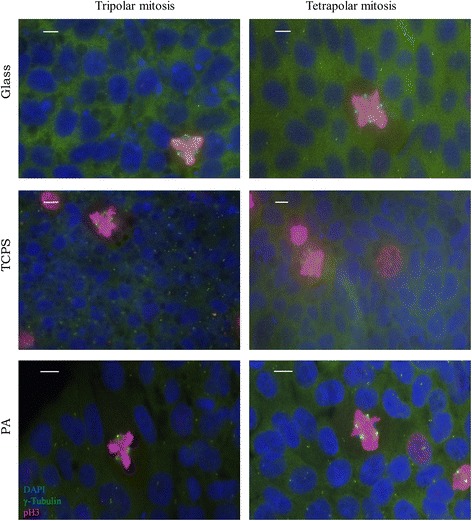
For this study, abnormal mitoses are defined as any prometaphases and metaphases that have 3 or more centrosomes or misaligned chromosomes (chromosomes separated from the rest of the chromosomes located at the metaphase plate). Abnormal mitotic spindles shown in Fig. 2 (Additional file 2: Figure S2 and Additional file 3: Figure S3) represent the different types of abnormalities that were pooled into the percentage of abnormal mitoses quantified in Figs. 3 and 4. Observed mitotic spindles with 3 or 4 centrosomes displaying triangular or square spindle morphology respectively are shown in Fig. 2. Other abnormalities observed include misaligned chromosomes, mitotic spindles with more than 4 centrosomes and potentially inactive centrosomes (Additional file 2: Figure S2 and Additional file 3: Figure S3). Abnormalities in anaphase such as lagging chromosomes and multipolar chromosome segregation during anaphase were also observed but not quantified (Additional file 4: Figure S4).
Fig. 2.

Commonly observed multipolar mitoses in 19-9-11 iPSCs cultured on substrates of varied stiffness. Nuclei are labeled in blue (DAPI), γ-tubulin is labeled in green while pH3 is labeled in red. The rows indicate the substrates human iPSCs were cultured on. Tripolar mitoses are characterized by 3 spindle poles (green γ-tubulin foci). Tetrapolar mitoses are characterized by 4 spindle poles. These types of abnormalities are included in the percentage of abnormal mitoses calculated in Figs. 3 and 4. Scale bars: 10 μm
Fig. 3.

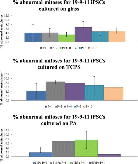
Frequency of abnormal mitoses for 19-9-11 iPSCs cultured on glass, TCPS and PA hydrogels. Bar plot for 19-9-11 iPSCs cultured on glass show percent of abnormal mitoses for 5 consecutive passages and passage 10. Plot for TCPS shows quantification for passages 1, 2, 4, 5 and 10. Passage 3 was unable to be quantified due to contamination. Quantification for PA hydrogels was done only for one passage. n = 26–132 mitoses per condition for 1–5 independent experiments done. Passage number since derivation for this cell line ranged from passage 34–55
Fig. 4.

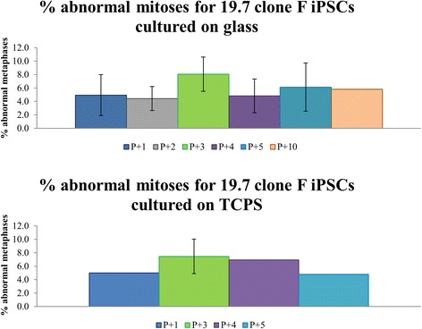
Frequency of abnormal mitoses for 19.7 clone F iPSCs cultured on glass and TCPS. Bar plot for 19.7 clone F iPSCs cultured on glass show percent of abnormal mitoses for 5 consecutive passages and passage 10. Plot for TCPS shows quantification for passages 1, 3, 4 and 5. Passage 2 was unable to be quantified due to contamination. n = 88–112 mitoses per condition for 1–5 independent experiments done. Passage number since derivation for this cell line ranged from passage 51–65
Summarized in Figs. 3 and 4 are the percentages of abnormal mitoses quantified from γ-tubulin (centrosome marker) and pH3 (mitotic marker) staining. Quantification of abnormal mitoses revealed that these occur at a frequency of up to 8.5 %. For the 19-9-11 cell line, iPSCs cultured on Matrigel-coated glass had a percentage of abnormal mitoses of 3.3 to 6.7 %. A range of 4.4 to 8.5 % abnormal mitoses was found for iPSCs cultured on TCPS. For cells grown on PA gels, 60 kPa PA had the lowest percentage abnormal mitoses with only 1 %. The percentage then increased with 1.9 % for 5 kPa, 7 % for 31 kPa and 7.6 % for 48 kPa hydrogels.
For the 19.7 clone F iPSC line, iPSCs cultured on Matrigel-coated glass coverslips had a percentage of abnormal mitoses of 4.4 to 8.1 %. A range of 4.8 to 7.5 % abnormal mitoses was found for iPSCs cultured on TCPS. Previous G-banding karyotype analysis of this line, supplied by WiCell, revealed that 20 % of the population was abnormal (data not shown).
These results are in discrepancy with the results published by Holubcová and colleagues [6] which report that human embryonic stem cells have a frequency of multicentrosomal mitosis of 10–23 %. A possible source of discrepancy is a cell line-specific susceptibility to centrosome amplification or substrate stiffness influence. Holubcová et al. found that different substrate-extracellular matrix (ECM) coating combinations altered the frequency of multicentrosomal mitosis. When comparing all substrates in the work reported here, the percent of abnormal mitoses did not follow any observable trend or bias with stiffness. Additionally, there were no differences between cell lines or passage number. This was confirmed by ANOVA and Tukey’s Honest Significant Difference test which found no statistically significant difference between the conditions. An explanation for these different results could be that cell-ECM interactions are influencing the frequency of multicentrosomal mitosis instead of cell-substrate interactions. Further studies are required to answer this question.
Alternatively, culture system differences (i.e. ECM coating, media, Rho-associated protein kinase (ROCK) inhibitor) may be the cause. The constant treatment with ROCK inhibitor in this prior study could be responsible since it has been shown that cells treated with ROCK inhibitor exhibit improper mitotic spindle positioning and assembly [28]. A future study of abnormal mitoses for iPSCs cultured under prolonged ROCK inhibition is warranted.
Despite the percentage of abnormal mitoses being lower than expected, the types of abnormalities found were varied. The presence of multipolar mitoses as a result of additional centrosomes may suggest a deregulation of the centrosome duplication cycle or failed cytokinesis particularly for cells that contain a large number of centrosomes such as those shown in Additional file 2: Figures S2A and S2B. Mechanisms that affect centrosome duplication have been mainly studied in the context of cancer and are reviewed in Meraldi et al. [29]. On the other hand, the presence of lagging and misaligned chromosomes may suggest altered microtubule dynamics such as incorrect chromosome-microtubule attachments or changes in motor proteins associated with microtubule positioning. Future studies addressing the roles of centrosome duplication errors or altered microtubule dynamics in the context of human PSC chromosome missegregation could provide insight into the mechanisms behind observed recurrent chromosomal abnormalities in these cells.
Acknowledgements
The authors would like to thank Prof. Beth A. Weaver, Max Salick, Chandler C. Benjamin, Carlos R. Marti-Figueroa, the Kamp/Lyons Lab Groups and the WiCell Cytogenetics department, specifically Erik McIntire, Karen Montgomery and Seth Taapken for helpful conversations. This research was supported with funds from the National Institutes of Health Grant K18 HL105504 from the Heart, Blood and Lung Institute and the Graduate School of the University of Wisconsin-Madison. SAA has been supported by the Graduate Engineering Research Scholars program of the University of Wisconsin-Madison and by the National Science Foundation Graduate Research Fellowship Program under Grant No. DGE-1256259.
Abbreviations
- PSCs
Pluripotent stem cells
- iPSCs
Induced pluripotent stem cells
- TCPS
Tissue culture polystyrene
- ANOVA
Analysis of variance
- PA
Polyacrylamide
- SCRO
Stem cell research oversight
- Sulfo-SANPAH
N-sulfosuccinimidyl-6-[4′-azido-2′-nitrophenylamino]
- BSA
Bovine serum albumin
- PBS
Phosphate buffered saline
- pH3
Phosphorylated histone 3
- DAPI
(4', 6-diamidino-2-phenylindole)
- ECM
Extracellular matrix
- ROCK
Rho-associated protein kinase
Additional files
Bar plot showing the mitotic index for 19-9-11 iPSCs cultured on substrates of varied stiffness. n = 197–1,132 total cells counted per condition for 2–3 independent experiments per culture substrate. (TIF 84 kb)
Examples of additional cell division abnormalities seen in 19-9-11 iPSCs on different substrates. Nuclei are labeled in blue (DAPI), γ-tubulin is labeled in green while pH3 is labeled in red. The rows indicate the substrates human iPSCs were cultured on. The left panels show representative images of multipolar mitoses where the centrosomes, indicated by γ-tubulin staining, have different morphology as those seen in Fig. 2. The right panels show misaligned chromosomes which are separated from the chromosomes aligned at the metaphase plate. These types of abnormalities are included in the percentage of abnormal mitoses calculated in Fig. 3. Scale bars: 10 μm. (TIF 4853 kb)
Abnormalities found in 19-9-11 iPSCs not common to all substrates. Nuclei are stained in blue (DAPI), γ-tubulin is labeled in green while pH3 is labeled in red. (A) Abnormal mitosis of an iPSC cultured on glass with 7 centrosomes indicated by bright γ-tubulin stain. (B) Multipolar mitosis of an iPSC cultured on TCPS with 6 centrosomes (C) A tripolar mitosis of an iPSC cultured on 31 kPa PA with a fourth potentially inactive centrosome. These types of abnormalities are included in the percentage of abnormal mitoses calculated in Fig. 3. Scale bars: 10 μm. (TIF 2417 kb)
Examples of abnormal anaphases in 19-9-11 iPSCs cultured on substrates of varied stiffness. Nuclei are stained in blue (DAPI), γ-tubulin is labeled in green while pH3 is labeled in red. Representative images of multipolar anaphases (left panels) and lagging chromosomes (right panels) detected in 19-9-11 iPSCs. Multipolar anaphases are cells that segregate their chromosomes improperly to more than two daughter cells. Lagging chromosomes are chromosomes that segregate separately from the rest of the chromosomes after anaphase onset, usually due to the chromosomes being attached to both spindle poles. The rows indicate the substrates human iPSCs were cultured on. These types of abnormalities are not included in the percentage of abnormal mitoses calculated in Fig. 3. Scale bars: 10 μm. (TIF 5065 kb)
Footnotes
Suehelay Acevedo-Acevedo and Wendy C. Crone contributed equally to this work.
Competing interests
The authors declare that they have no competing interests.
Authors’ contributions
SAA conducted the experiments, wrote the manuscript and participated in the statistical analysis and design of the study. WCC participated in the design and oversight of the study and drafting of the manuscript. Both authors read and approved the manuscript.
Contributor Information
Suehelay Acevedo-Acevedo, Email: suehelay.acv2@gmail.com.
Wendy C. Crone, Email: wendy.crone@wisc.edu
References
- 1.Catalina P, Montes R, Ligero G, Sanchez L, de la Cueva T, Bueno C, et al. Human ESCs predisposition to karyotypic instability: Is a matter of culture adaptation or differential vulnerability among hESC lines due to inherent properties? Mol Cancer. 2008;7:76. doi: 10.1186/1476-4598-7-76. [DOI] [PMC free article] [PubMed] [Google Scholar]
- 2.Draper JS, Smith K, Gokhale P, Moore HD, Maltby E, Johnson J, et al. Recurrent gain of chromosomes 17q and 12 in cultured human embryonic stem cells. Nat Biotechnol. 2003;22:53–54. doi: 10.1038/nbt922. [DOI] [PubMed] [Google Scholar]
- 3.Imreh MP, Gertow K, Cedervall J, Unger C, Holmberg K, Szöke K, et al. In vitro culture conditions favoring selection of chromosomal abnormalities in human ES cells. J Cell Biochem. 2006;99:508–516. doi: 10.1002/jcb.20897. [DOI] [PubMed] [Google Scholar]
- 4.Mitalipova MM, Rao RR, Hoyer DM, Johnson JA, Meisner LF, Jones KL, et al. Preserving the genetic integrity of human embryonic stem cells. Nat Biotechnol. 2005;23:19–20. doi: 10.1038/nbt0105-19. [DOI] [PubMed] [Google Scholar]
- 5.Mayshar Y, Ben-David U, Lavon N, Biancotti J-C, Yakir B, Clark AT, et al. Identification and Classification of Chromosomal Aberrations in Human Induced Pluripotent Stem Cells. Cell Stem Cell. 2010;7:521–531. doi: 10.1016/j.stem.2010.07.017. [DOI] [PubMed] [Google Scholar]
- 6.Holubcová Z, Matula P, Sedláčková M, Vinarský V, Doležalová D, Bárta T, et al. Human Embryonic Stem Cells Suffer from Centrosomal Amplification. STEM CELLS. 2011;29:46–56. doi: 10.1002/stem.549. [DOI] [PubMed] [Google Scholar]
- 7.Gisselsson D, Jin Y, Lindgren D, Persson J, Gisselsson L, Hanks S, et al. Generation of trisomies in cancer cells by multipolar mitosis and incomplete cytokinesis. Proc Natl Acad Sci. 2010;107:20489–20493. doi: 10.1073/pnas.1006829107. [DOI] [PMC free article] [PubMed] [Google Scholar]
- 8.Kocgozlu L, Rabineau M, Koenig G, Haikel Y, Schaaf P, Freund J-N, et al. The control of chromosome segregation during mitosis in epithelial cells by substrate elasticity. Biomaterials. 2012;33:798–809. doi: 10.1016/j.biomaterials.2011.10.024. [DOI] [PubMed] [Google Scholar]
- 9.Tse HTK, Weaver WM, Di Carlo D. Increased Asymmetric and Multi-Daughter Cell Division in Mechanically Confined Microenvironments. PLoS ONE. 2012;7:e38986. doi: 10.1371/journal.pone.0038986. [DOI] [PMC free article] [PubMed] [Google Scholar]
- 10.Borosilicate Glass. Matweb Material Property Data. Accessed: 05/2014. [http://www.matweb.com/index.aspx]
- 11.Callister WD. Fundamentals of Materials Science and Engineering: An Interactive Etext (5th Edition) New York: Wiley; 2001. [Google Scholar]
- 12.Chowdhury F, Li Y, Poh Y-C, Yokohama-Tamaki T, Wang N, Tanaka TS. Soft Substrates Promote Homogeneous Self-Renewal of Embryonic Stem Cells via Downregulating Cell-Matrix Tractions. PLoS ONE. 2010;5:e15655. doi: 10.1371/journal.pone.0015655. [DOI] [PMC free article] [PubMed] [Google Scholar]
- 13.Sun Y, Villa-Diaz LG, Lam RHW, Chen W, Krebsbach PH, Fu J. Mechanics Regulates Fate Decisions of Human Embryonic Stem Cells. PLoS ONE. 2012;7:e37178. doi: 10.1371/journal.pone.0037178. [DOI] [PMC free article] [PubMed] [Google Scholar]
- 14.Engler AJ, Sen S, Sweeney HL, Discher DE. Matrix elasticity directs stem cell lineage specification. Cell. 2006;126:677–689. doi: 10.1016/j.cell.2006.06.044. [DOI] [PubMed] [Google Scholar]
- 15.Eroshenko N, Ramachandran R, Yadavalli VK, Rao RR. Effect of substrate stiffness on early human embryonic stem cell differentiation. J Biol Eng. 2013;7:7. doi: 10.1186/1754-1611-7-7. [DOI] [PMC free article] [PubMed] [Google Scholar]
- 16.Lee J, Abdeen AA, Zhang D, Kilian KA. Directing stem cell fate on hydrogel substrates by controlling cell geometry, matrix mechanics and adhesion ligand composition. Biomaterials. 2013;34:8140–8148. doi: 10.1016/j.biomaterials.2013.07.074. [DOI] [PubMed] [Google Scholar]
- 17.Tse JR, Engler AJ. Stiffness Gradients Mimicking In Vivo Tissue Variation Regulate Mesenchymal Stem Cell Fate. PLoS ONE. 2011;6:e15978. doi: 10.1371/journal.pone.0015978. [DOI] [PMC free article] [PubMed] [Google Scholar]
- 18.Hazeltine LB, Simmons CS, Salick MR, Lian X, Badur MG, Han W, et al. Effects of Substrate Mechanics on Contractility of Cardiomyocytes Generated from Human Pluripotent Stem Cells. Int J Cell Biol. 2012;2012:1–13. doi: 10.1155/2012/508294. [DOI] [PMC free article] [PubMed] [Google Scholar]
- 19.Yu J, Vodyanik MA, Smuga-Otto K, Antosiewicz-Bourget J, Frane JL, Tian S, et al. Induced Pluripotent Stem Cell Lines Derived from Human Somatic Cells. Science. 2007;318:1917–1920. doi: 10.1126/science.1151526. [DOI] [PubMed] [Google Scholar]
- 20.Chen G, Gulbranson DR, Hou Z, Bolin JM, Ruotti V, Probasco MD, et al. Chemically defined conditions for human iPSC derivation and culture. Nat Methods. 2011;8:424–429. doi: 10.1038/nmeth.1593. [DOI] [PMC free article] [PubMed] [Google Scholar]
- 21.Tse JR, Engler AJ: Preparation of hydrogel substrates with tunable mechanical properties. Curr Protoc Cell Biol Editor Board Juan S Bonifacino Al. 2010, Chapter 10:Unit 10.16. doi: 10.1002/0471143030.cb1016s47. [DOI] [PubMed]
- 22.ASTM Standard D638-08: Standard test method for tensile properties of plastics. ASTM Int 2008.
- 23.Johnson B, Bauer JM, Niedermaier DJ, Crone WC, Beebe DJ. Experimental techniques for mechanical characterization of hydrogels at the microscale. Exp Mech. 2004;44:21–28. doi: 10.1007/BF02427971. [DOI] [Google Scholar]
- 24.Claxton NS, Ottenberg GK, Olenych SG, Griffin JD, Davidson MW: Staining Cells and Tissue Cryosections with Tubulin Primary Antibodies, Phallotoxins, and Synthetic Fluorophores. Olympus Microscopy Resource Center. Accessed: 09/2014. [http://www.olympusmicro.com/primer/techniques/confocal/applications/protocols/cellsandtissuestubulin.html]
- 25.R Core Team: R: A language and environment for statistical computing. R Found Stat Comput Vienna Austria 2012. [http://www.r-project.org/]
- 26.Matin MM. Specific Knockdown of Oct4 and 2-microglobulin Expression by RNA Interference in Human Embryonic Stem Cells and Embryonic Carcinoma Cells. Stem Cells. 2004;22:659–668. doi: 10.1634/stemcells.22-5-659. [DOI] [PubMed] [Google Scholar]
- 27.Hay DC, Sutherland L, Clark J, Burdon T. Oct-4 Knockdown Induces Similar Patterns of Endoderm and Trophoblast Differentiation Markers in Human and Mouse Embryonic Stem Cells. Stem Cells. 2004;22:225–235. doi: 10.1634/stemcells.22-2-225. [DOI] [PubMed] [Google Scholar]
- 28.Rosenblatt J, Cramer LP, Baum B, McGee KM. Myosin II-Dependent Cortical Movement Is Required for Centrosome Separation and Positioning during Mitotic Spindle Assembly. Cell. 2004;117:361–372. doi: 10.1016/S0092-8674(04)00341-1. [DOI] [PubMed] [Google Scholar]
- 29.Meraldi P, Nigg E. The centrosome cycle. FEBS Lett. 2002;521:9–13. doi: 10.1016/S0014-5793(02)02865-X. [DOI] [PubMed] [Google Scholar]


