In the past few years, I have become progressively more aware of the foundational importance of optimal mitochondrial function for health and the growing body of research showing that dysfunction is surprisingly common and associated with most chronic disease. In my IMCJ 12.5 editorial, Lara and I presented helpful clinical pearls on mitochondrial health from the Institute of Functional Medicine conference in Dallas last spring. In my IMCJ 13.1 editorial, I stated that glutathione is “… vital to mitochondrial function and maintenance of mitochondrial DNA (mtDNA).” I have found the mitochondrial connection to health so interesting and important that I have now developed a 90-minute lecture on the topic and think the time has come for an editorial.
As illustrated in Table 1, with such a long list of common diseases now caused by or aggravated by mitochondrial dysfunction, it is difficult to overstate its importance.
Table 1.
Partial List of Diseases Caused or Aggravated by Mitochondrial Dysfunction
|
How Much ATP Does the Body Produce Every Day?
I wish there were some way to cover up the answer—before you, dear reader, answer this question (please take a moment and guess how many g/d)—as I am sure everyone would guess wrong. Well, I should have said “almost everyone,” as we have some exceptionally knowledgeable readers. Having asked this question the 3 times I have given my mitochondrial lecture, no one was within 2 orders of magnitude of the correct answer. A healthy person at rest produces their body weight in adenosine triphosphate every day! At maximal exercise, this number can increase to 0.5 to 1.0 kg per minute—a truly remarkable indication of intense metabolic activity.
A large amount of adenosine triphosphate (ATP) must be produced by the mitochondria every second of every day because ATP cannot be stored. This function is so important that mitochondria can take up as much as 25% of the cell volume. Cells contain from 1000 to 2500 mitochondria.1 The average cell uses 10 billion ATP per day, which translates to the typical adult needing 3.0 × 1025 ATP (I am publishing these numbers with some trepidation as the research is surprisingly inconsistent about exactly how much ATP a cell needs).2 To accomplish this prodigious feat, each ATP needs to be recycled from ADP 1000 times per day. Because the body cannot store ATP, the mitochondria must function consistently all the time. At any given time there are about 250 g of ATP in the cells. This represents about 4.25 watts, the equivalent of the energy in an AA battery. Every day, a healthy person produces a remarkable 1200 watts!3 Because the brain uses 70% of ATP, this helps explain the strong correlation between mitochondrial dysfunction and neurodegeneration.
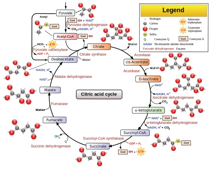
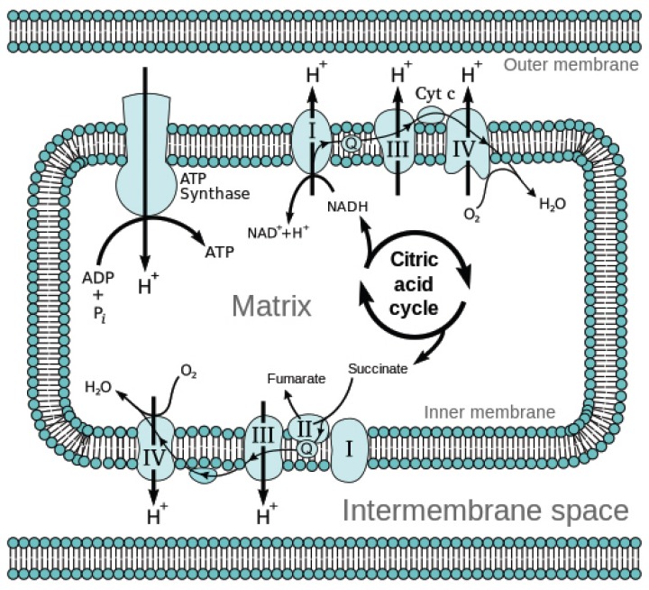
The production of ATP is quite a complex process, starting in the Krebs citric acid cycle, also known as the tricarboxylic acid cycle (TCA), as seen in Figure 1. But far more important than direct ATP production are the high-energy molecules the TCA provides the electron transport chain (ETC), as seen in Figure 2. This is where 90% of ATP are produced in aerobic metabolism.
Figure 1.
ATP Production From the Krebs Citric Acid Cycle4
Figure 2.
Citric Acid Cycle Provides High-Energy Molecules to ETC5
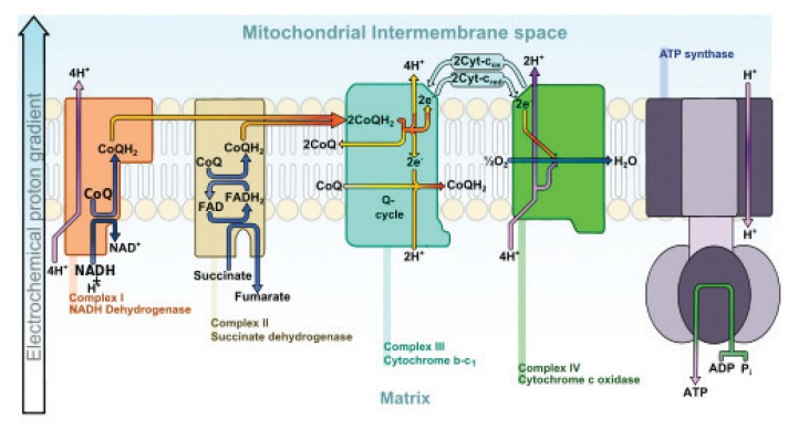
The ETC, as seen in Figure 3, is complex and quite fascinating. All the action occurs in and across the mitochondrial membranes and is highly dependent on the nutritional, toxic, and redox potential of the cell and mitochondria.
Figure 3.
ETC6
Mitochondrial Function and Protection
The mitochondria are especially susceptible to nutrient deficiencies, environmental toxins, and oxidative damage. Although many nutrients are necessary for ATP production, the most important are listed in Table 2.
Table 2.
Essential Nutrients for ATP Production
|
Research now shows that the primary source of oxidative stress in cells is leakage of oxygen and high-energy electrons from the mitochondria. This leakage increases when key nutrients/protective molecules are missing, such as the dose-dependent depletion of CoQ10 in patients taking statin drugs—a problem that has been known for a long time.7 Note in Figure 3 that the high-energy electrons are transported between the various mitochondrial complexes by CoQ10. Many factors are associated with increased damage to the mitochondria. The following list in Table 3 is by no means complete but is clinically relevant.
Table 3.
Factors Associated With Increased Mitochondrial Damage
|
Mitochondrial reactive oxygen leakage is a strong predictor across species for longevity—the better a species does protecting its mitochondria, the longer a species lives. Oxygen is essential for energy production and dangerous when not totally controlled. At rest, the body uses 1 kg of oxygen per day. During maximal exercise this can increase to 10 to 20 g per minute! As 1%–2% of oxygen is lost in “normal” mitochondria, this translates to 10 to 20 g O2 lost per day at rest and 200 mg per minute at maximal exercise. Obviously this represents huge oxidative stress, which explains why mitochondrial ROS production and leakage are the major source of intracellular oxidants. With age, mtDNA damage accumulates, resulting in even more oxygen and high-energy electron loss.8 This helps explain why people start “losing energy” once they reach around 55 years of age. The primary ways the mitochondria are protected from this intense oxidative stress are manganese-dependent superoxide dismutase, catalase, CoQ10, vitamin E, and glutathione (produced in cytoplasm and transported across the mitochondrial membrane).
Many prescription drugs damage mitochondria via a diverse range of mechanisms too complex to cover here. Among the worse are shown in Table 4. As you can see, these are all commonly prescribed.
Table 4.
Drugs That Damage Mitochondria
| Acetaminophen |
| Antibiotics |
| Aspirin |
| AZT |
| Cocaine |
| Grisepfulvin |
| Indomethacin |
| Methamphetamine |
| l-DOPA |
| NSAIDs |
| Statins |
These are all of significant concern, but particularly of concern are those prescribed frequently or taken for a long time. Some antibiotics, for example, not only impair ATP production; they also increase ROS production and damage mtDNA. The degree of mtDNA damage can be indirectly measured by 8-hydroxy-2′-deoxyguanosine (8-OHdG) in the urine. The numbers in Table 5 are cause for grave concern. This is new research from cell cultures and animals. It is reasonable to assume humans react similarly, though the research has yet to be published.9 Note especially the high level of mtDNA damage from ampicillin.
Table 5.
8-hydroxy-2′-deoxyguanosine (8-OHdG) in Urine
| After 4 Days | Ciplofloxan | Ampicillin | Kanamycin | Tetracycline |
|---|---|---|---|---|
| ATP production | −90% | −75% | −80% | −20% |
| ROS | +250% | +200% | +240% | +40% |
| MDA | +90% | +80% | +75% | +20% |
| 8-OHdG | +100% | +720% | +400% | +230% |
Another serious concern is the widespread and growing use of statin drugs. Many of the adverse events associated with these prescriptions are well explained by the mitochondrial dysfunction they induce, secondary to depletion of CoQ10. It is not surprising that statins increase all-cause mortality and most chronic disease.10 Recreational drugs are a problem as well. The research is too early on marijuana, but alcohol is clear: The more consumed, the greater the depletion of NADH needed for ATP production. In addition, ROS production is increased.11
Assessing Mitochondrial Function
At this time, there are no clinically available tests to directly measure mitochondrial ATP production. However, as several procedures are now being used in research laboratories, I think it is only a matter of time until 1 or more become commercially available. (Two years ago, I worked with a lab to try to make such a test available, but technical challenges resulted in it not being cost effective and the results not adequately reliable.)
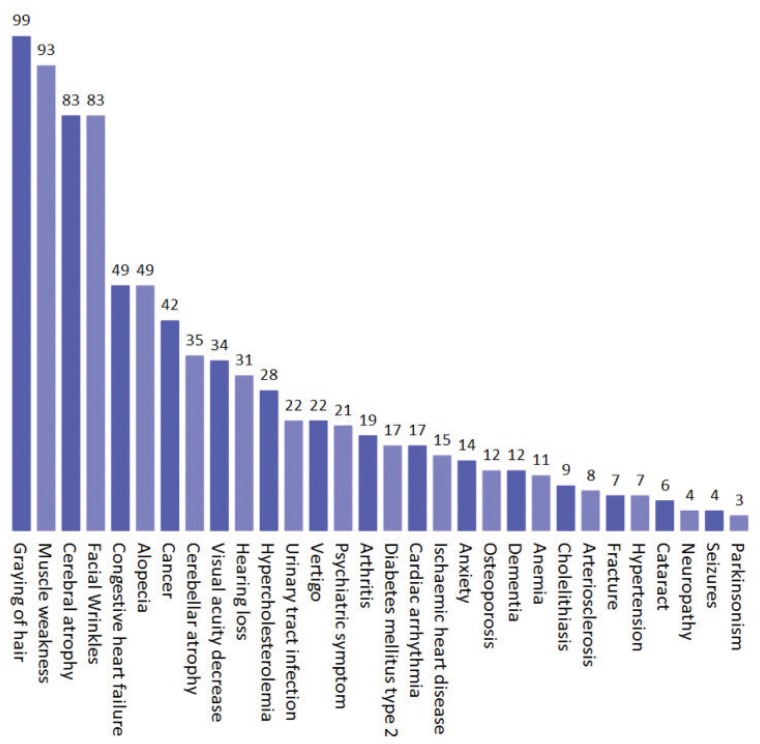
Fortunately, there are clinical assessment and indirect laboratory tests. Figure 4 shows the probability of mitochondrial dysfunction in various signs, symptoms, and diseases. There are several indirect measures of mitochondrial dysfunction.12 Most readily available are blood levels of lactate and pyruvate. However, these tests are quite susceptible to sampling error, recent exercise, and so forth, and they will only become abnormal when the mitochondrial dysfunction is so bad that anaerobic metabolism has to compensate. Urinary organic acid analysis can be quite useful for determining enzyme dysfunction in the citric acid cycle, which can pinpoint where the problem is. Finally, the amount of damage to the mtDNA can be estimated by 8-OHdG in the urine. Interestingly, this test also predicts cancer risk—not the type of cancer but rather the amount of DNA damage, which leads to cancer.13
Figure 4.
Probability of Mitochondrial Dysfunction in Common Signs, Symptoms, and Diseases14
Strategies to Improve Mitochondrial Function
Pick the right mother. Sorry, couldn’t resist, but starting out with optimal mtDNA certainly helps.
Optimize nutrient status to limit oxygen and high-energy electron leakage in the ETC. As the greatest source of oxidative stress and damage to mtDNA, this is perhaps the most powerful antiaging strategy we can provide our patients.
Decrease toxin exposure. This is obviously true for virtually every disease, but because of the huge metabolic activity of the mitochondria, they are especially susceptible.
Provide nutrients that protect the mitochondria from oxidative stress.
Utilize nutrients that facilitate mitochondrial ATP production.
Build muscle mass. Even those with mitochondrial damage, such as that found in Parkinson’s disease, can increase ATP production through strength training.15
Of course, the order of these priorities can be argued, but the basic need is clear and I recommend all of them, especially the critical natural health products at the dosages required to accomplish the goals. The foundation begins with a good multivitamin and mineral. They should be high quality with 2 to 3 times the recommended daily intake for most nutrients, especially the B vitamins. Of all the mitochondrial supportive nutrients, at the top of my list are (in order of my preference—which I am sure is debatable) CoQ10, α-lipoic acid plus acetyl-l-carnitine, resveratrol, NAC, and vitamin E. Also of value are coconut oil, pyrroloquinoline quinone (PQQ), Ginkgo biloba, proanthocyanidins, and melatonin.
CoQ10
There are many reasons to recommend CoQ10. As can be seen in Figure 3, CoQ10 carries the high-energy electrons through the ETC. Deficiency means not only decreased ATP production but also increased electron loss causing oxidative damage. CoQ10 is also an important intramitochondrial antioxidant right where those ROS are being produced. It is not surprising that research has shown a strong correlation between a species’ ability to produce CoQ10 and their longevity.16 Clinical research on supplemental CoQ10 is positive and growing17 (100 mg QD).
α-Lipoic Acid + Acetyl-l-Carnitine
These nutrients have been used together to increase mitochondrial ATP production in several animal models, including elderly animals in particular.18 Human research is starting to show the same benefits.19 (200 mg of α-lipoic acid and 500 mg of acetyl-l-carnitine BID.)
Resveratrol
The good news keeps coming on resveratrol—wine lovers rejoice! (Of course, there are several other “food” sources.) Resveratrol increases mitochondrial ATP production, protects from ROS, up-regulates sirtuin 1, and so forth.20 It even clears β-amyloid from Alzheimer’s disease cells.21 Human studies are now confirming animal studies showing improved mitochondrial functional at surprisingly reasonable dosages.22 (150 mg QD.)
N-Acetyl Cysteine (NAC)
As I discussed in my glutathione editorial in IMCJ 13.1, the key role of NAC is to increase intracellular glutathione, which is then pumped into the mitochondria. This glutathione is critical for protection of mitochondria from oxidative damage. (500 mg BID.)
Vitamin E
Not surprising to find extensive cell and animal research showing that the antioxidant vitamin E protects mitochondria from oxidative stress. The human research is not as strong and unfortunately is almost all with a single member of the vitamin E family. Nonetheless, there are promising early results.23 (Mixed tocopherols 500 IU QD.)
Summary
There is so much more that could be written about mitochondria from the perspective of an integrative medicine clinician. I think we are seeing only the tip of the iceberg of the role of mitochondrial dysfunction in our patients’ disease and ill health. As the environment becomes more polluted and drug prescribing increases, this problem will become even worse. The good news is that there is a lot we can do to help.
In This Issue
As I have mentioned several times before, one of the joys as editor of IMCJ is reading the reviews of associate editor Sid McDonald Baker, md. If it weren’t such a burden, I’d send him every submission for review. As has been the case before, his review of a paper was so insightful, I thought IMCJ readers would find value:
Authors writing on behalf of and for the integrative medicine community should feel obliged to avoid the fallacy of traditional medicine that diseases are entities that cause symptoms. This requires vigilance against old habits that come out when we say something like “compounding the impact of these conditions on individuals health systems and populations.” Comorbidity is another word we should all watch out for. It carries this silly notion that complex chronic illness is somehow the sum of the combined “attack” by each of several “disease entities.” Schizophrenia is a catchy word and it is tempting to use it in a title of an article about a dramatic clinical response. In the long-run, however, I think it would be safer to simply say hallucinations, night terrors, and abdominal pain to characterize the boy’s troubles.
Yes, conventional medicine’s standardization of disease, disease diagnosis, and disease treatment has facilitated critical advances in many areas. Unfortunately, this perspective has also greatly impaired recognizing the uniqueness of the patient who “has the disease” and has led to the symptomatic treatment—at the expense of curative care. This is at the heart of the health care crisis.
I really like our original research in this issue as it demonstrates that acupuncture can generate measurable physiological changes in the body. I think particularly of interest is that the research by Jeannette Painovich, daom, lac; Anita Phancao, md; Puja Mehta, md; Supurna Chowdhury, ms; Shivani Dhawan, ms; Ning Li, phd; Doris Taylor, phd; Yi Qiao, lac; Anna Brantman, daom, lac; Xiuling Ma, phd, lac; and C. Noel Bairey Merz, md, implies that acupuncture can have a regenerative effect.
Since my first editorial in IMCJ 1.1 over a decade ago, I have preached the gospel of collaboration providing our patients the best opportunity for optimal clinical outcomes. Ruthann Russo, phd, jd, mph, lac, explores the intriguing concept that with the growing public acceptance and research foundation for nonconventional interventions, informed consent could now legally mean that a conventional doctor must inform their patients of these other options. I find this especially interesting since some state licensing laws require CAM practitioners to inform their patients of conventional interventions. “The Times, They are a-Changin’ …”
Over 40 years ago as a naturopathic student, I was taught by Dr Bastyr the use of the “vag pack” and escharotics for the treatment of cervical dysplasia (there we go again—naming the disease). Specializing in natural childbirth, I successfully used this protocol to help a lot of women (along, of course, with optimizing their nutrition, cleaning up their diet, etc). So I am quite excited to see a good case study on the use of this protocol by Kimberly Windstar, med, nd; Corina Dunlap, ba; and Heather Zwickey, phd. This kind of research is critical for advancing the foundation of this medicine. Michael Murray, nd, and I had a chapter and appendix on cervical dysplasia and the vag pack way back in the first (1985) version of the Textbook of Natural Medicine. Since then, associate editor Tori Hudson, nd, has written about and taught this procedure to many integrative medicine clinicians.
Our interview this month is with Sarah Speck, md, and Dan Tripps, phd. I had an opportunity to tour their integrative cardiac function—not disease—assessment clinic in Seattle and was so impressed I thought IMCJ users would also be interested. Talking to them I developed a better understanding of the clinical significance of how various types of fitness training can imbalance a person’s anaerobic/aerobic capacity. This was really brought home to me recently when I prescribed strength training to an adolescent with a mitochondrial deficit. Based on my understanding as described above, I had already optimized his mitochondria as best I could with nutrition; now I wanted to increase his number of mitochondria. I was surprised—and impressed—when I heard back from the mother that the trainer wanted to know if I wanted to build lean or bulky muscle! Very good question. My assumption is that lean muscle would be the most aerobic, so that is what I prescribed. Reading this interview reminded me that now I no longer need to assume—Dr Speck and Dr Tripps can measure. So I just made the referral.
The keynote speaker we are featuring is Michael Greger, md, who will be covering “Reversing Chronic Disease Through Diet” at the American Association of Naturopathic Physicians Conference in August. Of course, all IMCJ readers are quite knowledgeable in this area. What I found most interesting is his candid comments about the intense politics involved in setting the USDA Dietary Guidelines.
I fully concur with John Weeks that our field owes a great debt of gratitude to Senator Tom Harkin (D-IA) for his adamant advocacy of integrative medicine at the federal level. The original Office of Alternative Medicine, the CAM Caucus, the White House Commission on Complementary and Alternative Medicine (on which I was privileged to serve), the National Center for Complementary and Alternative Medicine at the NIH— his remarkable leadership and accomplishments have been pivotal in the maturation and acceptance of this medicine. Congratulations to Patricia Herman, nd, phd, and Dugald Seely, nd, for publication of their seminal cost-effectiveness research. This is the kind of work we need to provide corporate decision makers the justification to explore other—and we believe better!—ways of promoting health in the workplace.
As usual, great BackTalk by Bill Benda, md. His comments about the hyping and marketing of medicine that drives us so crazy are right on. Thanks for the very kind compliment (I think), Dr Bill—but it is “Dr Joe” now …
Joseph Pizzorno, nd, Editor in Chief
drpizzorno@innovisionhm.com
References
- 1.No authors listed. Mitochondrian Wikipedia. [Accessed February 11, 2014]. http://en.wikipedia.org/wiki/Mitochondrion. Updated February 17, 2014.
- 2.Phillip M, Milo R. BioNumber of the Month. [Accessed February 12, 2014]. http://openwetware.org/wiki/BioNumber_Of_The_Month. Updated September 18, 2009.
- 3.Törnroth-Horsefield S, Neutze R. Opening and closing the metabolite gate. Proc Natl Acad Sci U S A. 2008;105(50):19565–19566. doi: 10.1073/pnas.0810654106. [DOI] [PMC free article] [PubMed] [Google Scholar]
- 4.Narayanese, WikiUserPedia, Yassine Mrabet, Toto Baggins, available at http://commons.wikimedia.org/wiki/File:Citric_acid_cycle_with_aconitate_2.svg.
- 5.Wikipedia Creative Commons, available at http://commons.wikimedia.org/wiki/File:Mitochondrial_electron_transport_chain%E2%80%94Etc4.svg#.
- 6.Wikipedia Creative Commons, available at http://commons.wikimedia.org/wiki/File:Electron_transport_chain.svg.
- 7.Langsjoen PH, Langsjoen AM. The clinical use of HMG CoA-reductase inhibitors and the associated depletion of coenzyme Q10. A review of animal and human publications. Biofactors. 2003;18(1–4):101–111. doi: 10.1002/biof.5520180212. [DOI] [PubMed] [Google Scholar]
- 8.Trifunovic A. Mitochondrial DNA and ageing. Biochim Biophys Acta. 2006;1757(5–6):611–617. doi: 10.1016/j.bbabio.2006.03.003. [DOI] [PubMed] [Google Scholar]
- 9.Kalghatgi S, Spina CS, Costello JC, et al. Bactericidal antibiotics induce mitochondrial dysfunction and oxidative damage in Mammalian cells. Sci Transl Med. 2013;5(192):192ra85. doi: 10.1126/scitranslmed.3006055. [DOI] [PMC free article] [PubMed] [Google Scholar]
- 10.Ray KK, Seshasai SR, Erqou S, et al. Statins and all-cause mortality in high-risk primary prevention: a meta-analysis of 11 randomized controlled trials involving 65,229 participants. Arch Intern Med. 2010;170(12):1024–1031. doi: 10.1001/archinternmed.2010.182. [DOI] [PubMed] [Google Scholar]
- 11.Manzo-Avalos S, Saavedra-Molina A. Cellular and mitochondrial effects of alcohol consumption. Int J Environ Res Public Health. 2010;7(12):4281–4304. doi: 10.3390/ijerph7124281. [DOI] [PMC free article] [PubMed] [Google Scholar]
- 12.Mitochondrial Medicine Society’s Committee on Diagnosis. Haas RH, Parikh S, et al. The in-depth evaluation of suspected mitochondrial disease. Mol Genet Metab. 2008;94(1):16–37. doi: 10.1016/j.ymgme.2007.11.018. [DOI] [PMC free article] [PubMed] [Google Scholar]
- 13.Valavanidis A, Vlachogianni T, Fiotakis C. 8-hydroxy-2′-deoxyguanosine (8-OHdG): a critical biomarker of oxidative stress and carcinogenesis. J Environ Sci Health C Environ Carcinog Ecotoxicol Rev. 2009;27(2):120–139. doi: 10.1080/10590500902885684. [DOI] [PubMed] [Google Scholar]
- 14.Scheibye-Knudsen M, Scheibye-Alsing K, Canugovi C, et al. A novel diagnostic tool reveals mitochondrial pathology in human diseases and aging. Aging. 2013;5(3):192–208. doi: 10.18632/aging.100546. [DOI] [PMC free article] [PubMed] [Google Scholar]
- 15.Kelly NA, Ford MP, Standaert DG, et al. Novel, high-intensity exercise prescription improves muscle mass, mitochondrial function, and physical capacity in individuals with Parkinson’s disease. J Appl Physiol. 1985 2014 Jan; doi: 10.1152/japplphysiol.01277.2013. [Epub ahead of print] [DOI] [PMC free article] [PubMed] [Google Scholar]
- 16.Lenaz G, D’Aurelio M, Merlo Pich M, et al. Mitochondrial bioenergetics in aging. Biochim Biophys Acta. 2000;1459:397–404. doi: 10.1016/s0005-2728(00)00177-8. [DOI] [PubMed] [Google Scholar]
- 17.Hargreaves IP. Coenzyme Q10 as a therapy for mitochondrial disease. Int J Biochem Cell Biol. 2014 Feb 2; doi: 10.1016/j.biocel.201401.020. [DOI] [PubMed] [Google Scholar]
- 18.Shenk JC, Liu J, Fischbach K, et al. The effect of acetyl-L-carnitine and R-alpha-lipoic acid treatment in ApoE4 mouse as a model of human Alzheimer’s disease. J Neurol Sci. 2009;283(1–2):199–206. doi: 10.1016/j.jns.2009.03.002. [DOI] [PMC free article] [PubMed] [Google Scholar]
- 19.McMackin CJ, Widlansky ME, Hamburg NM, et al. Effect of combined treatment with alpha-Lipoic acid and acetyl-L-carnitine on vascular function and blood pressure in patients with coronary artery disease. J Clin Hypertens (Greenwich) 2007;9(4):249–255. doi: 10.1111/j.1524-6175.2007.06052.x. [DOI] [PMC free article] [PubMed] [Google Scholar]
- 20.Lagouge M, Argmann C, Gerhart-Hines Z, et al. Resveratrol improves mitochondrial function and protects against metabolic disease by activating SIRT1 and PGC-1α. Cell. 2006;127(6):1109–1122. doi: 10.1016/j.cell.2006.11.013. [DOI] [PubMed] [Google Scholar]
- 21.Marambaud P, Zhao H, Davies P. Resveratrol promotes clearance of Alzheimer’s disease amyloid-beta peptides. J Biol Chem. 2005;280(45):37377–37382. doi: 10.1074/jbc.M508246200. [DOI] [PubMed] [Google Scholar]
- 22.Timmers S, Konings E, Bilet L, et al. Calorie restriction-like effects of 30 days of resveratrol supplementation on energy metabolism and metabolic profile in obese humans. Cell Metab. 2011;14(5):612–622. doi: 10.1016/j.cmet.2011.10.002. [DOI] [PMC free article] [PubMed] [Google Scholar]
- 23.Navarro A, Boveris A. Brain mitochondrial dysfunction in aging, neurodegeneration, and Parkinson’s disease. Front Aging Neurosci. 2010;2(pii):34. doi: 10.3389/fnagi.2010.00034. [DOI] [PMC free article] [PubMed] [Google Scholar]


