Abstract
The Drosophila visual system has been proved to be a powerful genetic model to study eye disease such as retinal degeneration. Here, we describe a genetic method termed “Rh1::GFP ey-flp/hid” that is based on the fluorescence of GFP-tagged major rhodopsin Rh1 in the eyes of living flies and can be used to monitor the integrity of photoreceptor cells. Through combination of this method and ERG recording, we examined a collection of 667 mutants and identified 18 genes that are required for photoreceptor cell maintenance, photoresponse, and rhodopsin synthesis. Our findings demonstrate that this “Rh1::GFP ey-flp/hid” method enables high-throughput F1 genetic screens to rapidly and precisely identify mutations of retinal degeneration.
Introduction
The Drosophila visual system has been shown to be proven to be a powerful genetic model for dissecting the molecular mechanisms underlying retinal degeneration and G-protein-coupled signaling cascades. Mutations in most genes that functions in phototransduction result in light-dependent photoreceptor cell death. Therefore, genetic screens in Drosophila could isolate mutations of many genes involved in retinal degeneration and could deepen our understanding of their counterpart genes in human diseases.
Drosophila phototransduction offers the opportunity to combine classical and modern genetic approaches to identify genes and proteins that function in phototransduction and/or that are required for photoreceptor cell survival [1–4]. Electroretinogram recordings (ERGs) are among the analysis tools that have driven the progress of Drosophila phototransduction research; this technique is simple enough to be used to perform genetic screens [3]. However, due to the requirement of fixing animals, flies can not survive after ERG assay, which makes it less suitable for mutagenesis F1 screens.
In addition, most retinal degeneration mutations in Drosophila were originally identified from photoresponse-based screens, which do not fully represent the complexity of retinal degeneration diseases in human. Moreover, classic screens of adult animals for aberrant phototransduction and eye morphology often cannot isolate essential genes involved in these pathways since such genes are often indispensable for organism viability. A few mosaic methods have been developed that make an entire eye homozygous for a mutation [5, 6]. Large scale screens for neurotransmission and phototransduction mutants have been conducted based on these methods [7, 8]. However, phototaxis in the F1 generation is not sensitive enough, and the ERG-based high throughput screening is time-consuming [8–10].
Given these nontrivial limitations, we were motivated to develop a fluorescence-based “Rh1::GFP ey-flp/hid” method for F1 screening of retinal degeneration mutants. This method is based on a modified EGUF/hid technique to generate eyes of homozygous mutations and uses GFP-tagged Rh1 (major rhodopsin) as a marker for photoreceptor cell integrity. Using this Rh1::GFP ey-flp/hid method, we screened the UCLA URCFG P-element recessive lethal collection, and identified several types of mutations affecting photoreceptor cell survival, phototransduction, and rhodopsin homeostasis.
Results
Development of the Rh1::GFP ey-flp/hid screening method
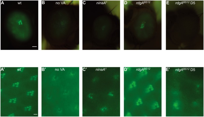
To monitor the integrity of photoreceptor cells in live animals, we generated Rh1::GFP transgenic flies, which express a GFP-tagged major rhodopsin Rh1 protein in R1-6 photoreceptor cells under the control of the ninaE (rh1) promotor. Under a fluorescence microscope, regular Rh1::GFP compound eyes showed an intensely green fluorescing deep pseudopupil. This fluorescence signal was markedly reduced in flies raised on vitamin A-free food as well as in ninaA 1 mutant flies with disrupted Rh1 biosynthesis. It was also reduced in the rdgA BS12 mutant background which caused a rapid retinal degeneration (Fig 1A–1E). Since the fluorescing Rh1-GFP pseudopupil can be observed in living flies and because it represents the Rh1 levels and/or rhabdomere structures, it is ideally suited for a use in a high throughput genetic screen. Photoreceptor cell integrity, indicated by the number of Rh1 GFP-tagged rhabdomeres, was further visualized at a higher resolution following cornea optical neutralization using fluorescence microscopy with oil-immersion objectives [11]. Compared with wild type, which had intensive GFP fluorescence for 6 rhabdomeres, the GFP signals were dramatically reduced in rhabdomeres of flies raised on vitamin A-free food and in ninaA 1 mutant (Fig 1A’–1C’). Rhabdomere structures were lost in the retina of rdgA BS12 flies at 5 days (Fig 1D’ and 1E’). Therefore, fluorescence of Rh1::GFP is a good marker for rhodopsin levels and is suitable for use in screens targeting mutants of retinal degeneration.
Fig 1. Rhodopsin levels and the integrity of photoreceptor cells using Rh1-GFP.
Representative images of the GFP fluorescence in intact eyes are shown. (A-E) The green fluorescing deep pseudopupil of flies with different genotypes expressing Rh1-GFP (upper panel). (A’-E’) GFP-fluorescence was detected in intact eyes after cornea optical neutralization by water immersion. (A, A’) wild type (Rh1::GFP), (B, B’) Rh1::GFP flies raised in vitamin A-free food, (C, C’) ninaA 1 (Rh1::GFP;ninaA 1), (D, D’) rdgA BS12 (rdgA BS12 Rh1::GFP), (E, E’) rdgA BS12 5 day-old. With the exception of the rdgA BS12 flies in E and E’, flies depicted in this figure were 1 day old. Scale bar on upper panels, 50 μm; on lower panel, 2 μm.
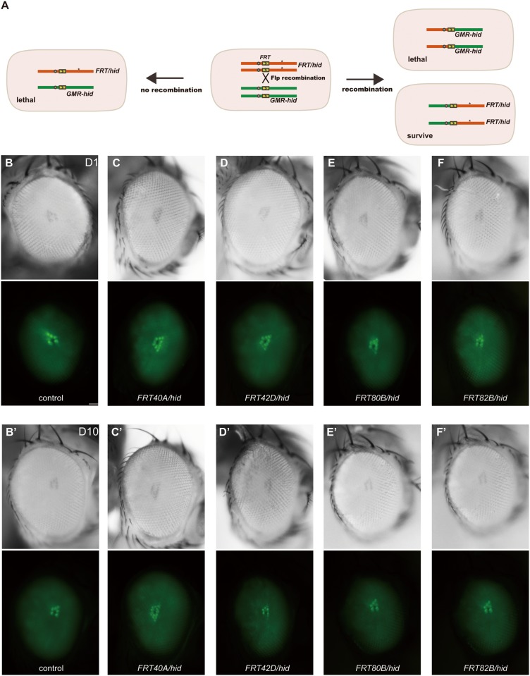
The EGUF/hid technique can generate eyes homozygous for a mutant allele in an otherwise heterozygous background [5]. This system employed the GAL4/UAS and FLP/FRT systems to induce mitotic recombination of FRT bearing chromosome arms specifically in the eye by combination of ey-GAL4/UAS-FLP, and the dominant photoreceptor-specific cell-lethal transgene GMR-hid and a recessive cell lethal (CL) mutation to eliminate all photoreceptors during development in which the desired chromosome arm has not been made homozygous. Thus, the mutant phenotype is recognized in the F1 generation, and mutations in essential genes can be recovered because the mutation is homozygous solely in the eye, making this screen potentially quite powerful. In combination with the Rh1::GFP transgene, this adult recombinant eye technique can facilitate high-throughput screening of candidate mutations. We also modified the technique by using ey-flp to express FLP directly under the ey promoter instead of ey-GAL4/UAS-FLP to simplify the manipulation. As well as the EGUF/hid system, normally-developed fully homozygous mutant eyes in otherwise heterozygous animals were generated with the ey-flp/hid system in 4 FRT arms, including FRT40A, FRT42D, FRT80B, and FRT82B (Fig 2). Moreover, upon introducing Rh1::GFP, fluorescing deep pseudopupils in mosaic animals were similar to those of the wild-type at both day1 and day 10, indicating that Rh1::GFP does not affect photoreceptor morphology (Fig 2).
Fig 2. Application of the ey-flp/hid method in Rh1::GFP background fly.
(A) Schematic diagram of mitotic recombination occurring in eye precursor cells with the Rh1::GFP ey-flp/hid method. Only retina cells that are homozygous for the FRT chromosome carrying a mutation (*) survive, because GMR-hid dominantly causes lethality of retina cells. (B-F) Drosophila eyes representative of the following genotypes are shown in light (upper panels) or fluorescence (lower panels) images: (B, B’) control: ey-flp Rh1::GFP;FRT40A, (C, C’) FRT40A/hid (ey-flp Rh1::GFP;FRT40A/GMR-hid CL FRT40A), (D, D’) FRT42d/hid (ey-flp Rh1::GFP;FRT42D/FRT42D GMR-hid CL), (E, E’) FRT80B/hid (ey-flp Rh1::GFP;FRT80B/GMR-hid CL FRT80B), (F, F’) FRT82B/hid (ey-flp Rh1::GFP;FRT82B/ FRT82B GMR-hid CL). 1 day-old flies were used in A-E. 10 day-old flies were used in A’-E’. Flies were raised under a 12 hr light/12 hr dark cycle. Scale bar, 50 μm.
Testing the Rh1::GFP ey-flp/hid method on mutants of rhodopsin homeostasis, retinal degeneration, and phototransduction
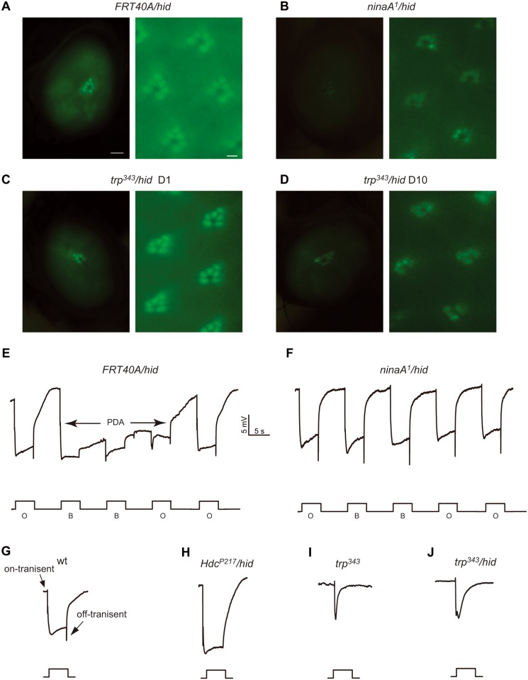
To determine whether the photoreceptors in the recombinant eyes generated via the ey-flp/hid system were capable of representing mutant phenotypes, we next generated recombinant eyes of a few mutant genes using the Rh1::GFP ey-flp/hid system and compared the recombinant mutant eyes with wild-type recombinant eyes. The GFP fluorescence was severely reduced in the ninaA 1 mutant recombinant eye (ninaA 1/hid), a result that correlates with the endogenous Rh1 levels [12, 13] (Fig 3A and 3B). We further checked the visual response by performing ERG recordings, which are extracellular recordings that measure the summed responses of all retinal cells to light. In response to orange light, wild-type flies display a rapid corneal negative response that quickly returns to baseline levels after cessation of the light stimulation. After exposure to blue light, there is a prolonged depolarization afterpotential (PDA) that is due to an excessive accumulation of Rh1 in an active form (Fig 3E). Thus, in ninaA 1 mutant flies, a PDA is not produced due to a large reduction of Rh1; and the same no PDA phenotype was recorded in the ninaA 1 /hid animals (Fig 3F).
Fig 3. Analysis of mutants of rhodopsin homeostasis, retinal degeneration, and phototransduction with the Rh1::GFP ey-flp/hid method.
(A-E) Detection of fluorescence in the deep pseudopupil (left panels) and by cornea optical neutralization (right panel). (A) FRT40A/hid, (B) ninaA 1 /hid (ey-flp Rh1::GFP;ninaA 1 FRT40A/GMR-hid CL FRT40A), (C) trp P343 /hid (ey-flp Rh1::GFP;FRT82B trp P343 / FRT82B GMR-hid CL), (D) trp P343 /hid 10 day-old. 1 day-old flies were used, with the exception of the trp P343 /hid flies, which were 10 day-old (D). Scale bar in right panels, 50 μm; in the left panels, 2μm. (E-H) ERG recordings of (E) wild type and (F) ninaA 1 /hid flies. Flies were exposed to 5 s pulses of orange light (O) or blue light (B), interspersed by 7 s, as indicated. A PDA was induced in the wild-type by blue light and terminated by orange light (arrows). (G-J) ERG response of (G) wild-type, (H) Hdc P217 /hid, (I) trp P343, and (J) trp P343 /hid flies in response to a 5-s orange light stimulus.
Given that Rh1-GFP also marks rhabdomeres and indicates the integrity of photoreceptor cells, we checked if the Rh1::GFP ey-flp/hid system could be used to monitor mutations that lead to retinal degeneration by generating recombinant eyes for a trp mutant (trp 343 /hid). The trp gene encodes a major ion channel in phototransduction, and mutations of trp result in transient receptor potential light responses and retinal degeneration [14, 15]. Similarly as in homozygous trp 343 mutant animals, the ERG phenotype of the trp 343 /hid flies was characterized by a transient response to light (Fig 3I and 3J). Moreover, the trp 343 /hid flies underwent an age-dependent loss of Rh1-GFP signals (Fig 3C and 3D). At the age of 10 days, the GFP signals were dramatically reduced in the trp 343 /hid eyes, and fewer fluorescing rhabdomeres were detected by cornea optical neutralization, which is consistent with light dependent retinal degeneration in trp 343 mutant animals (Fig 3D) [15].
Except the remained depolarization arising from responses of all retinal cells, the ERG diagram also includes on- and off-transients emanating from activity in the second-order neurons in the lamina (Fig 3F). The mutations with defective synaptic transmission or synapse development could result in preferential reduction in on- and off-transients. Mutations in the Hdc gene, which encodes a histidine decarboxylase for generating the photoreceptor cell neurotransmitter histamine, cause reduced histamine and loss of on- and off-transients in the ERG paradigm [16]. Although homozygous Hdc P217 eyes (Hdc P217 /hid) showed no Rh1-GFP fluorescence change, the Hdc P217 /hid flies lost on- and off- transients during light response (Fig 3H). These data suggested that the Rh1::GFP ey-flp/hid system can be used for high throughput screening for mutations that cause defects in rhodopsin homeostasis and retinal degeneration.
Genetic screen combining the Rh1::GFP ey-flp/hid method and ERG recording
To further test the Rh1::GFP ey-flp/hid system as an advanced genetic screen to isolate mutations regulating rhodopsin levels and photoreceptor cell integrity, we screened the P-element recessive lethal lines inserted in both arms of the second and third chromosomes from the UCLA URCFG collection [17]. This collection has been used for screening of genes required for photoreceptor cell development, cell survival, and rhodopsin localization [18–21]. Therefore, we only counted flies with normal morphology of compound eyes at 1 day old to avoid mutants affecting retina development and general cell survival. We analyzed insertion lines on the second or third chromosome carrying FRT sequences (667 independent lines) (S1 Table). By performing fluorescence deep pseudopupil assays and confirming with corneal optical neutralization at both day 1 and day 10, as well as ERG recording at day 1, we identified 18 lines with defects in Rh1 fluorescence at day 10 or defective ERG at day 1 (Table 1). Among these 18 lines, 14 lines showed loss of fluorescence deep pseudopupil at day 10, and these 14 retinal degeneration mutants included 4 lines with normal ERG and Rh1 levels, 1 line with Rh1 reduction, and 9 lines with ERG defect (Table 1). The remaining 4 lines only had ERG defects without Rh1-GFP fluorescence loss even at age of 20 days.
Table 1. Retinal degeneration and photoresponse defective mutants.
| Gene name | Phenotype | Function putative | ||
|---|---|---|---|---|
| Degeneration | ERG | Rh1 | ||
| CG30415 | degeneration | wt | decrease | unknown |
| Tps1 | degeneration | wt | wt | alpha, alpha-trehalose-phosphate synthase (UDP-forming) activity |
| dup | degeneration | wt | wt | DNA binding, glial cell development |
| Lis-1 | degeneration | wt | wt | dynein binding; enzyme regulator activity |
| dve | degeneration | wt | wt | sequence-specific DNA binding transcription factor activity |
| ifc | degeneration | small response | wt | sphingolipid delta-4 desaturase activity |
| sick | degeneration | small response | wt | ATP binding |
| srp54 | degeneration | small response | wt | mRNA binding |
| chn | degeneration | small response | wt | sequence-specific DNA binding transcription factor activity |
| aats-val | degeneration | no off-transient | wt | glutamate-tRNA ligase activity; valine-tRNA ligase activity |
| dmn | degeneration | no off-transient | wt | positive regulation of retrograde axon cargo transport |
| rab6 | degeneration | no off-transient | wt | regulation of postsynaptic membrane potential |
| Scox | degeneration | slow termination | wt | cytochrome-c oxidase activity, copper chaperone activity |
| porin | degeneration | slow termination | wt | regulation of anion transport on mitochondria |
| mmp2 | wt | small response | wt | metalloendopeptidase activity |
| milt | wt | no off-transient | wt | axon transport of mitochondrion |
| synj | wt | no off-transient | wt | inositol trisphosphate phosphatase activity |
| opa1-like | wt | no off-transient | wt | GTPase in mitofission |
Light-dependent retinal degeneration in the scox and porin mutant flies
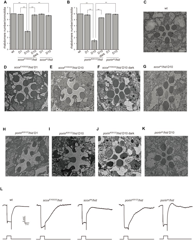
Among the 14 mutations with retinal degeneration phenotypes, two genes, including scox and porin, have putative functions in mitochondria. The scox gene encodes the Drosophila homologue of SCO (Synthesis of Cytochrome c Oxidase), which is a copper-donor chaperone required for the assembly of mitochondrial cytochrome c oxidase (COX) [22]. The scox EY05333 recombinant eye had normal Rh1-GFP fluorescence right after eclosure, but underwent an age- and light-dependent loss of fluorescence and rhabdomeres (Fig 4A). This age-dependent retinal degeneration was caused by disruption of scox by P-element EY05333, as precise excision of scox EY05333 (scox ex) rescued this loss of fluorescence (Fig 4A). The results obtained with the fluorescence deep pseudopupil and cornea optical neutralization methods were confirmed by examining the morphology of scox EY05333 retinas by transmission electron microscopy (Fig 4C–4G). The ommatidia from wild-type compound eyes contained the full set of seven intact rhabdomeres at all ages (Fig 4C). Few rhabdomeres were detected in 10-day-old scox EY05333 ommatidia, although seven intact rhabdomeres were present at day 1 (Fig 4D and 4E). Moreover, both the dark-raised scox EY05333 and scox ex flies raised under a normal light/dark cycle contained the full complement of seven rhabdomeres (Fig 4F and 4G). Since the human homologs of SCOX, Sco I, and Sco II are required for the assembly of mitochondrial respiratory complex IV, we tested the COX chaperon function of SCOX [23]. Using the hs-FLP/FRT system, we made homozygous scox mutant cell clones in heterozygous tissues that were marked by the absence of red fluorescent protein (RFP) [24]. By staining eye imaginal discs with anti-CoIV and anti-Tom20 antibodies, the cellular levels of COX and whole mitochondria were evaluated. As results, the levels of CoIV, but not Tom20, were reduced in the scox EY05333 mutant cells, indicating that the loss of scox in cells specifically reduced COX levels without affecting the overall amount of mitochondria (S1 Fig). It is therefore clear that the scox mutations disrupt cytochrome c oxidase assembly, thereby reducing the COX complex levels in the mutant cells.
Fig 4. The scox and porin mutations lead to light-dependent photoreceptor cell degeneration.
(A-B) Average rhabdomere numbers per ommatidia of (A) the scox mutant flies and (B) the porin mutant flies under the indicated conditions. Each data point was based on examination of >60 ommatidia from >3 flies. Error bars represent the SD. Asterisks indicate statistically-significant differences (one-way ANOVA and post-hoc Dunnett’s test, **p < 0.01). (C-K) Transmission electron microscopy sections of single ommatidia of fly compound eyes with the indicated genotype and conditions. (C) 10 day-old wild-type, (D) 1 day-old scox EY05333 /hid (ey-flp Rh1::GFP; scox EY05333 FRT40A/GMR-hid CL FRT40A), (E) 10 day-old scox EY05333 /hid, (F) 10 day-old scox EY05333 /hid under dark condition, (G) 10 day-old P-element excised scox ex /hid (ey-flp Rh1::GFP; scox ex FRT40A/GMR-hid CL FRT40A), (H) 1 day-old porin k05123 /hid (ey-flp Rh1::GFP; porin k05123 FRT40A/GMR-hid CL FRT40A), (I) 10 day-old porin k05123 /hid, (J) 10 day-old porin k05123 /hid under dark condition, (K) 10 day-old p-element excised porin ex /hid (ey-flp Rh1::GFP; porin ex FRT40A/GMR-hid CL FRT40A). Scale bar, 2 μm. With the exception of the dark-reared (F) scox EY05333 /hid and (J) porin k05123 /hid flies, flies were maintained under a 12 hr light/12 hr dark cycle. (L) ERG responses of wild-type, scox EY05333 /hid, scox ex /hid, porin k05123 /hid, and p-element excised porin ex /hid flies in response to a 10-s orange light stimulus as indicated. Flies used were less than 2 days old.
The Drosophila porin gene encodes a major Voltage-dependent anion channel (VDAC), which is an outer mitochondrial membrane component of mitochondrial permeability transition pores (PTP). Porin plays an important role in regulating energy metabolism and apoptosis by mediating the transport of ions and metabolites across the mitochondrial outer membrane [25]. porin k05123 mutant eyes gradually lost Rh1-GFP and rhabdomere fluorescence after eclosure, and remobilization of the P-element for the porin k05123 mutant (porin ex) rescued the age-dependent retinal degeneration phenotype (Fig 4B). The results obtained with the fluorescence deep pseudopupil and cornea optical neutralization methods were confirmed by examining the morphology of porin k05123 retinas with TEM (Fig 4H–4K). Both of the retinal degeneration phenotypes caused by the scox and porin mutations were light-dependent, as no significant fluorescence or morphological changes were detected in the dark-raised animals (Fig 4F and 4J). The scox and porin mutations may therefore affect phototransduction. It has been reported that the porin mutant animals displayed an inactivation ERG phenotype [26]. We next examined the ERG response of both scox and porin mutant eyes. Both scox EY05333 and porin k05123 mutant eyes displayed slower termination of the light response relative to wild-type, and scox ex and porin ex eyes reversed this slower termination phenotype, suggesting that mitochondria play an important role in light response termination (Fig 4L).
Dmn and Rab6 are required for synaptic transmission
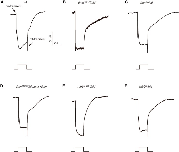
By performing ERG recording, we identified 13 mutants with impaired photoresponse without morphology changes in compound eyes. Among them the two new mutants, dmn K16109 and rab6 EP2397, reduced on- and off-transients. Drosophila dmn encodes p50/dynamitin, which is a subunit of dynactin complex and required for dynein-mediated transport [27]. The ERG of dmn K16109 showed a normal light response except that on- and off-transient spikes were largely reduced, indicating impaired photoreceptor synaptic transmission (Fig 5A and 5B). Both excision of the P element and expression dmn in dmn K16109 eye displayed a restored the on- and off- transient (Fig 5C and 5D). Similar as dmn K16109, the ERG of rab6 EP2397 diminished on- and off- transient spike, while precise excision of P-element rescued this phenotype (Fig 5E and 5F). These results indicate Dmn and Rab6 are required for photoreceptor synaptic transmission.
Fig 5. Reduced on- and off-transients in dmn and rab6 mutants.
ERG response of (A) control, (B) dmn K16109 /hid (ey-flp Rh1::GFP; FRT42D dmn K16109 / FRT42D GMR-hid CL), (C) precise p-element excised dmn ex /hid (ey-flp Rh1::GFP; FRT42D dmn ex / FRT42D GMR-hid CL), (D) dmn K16109 /hid;GMR>dmn (ey-flp Rh1::GFP; FRT42D dmn K16109 / FRT42D GMR-hid CL;GMR-gal4/UAS-dmn), (E) rab6 EP2397 /hid (ey-flp Rh1::GFP; rab6 EP2397 FRT40A/GMR-hid CL FRT40A), (F) precise p-element excised rab6 ex /hid (ey-flp Rh1::GFP; rab6 ex FRT40A/GMR-hid CL FRT40A) flies in response to a 2-s orange light stimuli.
The roh mutant is defective in the production of rhodopsin
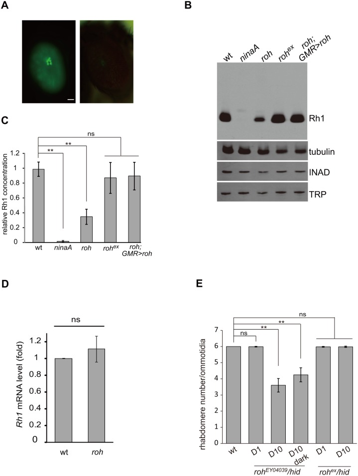
The maturation of rhodopsin is strictly regulated, and many mutations disrupting this process are known to cause reduced Rh1 accumulation [28, 29]. As we used Rh1-GFP to indicate Rh1 levels, we were able to target mutants that disrupt Rh1 homeostasis. The CG30415 EY04039 mutant eyes reduced Rh1-GFP fluorescence with normal morphology at one-day old (Fig 6A). We therefore named the CG30415 gene as roh (r eduction o f r h 1). We further checked if the mutation of roh specifically decreased Rh1 levels by performing western blots, and found that roh EY04039 reduced the amount of Rh1 by 70% but expressed normal levels of INAD and TRP, two rhabdomere specific proteins (Fig 6B and 6C). Using real-time PCR, we found that the rh1 mRNA levels were not changed in roh EY04039 mutant eyes, which suggested that this reduction of Rh1 resulting from mutation of roh was not due to decreased rh1 transcription (Fig 6D). Further, removal of the P-element and introduction of ROH back to photoreceptor cells by GMR-Gal4/UAS-roh restored the Rh1 levels of roh EY04039 mutant eyes (Fig 6C). We next checked whether the roh EY04039 mutant had altered localization of Rh1. Despite the reduced Rh1 signals, Rh1 exclusively localized to the rhabdomere in roh EY04039 mutant eyes, independent of the light condition, suggesting that ROH is not required for Rh1 localization (S2 Fig). We next examined if loss of ROH leads to eventual retinal cell death using cornea optical neutralization assays. The roh EY04039 mutant eye lost fluorescent rhabdomeres in an age-dependent manner, and remobilization of the P-element for the roh EY04039 mutant (roh ex) rescued this age-dependent retinal degeneration phenotype (Fig 6E). Moreover, the loss of rhabdomere fluorescence in the roh EY04039 mutant eye was light-independent, as the roh EY04039 mutant eye lost rhabdomeres in dark conditions as well (Fig 6E). These results suggest that ROH leads to retinal degeneration through downregulation of rhodopsin.
Fig 6. roh mutation results in Rh1 decrease.
(A) GFP fluorescence of one-day old wild-type (left panel) and roh EY04039 (right panel) flies showing a reduction in Rh1-GFP. Scale bar, 50 μm. (B) Western blotting showing decreased Rh1 levels in the roh EY04039 mutant (ey-flp Rh1::GFP; FRT42D roh EY04039 / FRT42D GMR-hid CL); the INAD and TRP levels are not affected compared to the wild-type. The reduction of Rh1 levels could be rescued in roh ex (ey-flp Rh1::GFP; FRT42D roh ex /FRT42D GMR-hid CL) and roh EY04039;GMR>roh (ey-flp Rh1::GFP; FRT42D roh EY04039 / FRT42D GMR-hid CL;GMR-gal4/UAS-roh) flies. Flies less than 1 day old were used. (C) Quantification of relative Rh1 level in various genotypes: wt, ninaA 1, roh EY04039, roh ex and roh EY04039;GMR>roh. The Rh1 levels were normalized to tubulin. (D) Quantitative real-time PCR of wild-type (ey-flp Rh1::GFP; FRT42D/ FRT42D GMR-hid CL) and roh EY04039 fly head with rh1-specific primers. The results are normalized to the expression level of gpdh. The graphs represent the means ± SD of three independent experiments. Error bars represent the SD. (E) Average rhabdomere numbers per ommatidia of the roh EY04039 mutant under the indicated conditions. Each data point was based on examination of >60 ommatidia from >3 flies. Error bars represent the SDs. Asterisks indicate statistically-significant differences (one-way ANOVA and post-hoc Dunnett’s test; ns: not significant, **p < 0.01).
Discussion
The Drosophila vision system has been served as important genetic system for searching the basis of and therapeutic treatments for human eye diseases. Two methods have been developed to make the entire eye homozygous for a mutation in otherwise heterozygous animals, satisfying the requirements for large scale genetic screening for essential genes [5, 6]. In one technique, with minute mutations integrated onto the marked FRT chromosome arms, which prevent the proliferation or survival of homozygous and heterozygous cells, the recombinant eyes are composed of more than 90% mutated chromosomes [6]. However, this method might not work well for some mutations that suppress growth rates. The EGUF/hid method generated flies with eyes composed exclusively of mutant clones by eliminating all photoreceptor cells not homozygous for the mutant chromosome arm. Here, we modified the EGUF/hid method by replacing EGUF with ey-flp, which abolished the fluorescence interruption by the pigmentation and simplified the genetic manipulation procedure [5]. It worth to mentioned that as the ey-flp/hid technique generates homozygous mutations in non-photoreceptor retinal cells as well, non-autonomous effects of these mutations on photoreceptor cell might be considered.
The time-consuming of ERG is a major concern for high-throughput F1 screen. Although rhabdomere and photoreceptor cell morphology based on the detection of simple deep pseudopupil (DPP) can be detected in living flies [30], the low sensitivity and the relatively transient lasting time prevent it widely to be used as a readout for high-throughput screen. Rh1-GFP introduced into flies, specifically marked the integrity and rhabdomeral morphology of photoreceptor cells and/or the amount of endogenous rhodopsin status. Through combining Rh1::GFP and ey-flp/hid technique, we can efficiently perform large scale screens for mutants of retinal degeneration and rhodopsin homeostasis.
The UCLA URCFG collection is a collection of P-element recessive lethal lines carrying FRT sequences, which has been successfully used by two retinal mosaic screenings for mutants of photoreceptor cell survival, eye development and rhodopsin [18–21]. One of the screenings used a two-color fluorescent imaging system to visualize the mosaic adult photoreceptor neurons with rhabdomere targeted Tomato, and identified multiple lines with defects in photoreceptor cell integrity including 4 mutations with apoptosis [18–21]. A similar retinal mosaic screening using Arrestin2::GFP to visualize endogenous Rh1 localization identified mutations of PIG-U (Phosphatidylinositol glycan anchor biosynthesis, class U) disrupting the transport of Rh1, along with several other mutations with rhabdomere morphological defects [18–21]. We screened UCLA URCFG collections and identified 14 retinal degeneration mutants, and 13 mutations with defective ERG. Among the 13 ERG mutants, 7 lines had defective phototransduction, and the other 6 lines only showed synaptic transmission defects. Importantly, 6 of 7 phototransduction mutants underwent retinal degeneration with the exception of mmp2 mutant, which is correlated with the theory that most phototransduction mutations ultimately result in photoreceptor cell death. Therefore, the Rh1::GFP ey-flp/hid” method could be used to screen for mutants of phototransduction.
Beyond their primary function of supplying ATP, mitochondria play important roles in cell signaling events and apoptosis in eukaryotic metabolic processes. We found that mutations of two mitochondrial proteins, SCOX and Porin, cause light-dependent retinal degeneration. Although loss of either SCOX or Porin did not cause immediate cell death and abnormal mitochondria, both mutations are associated with deficiencies in mitochondrial respiration and ATP supply in mammalian and flies [31–33]. The ATP concentration in the rhabdomere is expected to be critical for normal phototransduction termination. Eye-enriched PKC encoded by the inaC (inactivation nor afterpotential C) locus is required for deactivation of the visual cascade by ATP-dependent phosphorylation of signaling molecules including TRP channels [34, 35]. Moreover, it has been reported that depletion of ATP rapidly activated the TRP and TRPL channels in vivo, suggesting that an ATP-dependent process is required to close the channels in phototransduction [36]. Mutations of scox and porin have similar defects in the termination of photoresponse, which might be caused by reduction of ATP due to impairment of mitochondria function, rather than directly functioning in phototransduction [26]. Moreover, mutants that failed to terminate photoresponse likely keep excessive Ca2+ influx, and accumulate stable Rh1/Arr2 complexes during light responses, which potentially leads to retinal degeneration [37]
Rab6 is present in the Golgi apparatus and is involved in protein transport via the Golgi apparatus [38, 39]. A dominant negative Rab6 expression leading to a reduction of mature Rh1 levels suggested a role of Rab6 in rhodopsin transport [40]. However, only the synaptic function of Rab6 was reported in a recent loss function study of Rab6 [41]. Consistent with these results, we found that mutation of rab6 does not reduce Rh1 levels or alter its distribution, which forces questioning of the supposed role of Rab6 in rhodopsin maturation. Transport of neuronal vesicles along the microtubules is known to require Dynein as a molecular motor, and the dynactin complex functions as dynein cargo adaptor that participates in this neuronal vesicles transport process [42]. The mutations in p150 Glued, a large subunit of dynactin, caused defects in vesicular transport and neurotransmitter release in motor neurons of both mice and flies [43, 44]. Here, we observed that a mutation in the dmn gene that encodes the dynactin subunit 2, dynamitin, disrupted synaptic transmission in photoreceptor cells, providing evidence that the dynactin complex has a critical function in neuronal cell synaptic transmission. Rab6 has been reported to interact with the dynamitin complex and thus function in an important role in microtubule-dependent retrograde trafficking from the endosome to the Golgi and from the Golgi to the ER [45–47]. Given that both rab6 EP2397 and dmn K16109 display similar lack of on- and off- transients in the ERG recording profile, Rab6 might function with dynamitin in neuronal vesicle transport along the microtubules.
The first mutation identified to cause retinal degeneration was Drosophila ninaE, which encode the major rhodopsin, Rh1 [48–50]. Later evidence in human patients showed that mutations in rhodopsin and related genes are major causes of retinitis pigmentosa, the most common form of retinal degeneration disease [51, 52]. As our screening system takes advantage of Rh1-GFP as marker, the key factors affecting Rh1 synthesis and maturation can be targeted. We identified a new factor ROH related to Rh1 synthesis, and suggested that ROH is required for Rh1 biosynthesis and photoreceptor cell survival. However, as ROH does not contain any obvious protein motifs and exhibits no significant homology to any known protein in common databases, the mechanisms of how ROH affects Rh1 biosynthesis need to be further investigated.
The Drosophila vision system is a powerful model for the study of neural development, signal networks and photoresponse maintenance; studies based on this model system have provided important insights in the pathogenesis of several neurodegenerative diseases [53]. Several neurodegenerative diseases models, including Parkinson disease, Alzheimer's disease, Polyglutamine Diseases, and Amyotrophic Lateral Sclerosis disease were established in flies by inducing degenerative cell death of photoreceptor neurons in the compound eye [54–59]. Our method could represent a powerful tool for genetic screening of modifiers of neurodegenerative diseases.
Experimental Procedures
Fly Stocks
The following stocks were used: ey-flp Rh1::GFP;GMR-hid CL FRT40A/Cyo hs-hid, ey-flp Rh1::GFP; FRT42D GMR-hid CL/Cyo hs-hid, ey-flp Rh1::GFP;GMR-hid CL FRT80B/TM3 hs-hid, ey-flp Rh1::GFP;GMR-hid CL FRT80B/TM3 hs-hid, ey-flp Rh1::GFP; FRT82B GMR-hid CL /TM3 hs-hid, ey-flp Rh1::GFP;ninaA 1 40A, ey-flp Rh1::GFP;trp 343, ey-flp Rh1::GFP;Hdc P217, and hs-flp;ubi-RFP FRT40A. The P{ry[+t7.2] = ey-FLP.N}2 (ey-flp), longGMR-gal4, and UAS-dmn flies were obtained from the Bloomington stock center. The UCLA URCFG collections were obtained from the Kyoto Drosophila Genetic Resource Center. The Scox ex, porin ex, dmn ex, rab6 ex, and roh ex flies were generated by precise excision of the P-element lines scox EY05333, porin k05123, dmn K16109, rab6 EP2397 and roh EY04039, respectively. Briefly, P-element lines were mobilized by genetically introducing transposase using the Δ2–3 line, and precise excision lines were confirmed by DNA sequencing.
Generation of transgenic flies
To express GFP-labeled Rh1 under control of the ninaE (rh1) promoter, a GFP tag was added to the C terminus of the rh1 cDNA sequence and subsequently subcloned into the pcNX vector between the NotI and XbaI sites [60]. The w gene on the construct was subsequently knocked out by introducing a frame-shift mutation in its coding region. The construct was injected into w 1118 embryos, and transformants were identified on the basis of GFP fluorescence. The CG30415 cDNA molecule was amplified from the cDNA clone GH51119, and was subcloned into the puast-attB vector. The construct was injected into M{vas-int.Dm}ZH-2A;M{3xP3-RFP.attP}ZH-86Fb embryos, and transformants were identified on the basis of eye color.
Generation of the CoIV and Tom20 Antibodies
Polyclonal antibodies against Drosophila Cytochrome c Oxidase Subunit IV (CoIV) were generated by immunizing a rabbit with a synthetic peptide (IIDLEINPVTGLTSKWDYENKKW) conjugated to KLH. Polyclonal antibodies against Drosophila Tom20 were generated by immunizing a rat with a synthetic peptide (QEFGNRAAEGNDGPIVLGQS) conjugated to KLH. The specificity of the antibodies was testified by staining adult thoraxes (S1 Fig).
Fly imaging and optical neutralization assay
Flies were anaesthetized on a CO2 pad, and fluorescence and bright-field images were taken with Leica M165 FC Fluorescent Stereo Microscope. To perform optical neutralization assays, the heads of flies were cut off, and immerged into mineral oil in an orientation with eyes on the upper side. The samples were examined with a Nikon Ni-U fluorescent microscope.
Immunohistochemistry
Imaginal discs or adult thorax muscles were dissected in PBS solution (PH 6.8) and fixed in 4% paraformaldehyde in PBS buffer for 30 min. Eye discs or muscles were incubated in diluted primary antiserum, rabbit monoclonal anti-CoIV (1:200) and rat anti-Tom20 (1:200). Anti-rabbit and rat labeled secondary antibodies with Alexa 488, 568, or 647 (1:500) (Invitrogen) were used as secondary antibodies, and Phalloidin 650 (Thermo Scientific) was added to label actin filaments.
For eye section staining, semi-sectioned fly heads were fixed in 4% paraformaldehyde in PBS buffer on ice for 2 hours, and then embedded in LR White resin. Thin sections were prepared at a depth of ~30 μm and incubated with mouse monoclonal anti-Rh1 (1:200) and Rat anti-TRP (1:200) as primary antibodies and Alexa 647 or 750 labeled secondary antibodies (Invitrogen). Samples were examined and images were recorded using a Nikon confocal microscope. The acquired images were processed with Photoshop software.
Western blots
To perform western blots, fly heads were homogenized in SDS sample buffer with a Pellet Pestle (Kimble/Kontes). The proteins were fractionated by SDS-PAGE and transferred to Immobilon-P transfer membranes (Millipore) in Tris-glycine buffer. The blots were probed with mouse Tubulin primary antibodies (1:2000 dilution, Developmental Studies Hybridoma Bank), mouse Rh1 antibodies (1:2000 dilution, Developmental Studies Hybridoma Bank) and Rat anti-INAD (1:2000, C. Montell lab), followed by IRDye 680 goat anti-mouse IgG (LI-COR) and IRDye 800 goat anti-Rat IgG (LI-COR) as the secondary antibodies. The signals were detected with the Odyssey infrared imaging system (LI-COR).
Electroretinogram Recordings
ERG recordings were performed as described previously [61]. A Newport light projector (model 765) was used for stimulation. ERG signals were amplified with a Warner electrometer IE-210 and recorded with a MacLab/4 s A/D converter and the clampelx 10.2 program (Warner Instruments).
Transmission electron microscopy
Adult fly heads were dissected and fixed in paraformaldehyde/glutaraldehyde and osmium tetroxide solutions, dehydrated with an ethanol series, and embedded in LR White resin as described previously [62]. Thin sections (80 nm) were prepared at a depth of 30–40 μm and were examined using a transmission electron microscope (model 1230; JEOL). The images were acquired using a bottom-mount charge-coupled device camera (Ultrascan 1000; Gatan, Inc.). Digital Micrograph software (Gatan, Inc.) was used to convert images into TIFF files.
Supporting Information
(A) Verification of anti-CoIV and anti-Tom20 antibodies. Muscle fibers and mitochondria were organized into parallel stripes within indirect flying muscles in the thorax. Mitochondria of adult thoraxes were stained with anti-CoIV and anti-Tom20 antibodies, while muscle fibers were visualized by Phalloidin staining. Scale bar, 10 μm. (B) Imaginal disc staining of hs-flp; scox EY05333 FRT40A/ubi-RFP FRT40A by anti-CoIV (upper panel) and anti-Tom20 (lower panel) antibodies. RFP negative cells that are circled are scox homozygous mutant cells. Scale bar, 10 μm.
(EPS)
(A) adult eye sections were stained by anti-Rh1 (red) and anti-TRP (green) antibodies. One day-old roh EY04039 /hid (ey-flp Rh1::GFP; roh EY04039 FRT42D/GMR-hid CL FRT42D) and roh ex /hid (ey-flp Rh1::GFP; roh ex FRT42D/GMR-hid CL FRT42D) flies were raised either under a 12 hr light/12 hr dark cycle or in absolute dark conditions. Scale bar, 50 μm.
(EPS)
(XLS)
Acknowledgments
We thank Dr. Craig Montell, Dr. Nansi Colley, Dr. William Pak, the Bloomington Stock Center, and the Kyoto Drosophila Genetic Resource Center for fly stocks and reagents. We thank Xianglei Yu and Futing An for assistance with the genetic screen and thank Ying Wang for preparing sections for EM. We thank David O'Keefe for critical reading of the manuscript. This work was supported by a ‘973’ grant (2014CB849700) to T. W. from the Chinese Ministry of Science.
Data Availability
All relevant data are within the paper and its Supporting Information files.
Funding Statement
This work was supported by a ‘973’ grant (2014CB849700) to T.W. from the Chinese Ministry of Science. T.W. contributed to the study design, analysis, dicision to publish and preparation of the manuscript.
References
- 1. Benzer S. BEHAVIORAL MUTANTS OF Drosophila ISOLATED BY COUNTERCURRENT DISTRIBUTION. Proceedings of the National Academy of Sciences of the United States of America. 1967;58(3):1112–9. [DOI] [PMC free article] [PubMed] [Google Scholar]
- 2. Hotta Y, Benzer S. Genetic dissection of the Drosophila nervous system by means of mosaics. Proc Natl Acad Sci USA. 1970;67:1156–63. [DOI] [PMC free article] [PubMed] [Google Scholar]
- 3. Pak WL, Grossfield J, Arnold KS. Mutants of the visual pathway of Drosophila melanogaster . Nature. 1970;227(5257):518–20. Epub 1970/08/01. . [DOI] [PubMed] [Google Scholar]
- 4. Dolph PJ, Ranganathan R, Colley NJ, Hardy RW, Socolich M, Zuker CS. Arrestin function in inactivation of G protein-coupled receptor rhodopsin in vivo . Science. 1993;260:1910–6. [DOI] [PubMed] [Google Scholar]
- 5. Stowers RS, Schwarz TL. A genetic method for generating Drosophila eyes composed exclusively of mitotic clones of a single genotype. Genetics. 1999;152(4):1631–9. Epub 1999/08/03. [DOI] [PMC free article] [PubMed] [Google Scholar]
- 6. Newsome TP, Asling B, Dickson BJ. Analysis of Drosophila photoreceptor axon guidance in eye-specific mosaics. Development. 2000;127(4):851–60. Epub 2000/01/29. . [DOI] [PubMed] [Google Scholar]
- 7. Stowers RS, Megeath LJ, Gorska-Andrzejak J, Meinertzhagen IA, Schwarz TL. Axonal transport of mitochondria to synapses depends on milton, a novel Drosophila protein. Neuron. 2002;36(6):1063–77. Epub 2002/12/24. S0896627302010942 [pii]. . [DOI] [PubMed] [Google Scholar]
- 8. Verstreken P, Koh TW, Schulze KL, Zhai RG, Hiesinger PR, Zhou Y, et al. Synaptojanin is recruited by endophilin to promote synaptic vesicle uncoating. Neuron. 2003;40(4):733–48. . [DOI] [PubMed] [Google Scholar]
- 9. Ohyama T, Verstreken P, Ly CV, Rosenmund T, Rajan A, Tien AC, et al. Huntingtin-interacting protein 14, a palmitoyl transferase required for exocytosis and targeting of CSP to synaptic vesicles. The Journal of cell biology. 2007;179(7):1481–96. 10.1083/jcb.200710061 [DOI] [PMC free article] [PubMed] [Google Scholar]
- 10. Yamamoto S, Jaiswal M, Charng WL, Gambin T, Karaca E, Mirzaa G, et al. A drosophila genetic resource of mutants to study mechanisms underlying human genetic diseases. Cell. 2014;159(1):200–14. 10.1016/j.cell.2014.09.002 [DOI] [PMC free article] [PubMed] [Google Scholar]
- 11. Pichaud F, Desplan C. A new visualization approach for identifying mutations that affect differentiation and organization of the Drosophila ommatidia. Development. 2001;128(6):815–26. Epub 2001/02/27. . [DOI] [PubMed] [Google Scholar]
- 12. Schneuwly S, Shortridge RD, Larrivee DC, Ono T, Ozaki M, Pak WL. Drosophila ninaA gene encodes an eye-specific cyclophilin (cyclosporine A binding protein). Proc Natl Acad Sci USA. 1989;86(14):5390–4. [DOI] [PMC free article] [PubMed] [Google Scholar]
- 13. Shieh B-H, Stamnes M, Seavello S, Harris G, Zuker C. The ninaA gene required for visual transduction in Drosophila encodes ahomologue of cyclosporin A-binding protein. Nature. 1989;338:67–70. [DOI] [PubMed] [Google Scholar]
- 14. Montell C, Rubin GM. Molecular characterization of the Drosophila trp locus: a putative integral membrane protein required for phototransduction. Neuron. 1989;2(4):1313–23. Epub 1989/04/01. 0896-6273(89)90069-X [pii]. . [DOI] [PubMed] [Google Scholar]
- 15. Wang T, Jiao Y, Montell C. Dissecting independent channel and scaffolding roles of the Drosophila transient receptor potential channel. J Cell Biol. 2005;171(4):685–94. . [DOI] [PMC free article] [PubMed] [Google Scholar]
- 16. Burg MG, Sarthy PV, Koliantz G, Pak WL. Genetic and molecular identification of a Drosophila histidine decarboxylase gene required in photoreceptor transmitter synthesis. The EMBO journal. 1993;12(3):911–9. Epub 1993/03/01. [DOI] [PMC free article] [PubMed] [Google Scholar]
- 17. Chen J, Call GB, Beyer E, Bui C, Cespedes A, Chan A, et al. Discovery-based science education: functional genomic dissection in Drosophila by undergraduate researchers. PLoS Biol. 2005;3(2):e59 Epub 2005/02/19. 10.1371/journal.pbio.0030059 [DOI] [PMC free article] [PubMed] [Google Scholar]
- 18. Gambis A, Dourlen P, Steller H, Mollereau B. Two-color in vivo imaging of photoreceptor apoptosis and development in Drosophila. Dev Biol. 2011;351(1):128–34. Epub 2011/01/11. 10.1016/j.ydbio.2010.12.040 S0012-1606(10)01296-0 [pii]. [DOI] [PMC free article] [PubMed] [Google Scholar]
- 19. Satoh T, Inagaki T, Liu Z, Watanabe R, Satoh AK. GPI biosynthesis is essential for rhodopsin sorting at the trans-Golgi network in Drosophila photoreceptors. Development. 2013;140(2):385–94. Epub 2012/12/20. 10.1242/dev.083683 140/2/385 [pii]. . [DOI] [PubMed] [Google Scholar]
- 20. Dourlen P, Levet C, Mejat A, Gambis A, Mollereau B. The Tomato/GFP-FLP/FRT method for live imaging of mosaic adult Drosophila photoreceptor cells. J Vis Exp. 2013;(79):e50610 10.3791/50610 [DOI] [PMC free article] [PubMed] [Google Scholar]
- 21. Dourlen P. In vivo assessment of neuronal cell death in Drosophila. Methods Mol Biol. 2015;1254:351–8. 10.1007/978-1-4939-2152-2_25 . [DOI] [PubMed] [Google Scholar]
- 22. Porcelli D, Oliva M, Duchi S, Latorre D, Cavaliere V, Barsanti P, et al. Genetic, functional and evolutionary characterization of scox, the Drosophila melanogaster ortholog of the human SCO1 gene. Mitochondrion. 2010;10(5):433–48. Epub 2010/04/15. 10.1016/j.mito.2010.04.002 S1567-7249(10)00065-6 [pii]. . [DOI] [PubMed] [Google Scholar]
- 23. Leary SC, Kaufman BA, Pellecchia G, Guercin GH, Mattman A, Jaksch M, et al. Human SCO1 and SCO2 have independent, cooperative functions in copper delivery to cytochrome c oxidase. Hum Mol Genet. 2004;13(17):1839–48. Epub 2004/07/02. 10.1093/hmg/ddh197 ddh197 [pii]. . [DOI] [PubMed] [Google Scholar]
- 24. Xu T, Rubin GM. Analysis of genetic mosaics in developing and adult Drosophila tissues. Development. 1993;117(4):1223–37. Epub 1993/04/01. [DOI] [PubMed] [Google Scholar]
- 25. Colombini M. VDAC structure, selectivity, and dynamics. Biochim Biophys Acta. 2012;1818(6):1457–65. Epub 2012/01/14. 10.1016/j.bbamem.2011.12.026 S0005-2736(11)00463-9 [pii]. [DOI] [PMC free article] [PubMed] [Google Scholar]
- 26. Lee S, Leung HT, Kim E, Jang J, Lee E, Baek K, et al. Effects of a mutation in the Drosophila porin gene encoding mitochondrial voltage-dependent anion channel protein on phototransduction. Dev Neurobiol. 2007;67(11):1533–45. Epub 2007/05/26. 10.1002/dneu.20526 . [DOI] [PubMed] [Google Scholar]
- 27. Schroer TA. Dynactin. Annual review of cell and developmental biology. 2004;20:759–79. 10.1146/annurev.cellbio.20.012103.094623 . [DOI] [PubMed] [Google Scholar]
- 28. Montell C. Drosophila visual transduction. Trends in neurosciences. 2012;35(6):356–63. Epub 2012/04/14. 10.1016/j.tins.2012.03.004 S0166-2236(12)00037-9 [pii]. [DOI] [PMC free article] [PubMed] [Google Scholar]
- 29. Xiong B, Bellen HJ. Rhodopsin homeostasis and retinal degeneration: lessons from the fly. Trends in neurosciences. 2013;36(11):652–60. Epub 2013/09/10. 10.1016/j.tins.2013.08.003 S0166-2236(13)00148-3 [pii]. [DOI] [PMC free article] [PubMed] [Google Scholar]
- 30. Franceschini N, Kirschfeld K. [Pseudopupil phenomena in the compound eye of Drosophila]. Kybernetik. 1971;9(5):159–82. Epub 1971/11/01. . [DOI] [PubMed] [Google Scholar]
- 31. Anflous K, Armstrong DD, Craigen WJ. Altered mitochondrial sensitivity for ADP and maintenance of creatine-stimulated respiration in oxidative striated muscles from VDAC1-deficient mice. The Journal of biological chemistry. 2001;276(3):1954–60. Epub 2000/10/25. 10.1074/jbc.M006587200 M006587200 [pii]. . [DOI] [PubMed] [Google Scholar]
- 32. Sampson MJ, Decker WK, Beaudet AL, Ruitenbeek W, Armstrong D, Hicks MJ, et al. Immotile sperm and infertility in mice lacking mitochondrial voltage-dependent anion channel type 3. The Journal of biological chemistry. 2001;276(42):39206–12. Epub 2001/08/17. 10.1074/jbc.M104724200 M104724200 [pii]. . [DOI] [PubMed] [Google Scholar]
- 33. Park J, Kim Y, Choi S, Koh H, Lee SH, Kim JM, et al. Drosophila Porin/VDAC affects mitochondrial morphology. PLoS One. 2010;5(10):e13151 Epub 2010/10/16. 10.1371/journal.pone.0013151 e13151 [pii]. [DOI] [PMC free article] [PubMed] [Google Scholar]
- 34. Smith DP, Ranganathan R, Hardy RW, Marx J, Tsuchida T, Zuker CS. Photoreceptor deactivation and retinal degeneration mediated by a photoreceptor-specific protein kinase C. Science. 1991;254:1478–84. [DOI] [PubMed] [Google Scholar]
- 35. Popescu DC, Ham AJ, Shieh BH. Scaffolding protein INAD regulates deactivation of vision by promoting phosphorylation of transient receptor potential by eye protein kinase C in Drosophila . J Neurosci. 2006;26(33):8570–7. . [DOI] [PMC free article] [PubMed] [Google Scholar]
- 36. Agam K, von Campenhausen M, Levy S, Ben-Ami HC, Cook B, Kirschfeld K, et al. Metabolic stress reversibly activates the Drosophila light-sensitive channels TRP and TRPL in vivo. J Neurosci. 2000;20(15):5748–55. Epub 2000/07/26. 20/15/5748 [pii]. . [DOI] [PMC free article] [PubMed] [Google Scholar]
- 37. Wang T, Montell C. Phototransduction and retinal degeneration in Drosophila. Pflugers Arch. 2007;454(5):821–47. Epub 2007/05/10. 10.1007/s00424-007-0251-1 . [DOI] [PubMed] [Google Scholar]
- 38. Mayer T, Touchot N, Elazar Z. Transport between cis and medial Golgi cisternae requires the function of the Ras-related protein Rab6. The Journal of biological chemistry. 1996;271(27):16097–103. . [DOI] [PubMed] [Google Scholar]
- 39. Martinez O, Antony C, Pehau-Arnaudet G, Berger EG, Salamero J, Goud B. GTP-bound forms of rab6 induce the redistribution of Golgi proteins into the endoplasmic reticulum. Proceedings of the National Academy of Sciences of the United States of America. 1997;94(5):1828–33. [DOI] [PMC free article] [PubMed] [Google Scholar]
- 40. Shetty KM, Kurada P, O'Tousa JE. Rab6 regulation of rhodopsin transport in Drosophila . J Biol Chem. 1998;273(32):20425–30. . [DOI] [PubMed] [Google Scholar]
- 41. Tong C, Ohyama T, Tien AC, Rajan A, Haueter CM, Bellen HJ. Rich regulates target specificity of photoreceptor cells and N-cadherin trafficking in the Drosophila visual system via Rab6. Neuron. 2011;71(3):447–59. Epub 2011/08/13. 10.1016/j.neuron.2011.06.040 S0896-6273(11)00603-9 [pii]. [DOI] [PMC free article] [PubMed] [Google Scholar]
- 42. Kardon JR, Vale RD. Regulators of the cytoplasmic dynein motor. Nature reviews Molecular cell biology. 2009;10(12):854–65. 10.1038/nrm2804 [DOI] [PMC free article] [PubMed] [Google Scholar]
- 43. Laird FM, Farah MH, Ackerley S, Hoke A, Maragakis N, Rothstein JD, et al. Motor neuron disease occurring in a mutant dynactin mouse model is characterized by defects in vesicular trafficking. J Neurosci. 2008;28(9):1997–2005. Epub 2008/02/29. 10.1523/JNEUROSCI.4231-07.2008 28/9/1997 [pii]. . [DOI] [PMC free article] [PubMed] [Google Scholar]
- 44. Lloyd TE, Machamer J, O'Hara K, Kim JH, Collins SE, Wong MY, et al. The p150(Glued) CAP-Gly domain regulates initiation of retrograde transport at synaptic termini. Neuron. 2012;74(2):344–60. Epub 2012/05/01. 10.1016/j.neuron.2012.02.026 S0896-6273(12)00234-6 [pii]. [DOI] [PMC free article] [PubMed] [Google Scholar]
- 45. Matanis T, Akhmanova A, Wulf P, Del Nery E, Weide T, Stepanova T, et al. Bicaudal-D regulates COPI-independent Golgi-ER transport by recruiting the dynein-dynactin motor complex. Nat Cell Biol. 2002;4(12):986–92. Epub 2002/11/26. 10.1038/ncb891 ncb891 [pii]. . [DOI] [PubMed] [Google Scholar]
- 46. Short B, Preisinger C, Schaletzky J, Kopajtich R, Barr FA. The Rab6 GTPase regulates recruitment of the dynactin complex to Golgi membranes. Current biology: CB. 2002;12(20):1792–5. . [DOI] [PubMed] [Google Scholar]
- 47. Januschke J, Nicolas E, Compagnon J, Formstecher E, Goud B, Guichet A. Rab6 and the secretory pathway affect oocyte polarity in Drosophila. Development. 2007;134(19):3419–25. Epub 2007/09/11. 134/19/3419 [pii] 10.1242/dev.008078 . [DOI] [PubMed] [Google Scholar]
- 48. Stephenson RS, O'Tousa J, Scavarda NJ, Randall LL, Pak WL. Drosophila mutants with reduced rhodopsin content. Symp Soc Exp Biol. 1983;36:477–501. . [PubMed] [Google Scholar]
- 49. O'Tousa JE, Baehr W, Martin RL, Hirsh J, Pak WL, Applebury ML. The Drosophila ninaE gene encodes an opsin. Cell. 1985;40:839–50. [DOI] [PubMed] [Google Scholar]
- 50. Zuker CS, Cowman AF, Rubin GM. Isolation and structure of a rhodopsin gene from D. melanogaster . Cell. 1985;40:851–8. [DOI] [PubMed] [Google Scholar]
- 51. Hartong DT, Berson EL, Dryja TP. Retinitis pigmentosa. Lancet. 2006;368(9549):1795–809. Epub 2006/11/23. S0140-6736(06)69740-7 [pii] 10.1016/S0140-6736(06)69740-7 . [DOI] [PubMed] [Google Scholar]
- 52. Travis GH, Golczak M, Moise AR, Palczewski K. Diseases caused by defects in the visual cycle: retinoids as potential therapeutic agents. Annu Rev Pharmacol Toxicol. 2007;47:469–512. Epub 2006/09/14. 10.1146/annurev.pharmtox.47.120505.105225 [DOI] [PMC free article] [PubMed] [Google Scholar]
- 53. Charng WL, Yamamoto S, Bellen HJ. Shared mechanisms between Drosophila peripheral nervous system development and human neurodegenerative diseases. Curr Opin Neurobiol. 2014;27:158–64. Epub 2014/04/26. 10.1016/j.conb.2014.03.001 S0959-4388(14)00051-8 [pii]. [DOI] [PMC free article] [PubMed] [Google Scholar]
- 54. Jackson GR, Salecker I, Dong X, Yao X, Arnheim N, Faber PW, et al. Polyglutamine-expanded human huntingtin transgenes induce degeneration of Drosophila photoreceptor neurons. Neuron. 1998;21(3):633–42. Epub 1998/10/13. S0896-6273(00)80573-5 [pii]. . [DOI] [PubMed] [Google Scholar]
- 55. Warrick JM, Paulson HL, Gray-Board GL, Bui QT, Fischbeck KH, Pittman RN, et al. Expanded polyglutamine protein forms nuclear inclusions and causes neural degeneration in Drosophila . Cell. 1998;93(6):939–49. Epub 1998/07/11. S0092-8674(00)81200-3 [pii]. . [DOI] [PubMed] [Google Scholar]
- 56. Feany MB, Bender WW. A Drosophila model of Parkinson's disease. Nature. 2000;404(6776):394–8. Epub 2000/04/04. 10.1038/35006074 35006074 [pii]. . [DOI] [PubMed] [Google Scholar]
- 57. Wittmann CW, Wszolek MF, Shulman JM, Salvaterra PM, Lewis J, Hutton M, et al. Tauopathy in Drosophila: neurodegeneration without neurofibrillary tangles. Science. 2001;293(5530):711–4. Epub 2001/06/16. 10.1126/science.1062382 1062382 [pii]. . [DOI] [PubMed] [Google Scholar]
- 58. Li Y, Ray P, Rao EJ, Shi C, Guo W, Chen X, et al. A Drosophila model for TDP-43 proteinopathy. Proceedings of the National Academy of Sciences of the United States of America. 2010;107(7):3169–74. Epub 2010/02/06. 10.1073/pnas.0913602107 0913602107 [pii]. [DOI] [PMC free article] [PubMed] [Google Scholar]
- 59. Mizielinska S, Gronke S, Niccoli T, Ridler CE, Clayton EL, Devoy A, et al. C9orf72 repeat expansions cause neurodegeneration in Drosophila through arginine-rich proteins. Science. 2014;345(6201):1192–4. Epub 2014/08/12. 10.1126/science.1256800 science.1256800 [pii]. . [DOI] [PMC free article] [PubMed] [Google Scholar]
- 60. Wang T, Montell C. A phosphoinositide synthase required for a sustained light response. J Neurosci. 2006;26(49):12816–25. Epub 2006/12/08. 26/49/12816 [pii] 10.1523/JNEUROSCI.3673-06.2006 . [DOI] [PMC free article] [PubMed] [Google Scholar]
- 61. Wang T, Wang X, Xie Q, Montell C. The SOCS box protein STOPS is required for phototransduction through its effects on phospholipase C. Neuron. 2008;57(1):56–68. Epub 2008/01/11. 10.1016/j.neuron.2007.11.020 S0896-6273(07)00979-8 [pii]. [DOI] [PMC free article] [PubMed] [Google Scholar]
- 62. Kuo Y, Ren S, Lao U, Edgar BA, Wang T. Suppression of polyglutamine protein toxicity by co-expression of a heat-shock protein 40 and a heat-shock protein 110. Cell death & disease. 2013;4:e833 10.1038/cddis.2013.351 [DOI] [PMC free article] [PubMed] [Google Scholar]
Associated Data
This section collects any data citations, data availability statements, or supplementary materials included in this article.
Supplementary Materials
(A) Verification of anti-CoIV and anti-Tom20 antibodies. Muscle fibers and mitochondria were organized into parallel stripes within indirect flying muscles in the thorax. Mitochondria of adult thoraxes were stained with anti-CoIV and anti-Tom20 antibodies, while muscle fibers were visualized by Phalloidin staining. Scale bar, 10 μm. (B) Imaginal disc staining of hs-flp; scox EY05333 FRT40A/ubi-RFP FRT40A by anti-CoIV (upper panel) and anti-Tom20 (lower panel) antibodies. RFP negative cells that are circled are scox homozygous mutant cells. Scale bar, 10 μm.
(EPS)
(A) adult eye sections were stained by anti-Rh1 (red) and anti-TRP (green) antibodies. One day-old roh EY04039 /hid (ey-flp Rh1::GFP; roh EY04039 FRT42D/GMR-hid CL FRT42D) and roh ex /hid (ey-flp Rh1::GFP; roh ex FRT42D/GMR-hid CL FRT42D) flies were raised either under a 12 hr light/12 hr dark cycle or in absolute dark conditions. Scale bar, 50 μm.
(EPS)
(XLS)
Data Availability Statement
All relevant data are within the paper and its Supporting Information files.


