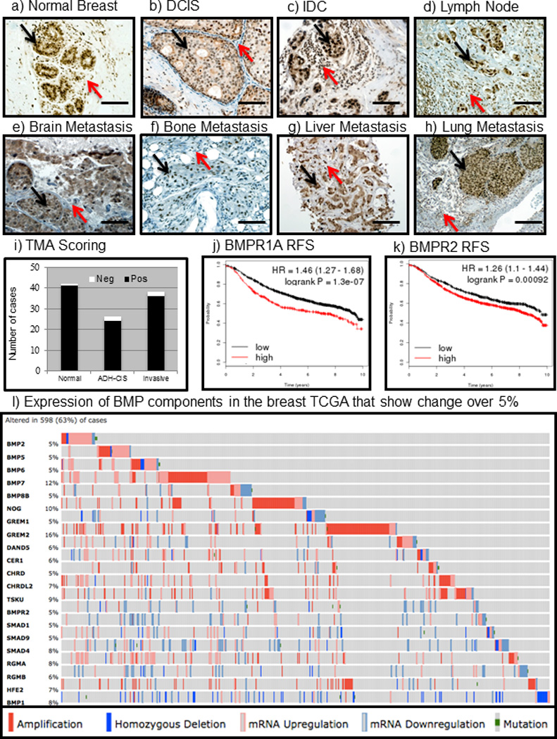
Figure 1. Bone Morphogenetic Protein signaling is active in human breast cancers and is rarely absent.

a) IHC for pSmad1/5/9 demonstrates that the BMP pathway is active in normal breast both in the epithelium (black arrow) and in the surrounding stroma (red arrow). b) In pre-cancerous DCIS lesions, heterogeneous staining showing BMP activation in both the epithelium (black arrow) as well as the surrounding stroma (red arrow). c) BMP signaling is quite strong and active in IDC not only in the primary tumor (black arrow) but also in the stromal infiltrates surrounding the tumor (red arrow). d–f) In metastases to the lymph node (d), brain (e), bone (f), liver (g), and lung (h) tumors exhibited strong staining for active BMP signaling in tumor cells (black arrows) as well as the tumor microenvironment (red arrows). i) IHC for pSmad1/5/9 was performed on two tissue microarrays purchased from US bio max catalog #’s 480 and 722 which contained normal breast, pre-cancerous hyperplasia's and invasive cancers. Scoring revealed that normal breast were 41/42 positive, ADH-CIS were 24/26 positive and Invasive cancers were 36/38 positive for pSmad1/5/9. j) BMP receptor IA (BMPR1A) was queried for correlation to overall survival of breast cancer patients using kmplot.com and found that high expression (red) correlated with poor survival (logrank P =1.3e-07). k) The type II BMP receptor BMPR2 high expression correlated with poor survival using kmplot.com (logrank P =0.00092). l) Using the cBio portal (cbioportal.org) to investigate BMP signaling components in the TCGA we found that in the provisional breast database consisting of 950 total samples 21 BMP related genes altered in greater than 5% of all patients. Solid dark red boxes indicate copy number amplification, light pink indicates mRNA upregulation, dark blue indicates homozygous deletion, light blue indicates mRNA downregulation and green boxes indicate mutations. Microscope scale bars = 100µM. Abbreviations: ADH, Atypical Ductal Hyperplasia. DCIS, Ductal Carcinoma In-Situ. IDC, Invasive Ductal Carcinoma. TMA, Tissue Micro-Array. RFS, Relapse Free Survival. TCGA, The Cancer Genome Atlas. IHC, Immunohistochemistry.
