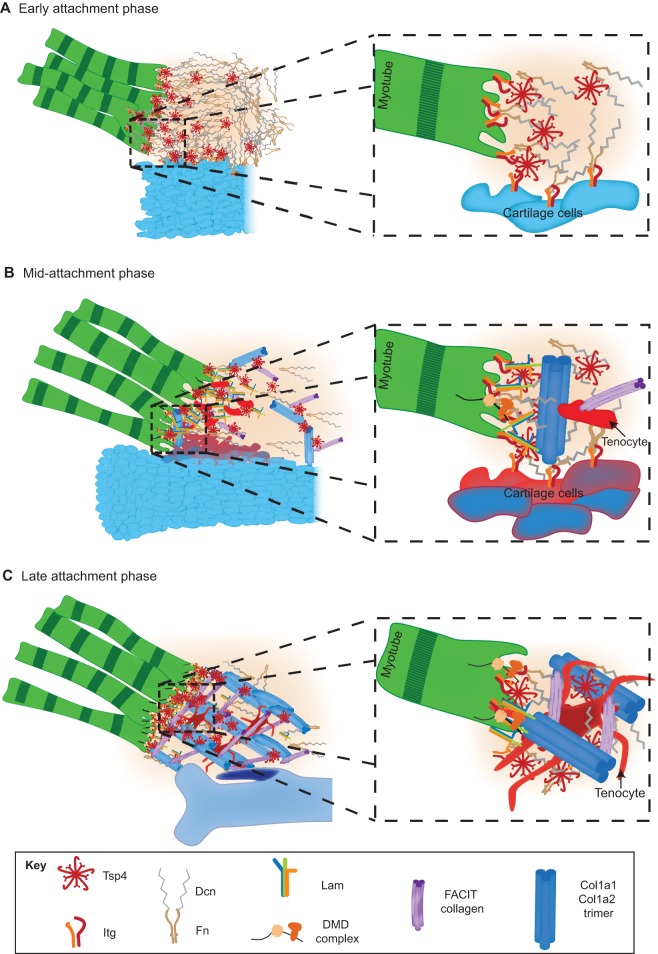
Fig. 4.
Maturation and assembly of the tendon ECM. Diagram illustrating progressive changes in the ECM at an MTJ as it matures. (A) In the early attachment phase, myoblasts (green) first extend towards a cartilage condensation (blue) and reorganize the local ECM by secreting Tsp4 (red), which interacts with Fn and Lam. A magnified view (right) of the boxed area illustrates how Tsp4 pentamers assemble Fn, Lam and Dcn and facilitate binding to Itgs on both muscle and cartilage cell surfaces, thereby promoting adhesion. (B) Following this, in the mid-attachment phase, linear collagen fibrils (Col1a1 trimers, dark blue) form, tenocytes (red) invade, and Sox9+/Scx+ progenitors (dark blue and orange) become detected at the future attachment site on the cartilage, the enthesis. The magnified view illustrates how Col1a1 trimers begin to align perpendicular to skeletal cells (enthesis, dark blue and orange). Dystrophin (DMD) complexes appear on muscle surfaces. (C) In the final late attachment phase, collagen fibrils become crosslinked into a lattice, with tenocytes (red cells) extending processes to surround fibrils, and entheses chondrifying (purple). The magnified view shows Col1a1 trimers becoming crosslinked by FACIT collagens and surrounded by tenocyte (red) processes, stabilizing the ECM and its interactions with Itgs on muscle and cartilage cells.

