Abstract
Susceptibility to scams is a significant issue among older adults, even among those with intact cognition. Age-related changes in brain macrostructure may be associated with susceptibility to scams; however, this has yet to be explored. Based on previous work implicating frontal and temporal lobe functioning as important in decision making, we tested the hypothesis that susceptibility to scams is associated with smaller grey matter volume in frontal and temporal lobe regions in a large community-dwelling cohort of nondemented older adults. Participants (N=327, mean age=81.55, mean education=15.30, 78.9% female) completed a self-report measure used to assess susceptibility to scams and an MRI brain scan. Results indicated an inverse association between overall grey matter and susceptibility to scams in models adjusted for age, education, and sex; and in models further adjusted for cognitive function. No significant associations were observed for white matter, cerebrospinal fluid, or total brain volume. Models adjusted for age, education, and sex revealed seven clusters showing smaller grey matter in the right parahippocampal/hippocampal/fusiform, left middle temporal, left orbitofrontal, right ventromedial prefrontal, right middle temporal, right precuneus, and right dorsolateral prefrontal regions. In models further adjusted for cognitive function, results revealed three significant clusters showing smaller grey matter in the right parahippocampal/hippocampal/fusiform, right hippocampal, and right middle temporal regions. Lower grey matter concentration in specific brain regions may be associated with susceptibility to scams, even after adjusting for cognitive ability. Future research is needed to determine whether grey matter reductions in these regions may be a biomarker for susceptibility to scams in old age.
Keywords: scam, cognition, brain volumetry, parahippocampus, hippocampus
Introduction
Adults over the age of 65 hold approximately a third of the U.S. household net worth (Laibson, 2011) and are at significant risk for financial scam and fraud (AARP, 1999). In a hearing before the Special Committee on Aging of the United States Senate, it was reported that more than $40 billion are lost annually to telemarketing fraud, more than $30 billion are lost annually to mail fraud and sweepstakes, and approximately 40% of older adults rank fraud a higher concern than health or terrorism threats (U.S. Senate, 2005). When an older adult is a victim of scam, the results can be catastrophic, resulting in a loss of wealth painstakingly accumulated over a lifetime, and a substantial reduction in independence and wellbeing (Templeton and Kirkman, 2007). Moreover, older adults are at a stage of life that is inherently marked by limited employment opportunities and declining abilities to recover from financial losses (Dessin, 2000; Jackson and Hafemeister, 2011). Thus, elucidating the mechanisms underlying susceptibility to scams in old age is a significant public health priority.
Data suggest that cognitive impairment in older adults results in proneness to poor decision making (Dessin, 2000; Tymula et al., 2013; Agarwal et al., 2009), and this in turn may result in greater susceptibility to scams. However, case studies from clinical and forensic reports suggest that cognitively intact older persons are also victims of scam (Templeton and Kirkman, 2007; Jackson and Hafemeister, 2011). These case studies suggest factors such as unscrupulous relatives and misplaced trust may be possible reasons for why cognitively intact older adults may show greater susceptibility to scams in old age; however, these implications are circumstantial and speculative at best. Hence the mechanisms that predispose an older adult towards greater susceptibility to scams remain enigmatic, and more research is needed to understand these complexities.
While cognitive functioning may not fully account for susceptibility to scams, age-related changes in grey matter of particular brain regions may represent another mechanism by which a person could become susceptible to scams in old age. Aging is associated with overall decreases in brain grey matter volume (Kennedy et al., 2009; Taki et al., 2011). Neuroimaging research supports a complex and interacting network of brain regions involved in valuation behaviors relevant to susceptibility to scams, many of which are localized to the frontal lobe and involve strategy switching, inhibitory function, self-control, abstraction, and value mapping (Coutlee and Huettel, 2012; Volz et al., 2006).
We investigated whether susceptibility to scams was associated with differences in grey matter volume in a sample of community-dwelling non-demented older adults from the Rush Memory and Aging Project. We primarily hypothesized that lower grey matter volume in frontal lobe regions would be associated with susceptibility to scams after controlling for the effects of age, education, and sex. We secondarily hypothesized that lower grey matter volume in medial temporal lobe regions would be associated with susceptibility to scams since these code for prospective decision outcomes by modulating frontal lobe valuation systems (Peters and Buchel, 2010; Peters and Buchel, 2012). Finally, because even cognitively intact older adults may be susceptible to scams, we explored whether brain grey matter associations with susceptibility to scams remained significant after further accounting for the effects of cognitive function. To our knowledge, this is the first systematic study of brain grey matter associations of susceptibility to scams in old age.
Methods
Participants
Participants came from the Rush Memory and Aging Project, a clinical-pathologic study of aging and dementia (Bennett et al., 2012). This study recruits participants from local residential facilities, including retirement homes, senior housing facilities, and community organizations in and around the greater Chicago metropolitan area. Most participants are English-speaking; however, recruitment materials and procedures are also offered in Spanish if preferred. Participants undergo detailed annual clinical evaluations as previously described (Bennett et al, 2012).
The Rush Memory and Aging Project began in 1997, and enrollment is ongoing. A decision making sub-study which had the scam susceptibility assessment was added in 2010. At the time of these analyses, 1671 participants had completed the baseline evaluation for the parent study; of those, 564 died, 83 refused further participation in the parent project before they were able to complete the baseline decision making battery, and 98 were not asked to participate due to severe difficulties with cognition, understanding, or having moved out of the geographical area. Of the remaining 926 potentially eligible persons, 802 (86.6%) completed the decision making battery, 71 had not yet completed the decision making battery, and 53 refused the decision making battery. Of the 802 participants who had completed the decision making battery, 260 had MRI contraindications leaving 542 eligible for scanning. Of these, 155 refused and 44 were still being scheduled, leaving 343 who had MRI scans acquired at the time of analysis. Of the 343, 8 had dementia and 8 failed quality control leaving 327 eligible for these analyses.
Assessment of Susceptibility to Scams
The susceptibility to scams scale is a five-item self-report measure in which participants rated their agreement to a statement according to a 7-point Likert scale (strongly agree to strongly disagree). The five statements included in the measure have been previously reported (James et al., 2014) and address topics such as telemarketing behaviors, older adults being targeted by con-artists, and suspiciousness of claims that seem too good to be true. The statements are:
I answer the phone whenever it rings, even if I do not know who is calling.
I have difficulty ending a phone call, even if the caller is a telemarketer, someone I do not know, or someone I did not wish to call me.
If something sounds too good to be true, it usually is.
Persons over the age of 65 are often targeted by con-artists.
If a telemarketer calls me, I usually listen to what they have to say.
Each question corresponds to a Likert scale and has a total possible range of 1 to 7 (1=strongly agree, 2=agree, 3=slightly agree, 4=neither agree or disagree, 5=slightly disagree, 6=disagree, 7=strongly disagree). The total score for susceptibility to scams was calculated by averaging the five items (with items 1, 2, and 5 reverse coded). The statements were based generally on findings from the AARP and the Financial Industry Regulatory Authority Risk Meter, a measure of poor and risky financial decision making that is widely used in finance studies (AARP, 1999; Financial Industry Regulatory Authority, 2013). The intraclass correlation coefficient for the measure was 0.63, and we previously demonstrated that responses to this measure were associated with factors commonly believed to correlate with susceptibility to scams, such as higher age, lower financial literacy, lower cognitive function, and lower psychological wellbeing (James et al., 2014). We also have demonstrated that this measure has been associated with cognitive decline among older adults without cognitive impairment (Boyle et al., 2012).
Assessment of Cognition
A battery of cognitive performance tests was administered by trained technicians supervised by a board-certified clinical neuropsychologist. Measures of cognitive function assessed a broad range of cognitive abilities (Bennett et al., 2012; Bennett et al., 2006) and included episodic memory measures (Word List Memory, Word List Recall and Word List Recognition from the procedures established by the CERAD; immediate and delayed recall of Logical Memory Story A and the East Boston Story), semantic memory measures (Verbal Fluency, Boston Naming, subsets of items from Complex Ideational Material, and the National Adult Reading Test), working memory measures (Digit Span subtests forward and backward of the Wechsler Memory Scale-Revised and Digit Ordering), perceptual speed measures (oral version of the Symbol Digit Modalities Test, Number Comparison, Stroop Color Naming, and Stroop Word Reading), and visuospatial ability measures (Judgment of Line Orientation and Standard Progressive Matrices). Raw scores on 19 tests were converted to z-scores using the mean and standard deviation from the baseline evaluation. A global cognition score was calculated by averaging the z-scores across these 19 measures of cognitive function as previously reported (Wilson et al., 2003). This was included as a covariate in some of the analyses. Diagnoses of dementia were determined in accordance with standard criteria by a clinician with expertise in aging as previously described (Bennett et al., 2012). First, an experienced neuropsychologist with expertise in Alzheimer’s disease (AD) and blinded to participant age, sex, and race reviewed all results of cognitive measures and rendered a clinical judgment as to cognitive impairment after reviewing data on education, sensory deficits, and motor deficits. Second, a physician with expertise in the diagnosis of AD reviewed all available participant information (brain scan, medical history, cognitive data, neurological exam) and rendered a clinical judgment as to whether the information was consistent with dementia according to NINCDS/ADRDA criteria (McKhann et al., 1984). These criteria of determining dementia have been used in multiple prior studies (Bennett et al., 2012; Boyle et al., 2010; Yu et al., 2012; Buchman et al., 2012; Wilson et al., 2011; James et al., 2011; Wilson et al., 2011).
Other Covariates
Age (based on date of birth and date of susceptibility to scams assessment), sex, and education (years of schooling) were self-reported and included as covariates.
Imaging Approach
All participants underwent neuroimaging within approximately 1 year of completing the clinical evaluation and susceptibility to scams assessment (mean=43 days; standard deviation=54 days). Magnetic resonance imaging (MRI) scans were conducted on a 1.5 Tesla clinical scanner (General Electric, Waukesha, WI) located within the community of the sample. High data quality was ensured through daily tests of the scanner’s performance and thorough quality control tests on the raw data. High-resolution T1-weighted anatomical images were collected with a 3D magnetization-prepared rapid acquisition gradient-echo (MPRAGE) sequence with the following parameters: TR = 6.3 ms; TE = 2.8 ms; preparation time = 1000 ms; flip angle = 8°; 160 sagittal slices; 1 mm slice thickness; field of view (FOV) = 24 cm × 24 cm; acquisition matrix 224 × 192, reconstructed to a 256 × 256 image matrix; scan time = 10 min and 56 secs. Two copies of the T1-weighted data were acquired on each subject, were coregistered, and averaged.
Structural scans were processed using the SPM8 software, where we utilized a voxel-based morphometric (VBM) approach as implemented in the VBM8 toolbox (http://dbm.neuro.uni-jena.de/vbm.html). Default parameters were used in the VBM toolbox. Images were bias-corrected, classified by tissue type, and registered into MNI space using linear and non-linear transformations. The tissue segmentation algorithm accounted for partial volume effects, and was based on adaptive maximum a posteriori estimations, a spatially adaptive non-local means denoising filter, as well as a hidden Markov random field model. This tissue classification was independent of tissue probability maps, thus acting as an additional safeguard against the potential influence of lesions and altered geometry. This yielded whole brain individual measurements of grey matter, white matter, and cerebrospinal fluid. Using affine registration and the non-linear DARTEL algorithm (Ashburner, 2007), the individual segments in native-space were then normalized to the MNI template supplied within the VBM8 toolbox. Next, analyses were performed on the voxel values of the normalized segments, which were multiplied by the Jacobian determinants derived from the spatial normalization step. Grey matter segments were not multiplied by the linear components of the registration in order to account for individual differences in brain size, alignment, and orientation. An absolute grey matter threshold of 0.15 (of a maximum value of 1) was used to avoid possible edge effects around the border between grey matter and white matter. Quality control was performed using tools from the VBM8 Toolbox and individual visual assessment, which yielded no artifacts or failed segmentation/normalization of the data. Lastly, the modulated grey matter volumes were smoothed with a Gaussian kernel of 12-mm full width at half maximum (FWHM). For visualization, a mean grey matter template from all subjects was created using normalized whole-brain images. This allowed for significant results from the statistical analysis to be directly superimposed on the participants’ mean anatomy for anatomic localization of significant changes in local gray matter concentration.
Statistical Analyses
Descriptive statistics were conducted for all variables of interest. Partial correlation models explored the association between susceptibility to scams and whole brain measures of grey matter, white matter, and CSF controlling for age, education, and sex. Additional analyses controlled for global cognition. Next, results of whole-brain VBM analyses were regressed with susceptibility to scams to identify specific regions of association, controlling for age, education, and sex, in the first model; and then age, education, sex, and global cognition in the second model. In order to interrogate brain regions associated with susceptibility to scams, VBM regional results were controlled for multiple comparisons by implementing a voxel-wise threshold of p<0.001, and a cluster-wise threshold of expected voxels per cluster according to random field theory (Worsley, 2011). Age, education, and sex have been implicated as demographic factors affecting financial outcomes (Chen and Sun, 2011; Barber and Odean, 2001; Cole et al., 2014) and volumetric results of VBM analyses (Good et al., 2001; Draganski et al., 2006; Curiati et al., 2009). These were therefore utilized as covariates in an initial analysis to implicate all brain regions associated with susceptibility to scams apart from demographic factors alone. This allowed us to ascertain all possible brain regions associated with susceptibility to scams. Results were further adjusted for global cognition in secondary models as this has been implicated in susceptibility to scams (James et al., 2014) and we were interested in determining if associations between brain regions and susceptibility to scams were independent of cognitive ability.
Results
Descriptives
Descriptive statistics for demographic, behavioral, cognitive, and neuroimaging whole brain values are provided in Table 1. Over 88% of the sample was White. Responses to the susceptibility to scams measure were inspected to ensure a normal distribution for subsequent analyses.
Table 1.
Demographic, cognitive, behavioral, and neuroimaging variables for the sample
| Imaging sample (n = 327) |
|
|---|---|
| Age (years) | |
| Mean (SD) | 81.55 (7.25) |
| Range | 60 – 100 |
| Education (years) | |
| Mean (SD) | 15.30 (2.91) |
| Range | 8 – 25 |
| Sex (% Female) | 74.10% (n = 258) |
| Race (% White) | 88.50% (n = 308) |
| MMSE (total score) | |
| Mean (SD) | 28.33 (1.79) |
| Range | 19 – 30 |
| Global Cognition Z-score | |
| Mean (SD) | 0.25 (0.51) |
| Range | −1.34 – 1.37 |
| Susceptibility to Scam | |
| Mean (SD) | 2.83 (0.66) |
| Range | 1.00 – 5.20 |
| Grey Matter (mm3) | |
| Mean (SD) | 414.53 (50.23) |
| Range | 272.60 – 586.49 |
| White Matter (mm3) | |
| Mean (SD) | 624.95 (91.80) |
| Range | 371.69 – 882.38 |
| Cerebrospinal fluid (mm3) | |
| Mean (SD) | 271.39 (43.05) |
| Range | 163.07 – 429.87 |
| Total Volume (mm3) | |
| Mean (SD) | 1310.87 (131.58) |
| Range | 897.46 – 1750.33 |
Global Volumetric Correlates of Susceptibility to Scams
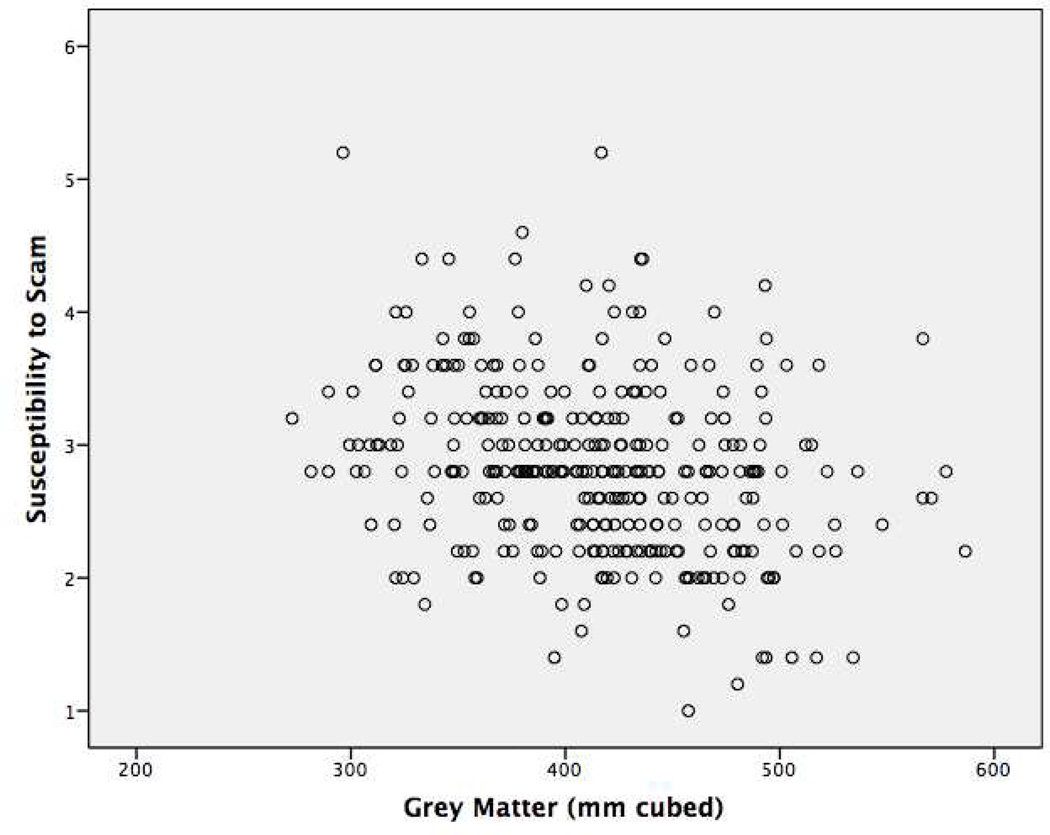
In order to determine whether there were associations between global measures of grey matter, white matter, and cerebrospinal fluid with susceptibility to scams, partial correlation models were conducted that controlled for the effects of age, education, and sex. Results revealed a significant association with only total grey matter volume such that lower total grey matter volume was associated with higher scores on the susceptibility to scams measure (Table 2). Total white matter volume, total cerebrospinal fluid, and total whole brain volume were not significantly associated. A second set of partial correlation models were then conducted to investigate whether these results persisted after adjustment for global cognition. Models of susceptibility to scams and global brain measures controlling for age, education, sex, and global cognition again revealed a significant correlation for total grey matter volume. Total white matter volume, total cerebrospinal fluid, and total whole brain volume were again not significantly associated with susceptibility to scams. A scatterplot of total grey matter values and responses to the susceptibility to scams measure is shown in Figure 1.
Table 2.
Partial correlation models of global brain volume measures with susceptibility to scams.
| Partial r (p-value) |
Model adjusted for age, education, sex |
Model adjusted for age, education, sex, and cognitive function |
||||||
|---|---|---|---|---|---|---|---|---|
| GM | WM | CSF | TV | GM | WM | CSF | TV | |
| Susceptibility to Scams | −0.153 (0.006) | 0.036 (0.519) | 0.039 (0.480) | −0.027 (0.627) | −0.133 (0.016) | 0.033 (0.557) | 0.024 (0.662) | −0.024 (0.662) |
GM=Grey Matter, WM=White Matter, CSF=cerebrospinal fluid, TV=total brain volume
Figure 1.
Scatterplot of global grey matter volume and the susceptibility to scams measure.
Specific Regional Grey Matter Correlates of Susceptibility to Scams
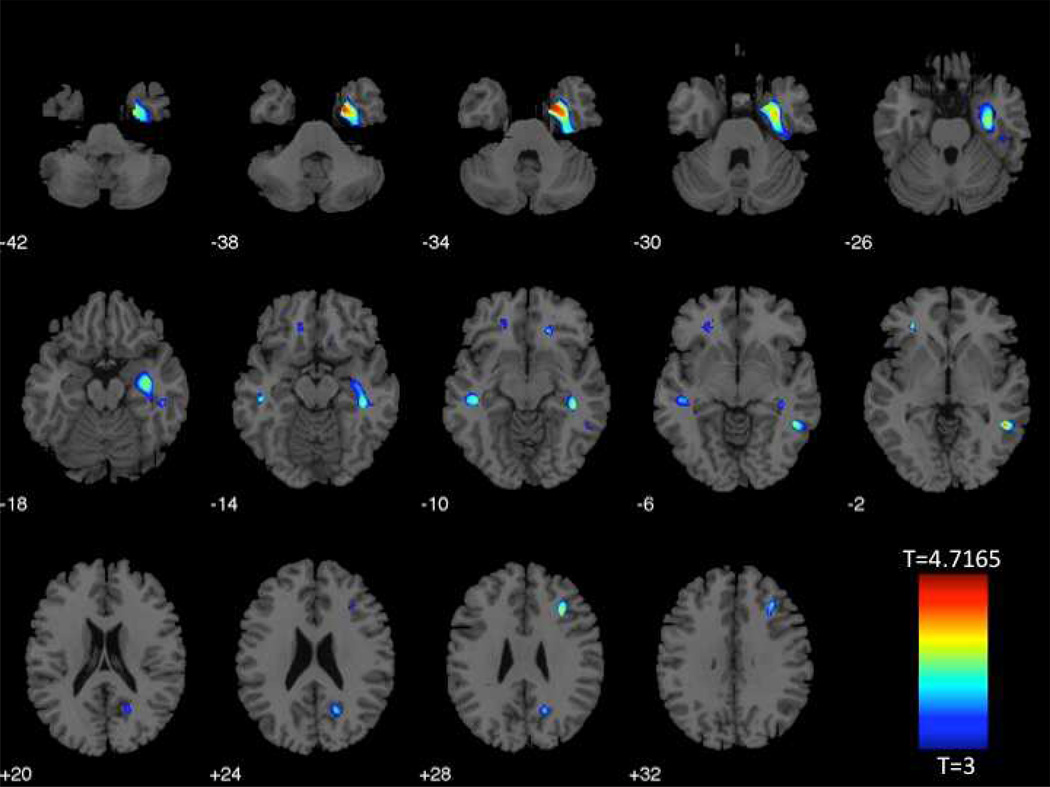
Because it was discovered that total grey matter estimations correlated with susceptibility to scams, we next conducted voxelwise whole-brain models to determine specifically where in the brain these associations occurred. Whole-brain models adjusted for age, education, and sex revealed seven clusters where lower grey matter concentration was significantly associated with higher susceptibility to scams. The first and largest cluster included portions of the right parahippocampal, hippocampal, and fusiform regions. Other clusters were observed in the left middle temporal region, the left orbitofrontal region, the right ventromedial prefrontal region, the right middle temporal region, the right precuneus region, and the right dorsolateral prefrontal region (Figure 2).
Figure 2.
Regions associated with susceptibility to scams, adjusting for age, education, and sex
| Cluster | Regions in Cluster | Maximum Intensity Voxel Coordinates (MNI) | Cluster Size (# of voxels) |
t-value | ||
|---|---|---|---|---|---|---|
| 1 | R parahippocampal/hippocampal/fusiform | 21 | −2 | −38 | 2995 | 4.7165 |
| 2 | L middle temporal | −44 | −27 | −11 | 298 | 3.9277 |
| 3 | L orbitofrontal | −29 | 38 | −3 | 298 | 4.0043 |
| 4 | R ventromedial frontal | 17 | 30 | −12 | 161 | 3.7844 |
| 5 | R middle temporal | 48 | −48 | −3 | 271 | 4.3822 |
| 6 | R precuneus | 18 | −62 | 26 | 247 | 3.6333 |
| 7 | R dorsolateral prefrontal | 32 | 23 | 29 | 272 | 4.0493 |
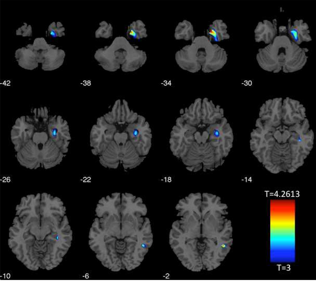
Susceptibility to scams is often fully attributed to poor cognitive functioning. To determine whether global cognition might fully account for these findings, we conducted subsequent whole-brain models further adjusting for the effects of global cognition. Results again revealed a significant negative correlation for total grey matter volume and susceptibility to scams. Voxelwise results revealed three significant clusters indicating lower grey matter concentration associated with higher susceptibility to scams. The first and largest cluster included portions of the right parahippocampal, hippocampal, and fusiform regions. The other two clusters were observed in the right hippocampal region and the right middle temporal region (Figure 3).
Figure 3.
Regions associated with susceptibility to scams, adjusting for age, education, sex, and global cognition.
| Cluster | Regions in Cluster | Maximum Intensity Voxel Coordinates (MNI) |
Cluster Size (# of voxels) |
t-value | ||
|---|---|---|---|---|---|---|
| 1 | R parahippocampal/hippocampal/fusiform | 21 | −2 | −38 | 1259 | 4.2613 |
| 2 | R hippocampal | 41 | −30 | −12 | 98 | 3.7138 |
| 3 | R middle temporal | 48 | −48 | −3 | 147 | 4.0538 |
Discussion
We explored grey matter associations with susceptibility to scams in a large group of non-demented community-dwelling older adults. Using voxel-based morphometric measures, we found an inverse association of total grey matter volume with susceptibility to scams in models adjusted for age, education, and sex, and in secondary models further adjusted for global cognition. Consistent with our hypotheses, we observed that lower grey matter concentration in multiple frontal and temporal lobe regions was associated with susceptibility to scams in voxel-level analyses adjusted for the effects of age, education, and sex. In models further adjusted for global cognition, clusters in right temporal lobe regions remained significant. Total white matter and cerebrospinal fluid were not associated, and no regions showed higher grey matter concentration associated with susceptibility to scams. These results suggest susceptibility to scams in old age may have relatively specific neuroanatomical grey matter correlates, and that the associations of these neural correlates with susceptibility to scams are above and beyond the effects of global cognition.
Neuroimaging research has identified a number of interacting brain regions in, or highly connected to, the frontal lobes as likely involved in economic decision making (Fellows and Farah, 2007; Bechara et al., 2000; Krawczyk, 2002; Kennerley et al., 2006), and we observed some of these same regions associated with susceptibility to scams in old age. Previous work has specifically implicated the ventromedial prefrontal cortex, orbitofrontal cortex, dorsolateral prefrontal cortex, and anterior cingulate cortex as each serving specific functions in a frontal lobe-mediated subnetwork contributing to financial or economic decision making outcomes. The ventromedial prefrontal cortex is believed to play a role in assessing the economic value of options in decision making (Fellows and Farah, 2007). The orbitofrontal cortex, according to the somatic marker hypothesis, serves to process the emotional qualities of potential options in an economic decision making context, and may be particularly sensitive to adverse or negative emotional qualities of a decision (Bechara et al., 2000). The dorsolateral prefrontal cortex appears to be involved in decisions that require active consideration of multiple different options due to its role in working memory processing (Krawczyk, 2002). The anterior cingulate appears to be critical for guiding voluntary choices based on the history of actions and their associated outcomes (Kennerley et al., 2006). Our results might suggest that as portions of the frontal lobe may deteriorate, the ability to assess economic values of options (ventromedial prefrontal cortex), process the emotional qualities of options (orbitofrontal cortex), and actively consider multiple different options at the same time (dorsolateral prefrontal cortex) may also deteriorate. Of particular note, the frontal lobe regions related to susceptibility to scams were not significant after adjusting for the effects of global cognition. This suggests that these potential frontal lobe functional contributions to susceptibility to scams might be mediated by cognitive ability, and as cognition declines, so may these specific functional abilities. However, more research is needed to demonstrate the validity of these statements.
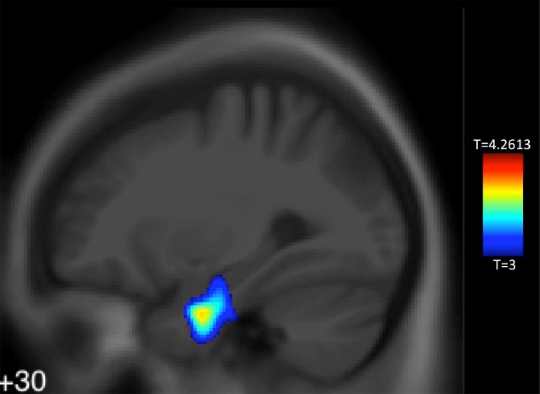
While frontal lobe contributions to susceptibility to scams may be mediated by cognitive abilities, temporal lobe regions may have an independent association to susceptibility to scams in old age (see Figure 4). Temporal lobe regions, particularly medial structures implicated in prospective memory processing, have garnered increasing attention in recent years for their importance in decision making (Peters and Buchel, 2010; Peters and Buchel, 2012; Delazer et al., 2010; Guitart-Masip et al., 2013). Recent work has found that future thinking reduces the tendency of humans to discount the value of future rewards for more immediate smaller rewards, and this has been attributed to the functional coupling between medial temporal lobe structures and the anterior cingulate cortex (Peters and Buchel, 2010; Guitart-Masip et al., 2013). As described above, the anterior cingulate cortex guides voluntary choices exerted through frontal lobe networks based on the history of actions and their associated outcomes. The medial temporal lobe structures are important for accurately representing this history of actions and outcomes, and these interact with the anterior cingulate cortex to modulate downstream frontal lobe structures (e.g., ventromedial prefrontal cortex, orbitofrontal cortex, dorsolateral prefrontal cortex). We also observed middle temporal lobe and precuneus associations with susceptibility to scams in demographically adjusted models. These regions have also been implicated in imagery or prospective processing (Buckner et al., 2008), and together with the medial temporal lobes constitute a potential prospective functional brain network important for decision making (Peters and Buchel, 2012). If this network should functionally decline, adults may not be able to prospectively consider the negative consequences of choices associated with scam situations and thus may become susceptible to scams. It is important to note that medial temporal lobe regional grey matter associations remained significant above and beyond the effects of global cognition. The implication is that grey matter differences in this region may be a sensitive biomarker for susceptibility to scams.
Figure 4.
Cluster indicating less grey matter in medial temporal lobe associated with susceptibility to scams
Cluster indicating less grey matter in medial temporal lobe associated with susceptibility to scams, adjusting for age, education, sex, and cognitive function. Maximum intensity voxel MNI coordinates x=21, y=−2, z=−38, 1259 voxels, t-value=4.2613, superimposed on an MNI template.
We previously showed in the same cohort of non-demented participants that susceptibility to scams was associated with higher age and lower cognitive functioning (James et al., 2014). We also showed that poorer decision making is a consequence of cognitive decline among older adults without dementia or mild cognitive impairment, suggesting that even very subtle changes in cognitive functioning impact decision making (Boyle et al., 2012). This study extends our previous findings by suggesting that lower grey matter concentration in specific brain regions implicated in prospective encoding of decisional outcomes may be a neuroanatomical basis for susceptibility to scams in old age independent of cognitive functioning. Future work is needed to determine if disproportionate longitudinal declines in the grey matter of these regions may predispose an older adult to scams susceptibility. Research is also needed to determine whether other factors associated with susceptibility to scams, such as financial literacy and psychological wellbeing (James et al., 2014), may also have specific brain region grey matter correlates in old age.
This study has a number of strengths and limitations. The association of this measure to any specific amounts of financial losses is unclear because we have not associated this measure with actual instances of scam. Future efforts will focus on documenting scam victimization in the cohort. Another limitation is the limited racial representation of the cohort. Future efforts will focus on investigating whether these trends are also observed in other racial groups. The exploratory nature of the VBM approach is an additional limitation and future studies are needed to support the present findings. The ICC for the susceptibility to scams measure suggests there may be some variability in how participants respond to the measure. This variability could affect how generalizable our findings are to other populations. Strengths of the study include the utilization of a large, well-characterized, community-dwelling cohort of non-demented older adults, a broad battery of neurocognitive measures, a measure of susceptibility to scams informed by research on victimized older adults, and a sensitive method to ascertain local grey matter concentration from brain MRI scans. The implications of this study are far-reaching. If future studies support our finding that lower grey matter concentration in specific regions of the brain may be associated with susceptibility to scams, then this neuroimaging biomarker may be used as an additional screening measure in order to help older adults protect and ensure their wellbeing and independence into old age.
Supplementary Material
Acknowledgements
This research was supported by National Institute on Aging grants R01AG017917, R01AG033678, K23AG040625, the American Federation for Aging Research, and the Illinois Department of Public Health. The authors gratefully thank the Rush Memory and Aging Project staff and participants.
Footnotes
Disclosure Statement
S. Duke Han, Patricia A. Boyle, Lei Yu, Konstantinos Arfanakis, Bryan D. James, Debra Fleischman, and David A. Bennett declare no conflicts of interests.
Ethical Statement
All procedures followed were in accordance with the ethical standards of the responsible committee on human experimentation (institutional and national) and with the Helsinki Declaration of 1975, and the applicable revisions at the time of the investigation. Informed consent was obtained from all patients for being included in the study.
References
- 1.AARP. AARP poll: Nearly one in five Americans report they’ve been victimized by fraud. Washington, DC: Author; 1999. [Google Scholar]
- 2.Agarwal S, Driscoll JC, Gabaix X, Laibson D. The age of reason: financial decisions over the lifecycle with implications for regulation. Brookings Papers on Economic Activity. 2009;2:51–117. [Google Scholar]
- 3.Ashburner J. A fast diffeomorphic image registration algorithm. NeuroImage. 2007;38:95–113. doi: 10.1016/j.neuroimage.2007.07.007. [DOI] [PubMed] [Google Scholar]
- 4.Barber BM, Odean T. Boys will be boys: Gender, overconfidence, and common stock investment. The Quarterly Journal of Economics. 2001;116:261–292. [Google Scholar]
- 5.Bechara A, Damasio H, Damasio AR. Emotion, decision making, and the orbitofrontal cortex. Cerebral Cortex. 2000;10:295–307. doi: 10.1093/cercor/10.3.295. [DOI] [PubMed] [Google Scholar]
- 6.Bennett DA, Schneider JA, Arvanitakis Z, Kelly JF, Aggarwal NT, Shah RC, et al. Neuropathology of older person without cognitive impairment from two community-based studies. Neurology. 2006;27:1837–1844. doi: 10.1212/01.wnl.0000219668.47116.e6. [DOI] [PubMed] [Google Scholar]
- 7.Bennett DA, Schneider JA, Buchman AS, Barnes LL, Boyle PA, Wilson RS. Overview and findings from the Rush Memory and Aging Project. Curr Alzheimer Res. 2012;9(6):646–663. doi: 10.2174/156720512801322663. [DOI] [PMC free article] [PubMed] [Google Scholar]
- 8.Boyle PA, Buchman AS, Barnes LL, Bennett DA. Effect of a purpose in life on risk of incident Alzheimer’s disease and mild cognitive impairment in community-dwelling older persons. Arch Gen Psychiatry. 2010;67:304–310. doi: 10.1001/archgenpsychiatry.2009.208. [DOI] [PMC free article] [PubMed] [Google Scholar]
- 9.Boyle PA, Yu L, Wilson RS, Gamble K, Buchman AS, Bennett DA. Poor decision making is a consequence of cognitive decline among older persons without Alzheimer’s disease or mild cognitive impairment. Plos One. 2012;7:e43647. doi: 10.1371/journal.pone.0043647. [DOI] [PMC free article] [PubMed] [Google Scholar]
- 10.Brier MR, Thomas JB, Snyder AZ, Wang L, Fagan AM, Benzinger T, et al. Unrecognized preclinical Alzheimer disease confounds rs-fcMRI studies of normal aging. Neurology. 2014;83:1613–1619. doi: 10.1212/WNL.0000000000000939. [DOI] [PMC free article] [PubMed] [Google Scholar]
- 11.Buchman AS, Boyle PA, Yu L, Shah RC, Wilson RS, Bennett DA. Total daily physical activity and the risk of cognitive decline in older adults. Neurology. 2012;78:1323–1329. doi: 10.1212/WNL.0b013e3182535d35. [DOI] [PMC free article] [PubMed] [Google Scholar]
- 12.Buckner RL, Andrews-Hanna JR, Schacter DL. The brain’s default network: Anatomy, function, and relevance to disease. Ann NY Acad Sci. 2008;1124:1–38. doi: 10.1196/annals.1440.011. [DOI] [PubMed] [Google Scholar]
- 13.Chen Y, Sun Y. Age differences in financial decision-making: using simple heuristics. Educational Gerontology. 2011;29:627–635. [Google Scholar]
- 14.Cole S, Paulson A, Shastry GK. Smart money? The effect of education on financial outcomes. The Review of Financial Studies. 2014;27:2022–2051. [Google Scholar]
- 15.Coutlee CG, Huettel SA. The functional neuroanatomy of decision making: Prefrontal control of thought and action. Brain Research. 2012;1428:3–12. doi: 10.1016/j.brainres.2011.05.053. [DOI] [PMC free article] [PubMed] [Google Scholar]
- 16.Curiati PK, Tamashiro JH, Squarzoni P, Duran FLS, Santos LC, Wajngarten M, Leite CC, Vallada H, Menezes PR, Scazufca M, Busatto GF, Alves TCTF. Brain structural variability due to aging and gender in cognitive healthy elders: Results from the Sao Paulo Ageing and Health Study. AJNR. 2009;30:1850–1856. doi: 10.3174/ajnr.A1727. [DOI] [PMC free article] [PubMed] [Google Scholar]
- 17.Delazer M, Zamarian L, Bonatti E, Kuchukhidze G, Koppelstater F, Bodner T, et al. Decision making under ambiguity and under risk in mesial temporal lobe epilepsy. Neuropsychologia. 2010;48:194–200. doi: 10.1016/j.neuropsychologia.2009.08.025. [DOI] [PubMed] [Google Scholar]
- 18.Dessin CL. Financial abuse of the elderly. Idaho Law Review. 2000;36(2):203–226. [Google Scholar]
- 19.Draganski B, Gaser C, Kempermann G, Kuhn HG, Winkler J, Buchel C, May A. Temporal and spatial dynamics of brain structure changes during extensive learning. Journal of Neuroscience. 2006;26:6314–6317. doi: 10.1523/JNEUROSCI.4628-05.2006. [DOI] [PMC free article] [PubMed] [Google Scholar]
- 20.Fellows LK, Farah MJ. The role of the ventromedial prefrontal cortex in decision making: Judgment under uncertainty or judgment per se? Cerebral Cortex. 2007;17:2668–2674. doi: 10.1093/cercor/bhl176. [DOI] [PubMed] [Google Scholar]
- 21.Financial Industry Regulatory Authority. Financial Industry Regulatory Authority risk meter. 2013 Retrieved from http://apps.finra.org/meters/1/riskmeter.aspx. [Google Scholar]
- 22.Good CD, Johnsrude IS, Ashburner J, Henson RNA, Friston KJ, Frackowiak RSJ. A voxel-based morphometric study of ageing in 465 normal adult human brains. NeuroImage. 2001;14:21–36. doi: 10.1006/nimg.2001.0786. [DOI] [PubMed] [Google Scholar]
- 23.Gordon BA, Blazey T, Benzinger TLS. Regional variability in Alzheimer’s disease biomarkers. Future Neurology. 2014;9:131–134. doi: 10.2217/fnl.14.9. [DOI] [PMC free article] [PubMed] [Google Scholar]
- 24.Guitart-Masip M, Barnes GR, Horner A, Bauer M, Dolan RJ, Duzel E. Synchronization of medial temporal lobe and prefrontal rhythms in human decision making. J Neurosci. 2013;33:442–451. doi: 10.1523/JNEUROSCI.2573-12.2013. [DOI] [PMC free article] [PubMed] [Google Scholar]
- 25.Jackson SL, Hafemeister TL. Financial abuse of elderly people vs. other forms of elder abuse: Assessing their dynamics, risk factors, and society’s response. Final Report Presented to the National Institute of Justice. 2011 [Google Scholar]
- 26.James BD, Boyle PA, Bennett DA. Correlates of susceptibility to scams in older adults without dementia. Journal of Elder Abuse and Neglect. 2014;26:107–122. doi: 10.1080/08946566.2013.821809. [DOI] [PMC free article] [PubMed] [Google Scholar]
- 27.James BD, Boyle PS, Buchman AS, Barnes LL, Bennett DA. Life space and risk of Alzheimer’s disease, mild cognitive impairment, and cognitive decline in old age. Am J Geriatr Psychiatry. 2011;19:961–969. doi: 10.1097/JGP.0b013e318211c219. [DOI] [PMC free article] [PubMed] [Google Scholar]
- 28.Kennedy KM, Erickson KI, Rodrigue KM, Voss MW, Colcombe SJ, Kramer AF, et al. Age-related differences in regional grey matter volumes: A comparison of optimized voxel-based morphometry to manual volumetry. Neurobiology of Aging. 2009;30:1657–1676. doi: 10.1016/j.neurobiolaging.2007.12.020. [DOI] [PMC free article] [PubMed] [Google Scholar]
- 29.Kennerley SW, Walton ME, Behrens TEJ, Buckley MJ, Rushworth MFS. Optimal decision making and the anterior cingulate cortex. Nature Neuroscience. 2006;9:940–947. doi: 10.1038/nn1724. [DOI] [PubMed] [Google Scholar]
- 30.Krawczyk DC. Contributions of the prefrontal cortex to the neural basis of decision making. Neuroscience and Biobehavioral Reviews. 2002;26:631–664. doi: 10.1016/s0149-7634(02)00021-0. [DOI] [PubMed] [Google Scholar]
- 31.Laibson D. Age of Reason. Closing keynote presentation at the 23rd annual Morningstar Investment Conference; Chicago, IL. 2011. [Google Scholar]
- 32.McKhann G, Drachman D, Folstein M, Katzman R, Price D, Standlan E. Clinical diagnosis of Alzheimer’s disease: Report of the NINCDS/ADRDA Work Group under the auspices of Department of Health and Human Services Task Force on Alzheimer’s Disease. Neurology. 1984;34:939–944. doi: 10.1212/wnl.34.7.939. [DOI] [PubMed] [Google Scholar]
- 33.Peters J, Buchel C. Episodic future thinking reduces reward delay discounting through an enhancement of prefrontal-mediotemporal interactions. Neuron. 2010;66:138–148. doi: 10.1016/j.neuron.2010.03.026. [DOI] [PubMed] [Google Scholar]
- 34.Peters J, Buchel C. The neural mechanisms of inter-temporal decision-making: understanding variability. Trends in Cognitive Sciences. 2012;15:207–239. doi: 10.1016/j.tics.2011.03.002. [DOI] [PubMed] [Google Scholar]
- 35.Taki Y, Kinomura S, Sato K, Goto R, Kawashima R, Fukuda H. A longitudinal study of grey matter volume decline with age and modifying factors. Neurobiology of Aging. 2011;32:907–915. doi: 10.1016/j.neurobiolaging.2009.05.003. [DOI] [PubMed] [Google Scholar]
- 36.Templeton VH, Kirkman DN. Fraud, vulnerability, and aging. Alzheimer’s Care Today. 2007;8(3):265–277. [Google Scholar]
- 37.Tymula A, Rosenberg Belmaker LA, Ruderman L, Glimcher PW, Levy I. Like cognitive function, decision making across the life span showed profound age-related changes. PNAS. 2013;110(42):17143–17148. doi: 10.1073/pnas.1309909110. [DOI] [PMC free article] [PubMed] [Google Scholar]
- 38.U.S. Senate. Washington, DC: U.S. Government Printing Office; 2005. Old Scams-New Victims: Breaking the Cycle of Victimization (109th Congress, First Session, Serial No. 109–113) http://www.gpo.gov/fdsys/pkg/CHRG-109shrg25878/html/CHRG-109shrg25878.htm. [Google Scholar]
- 39.Volz KG, Schubotz RI, Yves von Cramon D. Decision-making and the frontal lobes. Curr Opin Neurol. 2006;19:401–406. doi: 10.1097/01.wco.0000236621.83872.71. [DOI] [PubMed] [Google Scholar]
- 40.Wilson RS, Barnes LL, Bennett DA. Assessment of lifetime participation in cognitively stimulating activities. J Clin Exp Psychol. 2003;25:634–642. doi: 10.1076/jcen.25.5.634.14572. [DOI] [PubMed] [Google Scholar]
- 41.Wilson RS, Boyle PA, Buchman AS, Yu L, Arnold SE, Bennett DA. Harm avoidance and risk of Alzheimer’s disease. Psychosomatic Medicine. 2011;73:690–696. doi: 10.1097/PSY.0b013e3182302ale. [DOI] [PMC free article] [PubMed] [Google Scholar]
- 42.Worsley K. Random field theory. Statistical Parametric Mapping: The Analysis of Functional Brain Images: The Analysis of Functional Brain Images. 2011 [Google Scholar]
- 43.Yu L, Boyle PA, Wilson RS, Segawa E, Leurgans S, De Jager PL, Bennett DA. A random change point model for cognitive decline in Alzheimer’s disease and mild cognitive impairment. Neuroepidemiology. 2012;39:73–83. doi: 10.1159/000339365. [DOI] [PMC free article] [PubMed] [Google Scholar]
Associated Data
This section collects any data citations, data availability statements, or supplementary materials included in this article.


