

Abstract
Background:
Human Wharton’s jelly mesenchymal stem cells (HWJMSCs) express liver-specific markers such as albumin, alpha-fetoprotein, cytokeratin-19, cytokeratin-18, and glucose-6-phosphatase. Therefore, they can be considered as a good source for cell replacement therapy for liver diseases. This study aimed to evaluate the effects of various culture systems on the hepatocyte-specific gene expression pattern of naïve HWJMSCs.
Methods:
HWJMSCs were characterized as MSCs by detecting the surface CD markers and capability to differentiate toward osteoblast and adipocyte. HWJMSCs were cultured in 2D collagen films and 3D collagen scaffolds for 21 days and were compared to control cultures. Real time RT-PCR was used to evaluate the expression of liver-specific genes.
Results:
The HWJMSCs which were grown on non-coated culture plates expressed cytokeratin-18 and -19, alpha-fetoprotein, albumin, glucose-6-phosphatase, and claudin. The expression of the hepatic nuclear factor 4 (HNF4) was very low. The cells showed a significant increase in caludin expression when they cultured in 3D collagen scaffolds compared to the conventional monolayer culture and 2D collagen scaffold.
Conclusion:
Various culture systems did not influence on hepatocyte specific marker expression by HWJMSCs, except for claudin. The expression of claudin showed that 3D collagen scaffold provided the extracellular matrix for induction of the cells to interconnect with each other.
Keywords: Wharton jelly, Mesenchymal stem cell, Collagen, Scaffold, Hepatocyte
What Known
Collagen scaffolds accelerated the expression of liver-specific markers by MSCs exposed to hepatogenic medium.
According to the literature, it seems that collagen scaffolds exerts a dual effect on the cells according to the degree of differentiation.
What’s New
HWJMSCs showed different behaviors in 2D and 3D culture systems.
Yet, HWJMSCs, as a good source for cell therapy and fabricating tissue-engineered organs in liver diseases, need to continue.
2D collagen films may be recommended for the initial step of cell differentiation because the cells could express a higher level of HNF4.
3D culture system seems to be more suitable for the second step of hepatogenic differentiation.
Introduction
Thomas Wharton was the first scientist who described umbilical cord as a primitive connective tissue in 1656.1 Human umbilical cord is considered as an extra-embryonic tissue, which is naturally disposed after birth.2 This tissue can be an available source of mesenchymal stem cells (MSCs) with little ethical concern and large expansion capacity for use in regenerative medicine, which was introduced by McElreavey et al.3 Human Wharton’s jelly MSCs (HWJMSCs) can be regarded as multipotent stem cells with self-renewal and immunoregulatory potential. They are also able to differentiate toward adipocytes,4 chondrocytes, osteocytes,5,6 cardiomyocytes,7 skeletal myocytes,8 neuron-like cells,9 insulin-producing cells,10 and hepatocytes11,12 under suitable conditions. Up to now, most investigations have been focused on isolation, differentiation potential, and clinical therapeutic application of HWJMSCs.13 The chromosomal stability, mechanism of telomere activation, and cell cycle regulated gene expression in these cells has also been studied under long-term culture conditions.14
HWJMSCs have a vast gene expression profile. They can express the embryonic as well as adult stem cell markers.15 These cells showed a positive reaction to MSC markers, such as CD10, CD13, CD29, CD44, and CD90 and a negative reaction to CD14, CD33, CD56, CD31, CD34, CD45, and HLA-DR.16,17 In addition, they express a number of genes found in undifferentiated embryonic stem cells (ESCs), such as the markers involved in pluripotency. Moreover, some markers of three germ layers; i.e., endoderm, ectoderm, and mesoderm, were demonstrated to be expressed by HWJMSCs.18 HWJMSCs population has been found to express the early hepatic markers, such as albumin, alpha-fetoprotein (AFP), cytokeratin-19 (CK-19), some mid- and late- hepatic markers, including CK18 and glucose 6-phosphatase (G6P),19,20 as well as a low level of hematopoietic markers, such as c-kit and phosphoenolpyruvate carboxykinase (PEPCK). Nonetheless, expression of the hepatic nuclear factor-4 (HNF 4) as an endodermal marker and a key regulator of hepatogenic fate is controversial.21,22 Claudins as a marker of tight junction, showed a pivotal role in morphogenesis and embryo development.23 Claudins also plays a critical role in remodeling of bile duct during liver morphogenesis.24 Furthermore, it has been demonstrated that claudin expression influence bile canaliculi formation.25 Hence, HWJMSCs have been shown capable of producing hepatocyte markers and they can be considered as a very attractive cell source for the treatment of liver diseases.26 Also, in vivo and in vitro studies showed that their differentiation ability to hepatic lineage can be improved by adding hepatogenic factors to culture media.11
Stem cell three dimensional (3D) culture methods, such as cell-seeded in a collagen scaffold, can enhance intercellular contacts, improve cell migration, and mimic an in vivo environmental condition. The 3D culture system also improves the cell stability and influences gene expression pattern. HWJMSCs showed different behaviors in 2D and 3D culture systems.8 Yet, HWJMSCs, as a good source for cell therapy and fabricating tissue-engineered organs in liver diseases, need to continue and enhance the expression of hepatic markers in 3D scaffolds as well as in 2D culture systems. Therefore, the current study aims to evaluate and compare the ability of HWJMSCs to express hepatic markers cultured in 2D and 3D conditions.
Materials and Methods
Time and Setting
The experiment was performed in the Laboratory for Stem Cell Research of Anatomy Department at Shiraz University of Medical Sciences from 2011 to March 2014.
Isolation and Culture of HWJMSCs
Human umbilical cords were obtained from 10 full-term infants born through caesarian delivery at Shafa and Hafez Hospitals after getting informed permission from their parents. All specimens were prepared in accordance with the guidance of the Ethic Committee of Shiraz University of Medical Sciences. The umbilical cords were collected in cold phosphate buffered saline (PBS) containing 100 μg/mL penicillin, 100 U/mL streptomycin (Sigma, USA). Then the arteries were discarded and the endothelium of the vein and amniotic epithelium were scraped. Afterward, small explant pieces (about 4×5 mm) were prepared from umbilical cord matrix. The α-MEM (Sigma, USA) containing 10% fetal bovine serum (FBS) (Gibco, Germany), 1% L-glutamine, and 100 μg/mL penicillin, 100 U/mL streptomycin were added after 15 min. The MSCs reached 70-80% confluence after about 15 days.
Flow Cytometric Analysis for Cell Surface Markers
To stain the HWJMSCs, the cells at passage 3 were detached from the culture surface. Afterward, 1×10⁶ cells/mL were washed in cold PBS containing 10% FBS as blocking solution for 20 min. Then, the cells were incubated with FITC-conjugated anti-CD44 and CD144, phycoerythrin-conjugated anti-CD34 and CD106, and PerCP-conjugated anti-CD105 antibodies (all from Abcam, Cambridge, UK) for 30 min. After the washing phase, the cells were re-suspended in PBS containing 10% FBS. Then, the cells were subjected to flow cytometry (BD, USA) and the data were analyzed using FlowJo™ software (TreeStar, Ashland, OR, USA).
Adipogenic and Osteogenic Differentiation of HWJMSCs
HWJMSCs were induced to differentiate into osteocytes and adipocytes by being exposed to osteogenic (MACS, Germany) and adipogenic media (Stem Cell Technologies Inc., Canada) for 4 and 3 weeks, respectively. The culture media were replaced twice a week. Then, the HWJMSCs differentiated toward osteogenic lineage were fixed with 4% paraformaldehyde and stained with Alizarin Red S (Sigma, USA). To demonstrate the adipogenic differentiation, the cells were stained by oil red O (Sigma, USA).
Experimental Design
The cells were divided into three groups. One group was seeded on the surface of the culture plate without coating and considered as conventional monolayer culture condition (control). The second group was cultured on the surface of the collagen film (2D culture), and the last group was allowed to grow in the 3D collagen scaffolds (3D culture). All cells were cultured for 21 days without passaging.
2D Culture
A stock solution of collagen type I from Rat Tail (Gibco, A10483-01) at a concentration of 3 mg/mL was diluted with 10X DMEM (Sigma, USA) at the ratio of 8:1 on ice immediately before use. Each well was coated with 125 μL of the collagen solution, which was polymerized at 37°C for 1 h. After polymerization, 1×10³ cells were seeded per well in a 24-well plate. Half of the culture media was changed every 3 days. The cells were kept in this condition for 21 days. In order to obtain the basic-level of the hepatic markers expression by HWJMSCs, some cells were also cultured on plastic culture dishes until they reached confluency (control).
3D Culture
The collagen solution was prepared using the method mentioned above. Wharton’s jelly-derived MSCs were harvested and seeded at a density of 7×10⁵ cell/mL of collagen. The final concentration of the collagen was 1 mg/mL. The collagen gel was allowed to polymerize for 1 h at 37°C. Then, DMEM (low glucose) supplemented with 5% FBS was added. The cells were kept in this condition for 21 days.
Some cell-containing collagen scaffolds were fixed in Bouin’s fixative, embedded in paraffin, and sectioned at 10 µm. The slides were then stained using hematoxylin and eosin (H&E) staining method.
Real Time RT_PCR
Biazol isolation reagent (Bioflux) was used to extract total RNA from HWJMSCs according to the manufacturer’s instructions. The quantity of RNA was estimated at 260/280 nm wavelength by Biophotometer (Eppendorf, Germany). Additionally, cDNA (ABI cDNA kit, USA) was made of 5 µg RNA according to the manufacturer’s instructions.
The cDNA (2 μL) was used as the template for real-time PCR reactions. The SYBR green PCR master mix (Applied Biosystems, Rotkreuz, Switzerland) was used for real-time PCR and the cDNA was amplified for 50 cycles. The initial denaturation temperature was 95°C for 10 min, continued by cycling denaturation in 95°C for 15 seconds and annealing at 60°C for 1 min. After amplification, the melting curve was analyzed to ensure that no primer dimers were produced in samples and the accuracy of the reactions was confirmed. The data were normalized by comparing them with housekeeping gene, 18S. The following equation was applied to each data point: 2-∆∆Ct
Primer-BLAST online program was used to design the primers based on the human DNA sequences found in the genebank27 (table 1).
Table 1.
The primer sequences used for quantities real time PCR
| Primer | Sequence | Size (base pair) |
|---|---|---|
| Albumin | 5’-ACAGAGACTCAAGTGTGCCAGT-3’ 5’-GCAAGGTCCGCCCTGTCATC-3’ |
198 bp |
| Alpha-fetoprotein | 5’-TTCATATGCCAACAGGAGGC-3’ 5’-TGAGAAACTCTTGCTTCATCGT-3’ |
152 bp |
| Cytokeratin-18 | 5’-AAATCCGGGAGCACTTGGAG-3’ 5’-CAATCTGCAGAACGATGCGG3 |
132 bp |
| Cytokeratin-19 | 5’-ACTACACGACCATCCAGGAC-3’ 5’-CCGTCTCAAACTTGGTTCGGA3 |
126 bp |
| CYP2B6 | 5’-TTCTTCCGGGGATATGGTGT-3’ 5’-TCCCGAAGTCCCTCATAGTG-3’ |
91 bp |
| Hepatic nuclear factor 4 | 5’-AAGAAATGCTTCCGGGCTGG-3’ 5’-GACGGGGGAGGTGATCTGTC-3’ |
156 bp |
| Glucose-6-phosphatase | 5’-CGACGAAGCGCAGACAG-3’ 5’-GTATCCGACTGATGGAAGGC-3’ |
113 bp |
| Cluadin | 5’-CTTCTTGCAGGTCTGGCTAT-3’ 5’-AGGTTGTTTTTCGGGGACAG-3’ |
196 bp |
| 18S | 5’-GTTGATTAAGTCCCTGCCCT-3’ 5’-TCCGAGGGCCTCACTAAACC-3’ |
75 bp |
Statistical Analyses
All the experiments were done in triplicate. The data were entered into the GraphPad Prism software and analyzed using Mann-Whitney test. Besides, P values less than 0.05 were considered as statistically significant.
Results
Culture and Characterization of HWJMSCs
HWJMSCs were characterized by flow cytometry and showed that they expressed CD44 (95.5%), CD106 (32.7%), and CD105 (80.6%). However, the cells were negative for hematopoietic stem cell (CD34, 1.51%) and endothelial cell (CD144, 0.84%) markers (figure 1). Furthermore, the alizarin red S and oil red O staining revealed the capability of the cells to differentiate toward osteogenic and adipogenic cell lineages, respectively (figure 1).
Figure 1.
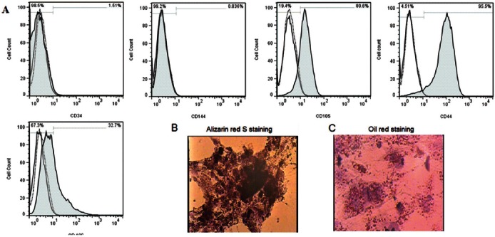
The flow cytometry showed the CD marker expression pattern of Wharton’s jelly mesenchymal stem cells (A). The cells differentiated into osteogenic (B) and adipogenic cell lineages (C).
Morphological Observation
HWJMSCs showed different morphologies in 2D and 3D culture systems. The morphology of the primary cells in the conventional culture condition (control) and in the 2D collagen films was a typical characteristic of MSCs; i.e., fibroblast-like and spindle shape. However, the morphology of the cells cultured in 3D collagen scaffolds was modified into a star and sometimes round shape (figure 2). In addition, a few cells, which were cultured in 3D collagen scaffolds showed a tendency to aggregate into a tubular shape with a lumen at the center (figure 3).
Figure 2.
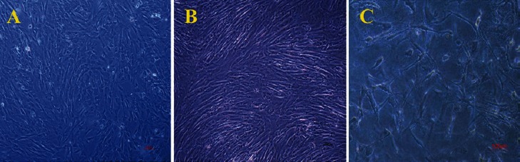
The inverted microscopy showed that the cells cultured on conventional monolayer system (A) and 2D collagen gel (B) had fibroblast-like morphology. The cells, which were cultured on 3D collagen scaffolds (C) showed a star-like morphology; however, some round cells were also observed.
Figure 3.
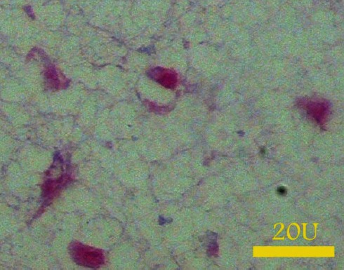
A micrograph from the section of the 3D collagen scaffolds containing Wharton’s jelly mesenchymal stem cells. Some cells came together and formed a tubular-shape aggregate H&E.
Quantitative Real-Time RT-PCR
After 21 days, the expressions of albumin, AFP, CK18, CK19, G6P, claudin, HNF4, and CYP2B6 mRNA were evaluated in the cells cultured on the 2D collagen films and in the 3D collagen scaffolds and compared to the control group. According to the results, the HWJMSCs could express different levels of some liver-specific genes. In conventional monolayer culture condition, these stem cells expressed some secretory products of the hepatocytes, such as albumin, AFP and G6P at a low level. Culturing the cells on the 2D collagen film or 3D collagen scaffold did not alter the expression level of secretory products of the hepatocytes significantly.
The cytoskeletal elements, such as CK-18 and 19 also expressed by HWJMSCs in 2D conventional culture media and culturing the cells on 2D collagen film or 3D collagen scaffold for a long period of time did not influence the expression level of these genes.
Tight junction marker, claudin, also showed a significant increase (P=0.017) in expression when the cells were grown in 3D collagen scaffolds compared with those in the cells that cultured in conventional monolayer condition or 2D collagen films.
The expression of the HNF4 was negligible in the HWJMSCs cultured in all conditions; however, the cells cultured on 2D collagen film showed a non-significant increase in HNF4 expression compared to those cultured in conventional monolayer or 3D collagen scaffold (figure 4). The expression of the CYP2B6, as a late liver-specific marker, was also very low. Culturing the cells on the collagen film or in the scaffolds led to a non-significant decrease in CYP2B6 expression compared to the conventional culture condition.
Figure 4.
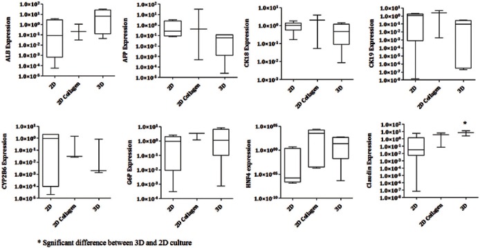
Real time RT-PCR showed the expression pattern of liver-specific markers in conventional monolayer system, 2D culture films, and 2D collagen scaffolds.
After gel running, the band of the PCR product was formed at the appropriate position (data not shown).
Discussion
Several previous studies have suggested that naïve HWJMSCs to be a good candidate for hepatocyte differentiation11 because they have been shown to express some hepatic markers, such as albumin, AFP, CK18, CK19, G6P, and claudin.11,28 The expression of some liver-specific markers has also been reported in naïve adipose-derived MSCs.29 The current investigation also showed that HWJMSCs expressed early- and mid-liver-specific genes, such as CK19, CK18, albumin, and AFP when they were cultured for long-term without any hepatogenic supplementation. These results were in agreement with the findings of the previous research.11 HNF4 can be considered as a gene with pivotal role in hepatocyte differentiation.30 Yet, there is a controversy regarding the expression of HNF4 in HWJMSCs. Some investigations have reported the capability of the naïve HWJMSCs to express HNF4,11,21 while some others have shown that HNF4 could not be expressed by these cells.22 In addition, the presence of CYP2B6 in naïve HWJMSCs was not detected by real time RT-PCR.11 The results of this study showed that a negligible amount of HNF4 and a low level of CYP2B6 expression in naïve HWJMSCs grown on non-coated culture plates.
In general, various culture systems influence the cells’ gene expression pattern.31 It is important to optimize the culture system for expression of the liver-specific markers by HWJMSCs, as an appropriate source for liver cell therapy. The culturing of ESCs within 3D alginate scaffold in the presence of the hepatogenic medium has been shown to improve the hepatocyte differentiation and function.32 Collagen coated plate also improved cell attachment, hepatocyte phenotype,33 and maturation of the hepatocytes differentiated from human ESCs.34
The real time RT-PCR detected a low level of the expression of HNF4 by HWJMSCs cultured on 2D collagen films. However, the expression of the other liver-specific marker, claudin, was increased significantly in the presence of collagen film. Significant increase in claudin in the cells grown in 3D collagen scaffold may show improvement in the cell-cell junction. It has been demonstrated that embedding the hepatocytes into a 3D collagen gel improved the bile canaliculi formation, which is accompanied with the formation of tight junction between the cells.35 Claudins were started to express in epiblast after neurulation and also expressed in endoderm24 and liver.25 Claudins are required for canaliculi and polarity formation in hepatocytes.25 Establishing apicobasal polarity in hepatocytes by tight junction formation is important in later stage of liver development. Claudin knockout mice have been shown to contain fewer polarized hepatocytes.23 Our results showed a significant increase in the claudin expression in 3D culture condition compared with 2D culture on collagen film or conventional culture condition. Recent study showed endodermal specific genes, such as GATA, enhance the claudin expression during cell differentiation.36 Several endodermal markers have been detected to express in postnatal liver progenitor cells cultured in collagen 3D condition.37 The higher level of endodermal markers in 3D collagen scaffold36 may lead to increase in claudin expression as our data indicated.
MSCs lose their stemness properties when they are grown in a conventional 2D culture and they differentiate with the progress of time.38 On the other hand, in vitro culturing of adult human hepatocytes on the collagen led to an increase in the amount of intermediate filament, such as CK18 and 1939 and albumin.40 Collagen scaffolds also accelerated the expression of liver-specific markers by MSCs exposed to hepatogenic medium.11 According to the literature, it seems that collagen scaffolds exerts a dual effect on the cells according to the differentiation degree. In adult hepatocyte and the stem cells induced by hepatogenic medium, collagen scaffolds caused an increase in the expression of downstream genes, such as CKs, and synthesis of secretory products. They also increased the pluripotency genes expression in naïve stem cells. However, the data from current study showed culturing the naïve HWJMSCs in the presence of collagen led to a significant increase in some liver-specific markers, claudin but not CKs, albumin, AFP or HNF4. The increase in claudin expression may be attributed to the improvement of the cell-cell contact in a 3D environment.
It has been demonstrated that 3D culture in collagen scaffold of postnatal liver progenitor cells led to an increase in hepatic differentiation markers, such as albumin and AFP, compared with 2D collagen film; however, the expression of the CK19 decreased.36 Our results indicated a non-significant decrease in albumin, AFP and CK19 in the HWJMSCs cultured in 3D collagen scaffolds. Although the current study showed HWJMSCs behave like oval cells in CK19 expression level, the expression of the albumin and AFP is not in accordance with the previous study36 and it seems that the HWJMSCs showed a different behavior compared with postnatal liver progenitor cells in 3D culture condition.
Conclusion
The naïve HWJMSCs expressed liver-specific markers, except for HNF4, when they were cultured on the conventional monolayer culture system. 3D collagen scaffolds led to a significant increase in tight junction marker, such as claudin. However, the expression of CK18, 19 and AFP did not change after embedding the cells into the collagen scaffolds. Nevertheless, the expression of HNF4 started to increase in the cells cultured on 2D collagen films. 2D collagen films may be recommended for the initial step of cell differentiation because the cells could express a higher level of HNF4.
Acknowledgment
The authors wish to thank the Research Vice-chancellor of Shiraz University of Medical Sciences for financially supporting the study [grant No. 6205]. They are also grateful for Ms. M. Salmannjad and M. Sani for their excellent technical support. Thanks also go to Ms. A. Keivanshekouh at the Research Improvement Center of Shiraz University of Medical Sciences for improving the use of English in the manuscript. The present study was done in fulfillment of the requirements for PhD degree awarded to Z. Khodabandeh.
Conflict of Interest: None declared.
References
- 1.Wharton T, Freer S. Thomas Wharton’s Adenographia. Oxford, UK: Oxford University Press; 1996. pp. 242–8. [Google Scholar]
- 2.Marcus AJ, Woodbury D. Fetal stem cells from extra-embryonic tissues: do not discard. J Cell Mol Med. 2008;12:730–42. doi: 10.1111/j.1582-4934.2008.00221.x. [ PMC Free Article] [DOI] [PMC free article] [PubMed] [Google Scholar]
- 3.McElreavey KD, Irvine AI, Ennis KT, McLean WH. Isolation, culture and characterisation of fibroblast-like cells derived from the Wharton’s jelly portion of human umbilical cord. Biochem Soc Trans. 1991;19:29s. doi: 10.1042/bst019029s. [DOI] [PubMed] [Google Scholar]
- 4.La Rocca G, Anzalone R, Corrao S, Magno F, Loria T, Lo Iacono M, et al. Isolation and characterization of Oct-4+/HLA-G+mesenchymal stem cells from human umbilical cord matrix: differentiation potential and detection of new markers. Histochem Cell Biol. 2009;131:267–82. doi: 10.1007/s00418-008-0519-3. [DOI] [PubMed] [Google Scholar]
- 5.Fong CY, Subramanian A, Gauthaman K, Venugopal J, Biswas A, Ramakrishna S, et al. Human umbilical cord Wharton’s jelly stem cells undergo enhanced chondrogenic differentiation when grown on nanofibrous scaffolds and in a sequential two-stage culture medium environment. Stem Cell Rev. 2012;8:195–209. doi: 10.1007/s12015-011-9289-8. [DOI] [PubMed] [Google Scholar]
- 6.Gauthaman K, Fong CY, Venugopal JR, Biswas A, Ramakrishna S, Bongso A. Propagation and differentiation of human Wharton’s jelly stem cells on three-dimensional nanofibrous scaffolds. Methods Mol Biol. 2013;1058:1–23. doi: 10.1007/7651_2012_1. [DOI] [PubMed] [Google Scholar]
- 7.Hollweck T, Hartmann I, Eblenkamp M, Wintermantel E, Reichart B, Überfuhr P, et al. Cardiac differentiation of human Wharton’s jelly stem cells—experimental comparison of protocols. Open Tissue Eng Regen Med J. 2011;4:95–102. [Google Scholar]
- 8.Wang L, Ott L, Seshareddy K, Weiss ML, Detamore MS. Musculoskeletal tissue engineering with human umbilical cord mesenchymal stromal cells. Regen Med. 2011;6:95–109. doi: 10.2217/rme.10.98. [ PMC Free Article] [DOI] [PMC free article] [PubMed] [Google Scholar]
- 9.Yan M, Sun M, Zhou Y, Wang W, He Z, Tang D, et al. Conversion of human umbilical cord mesenchymal stem cells in Wharton’s jelly to dopamine neurons mediated by the Lmx1a and neurturin in vitro: potential therapeutic application for Parkinson’s disease in a rhesus monkey model. PLoS One. 2013;8:e64000. doi: 10.1371/journal.pone.0064000. [ PMC Free Article] [DOI] [PMC free article] [PubMed] [Google Scholar] [Retracted]
- 10.Wang HW, Lin LM, He HY, You F, Li WZ, Huang TH, et al. Human umbilical cord mesenchymal stem cells derived from Wharton’s jelly differentiate into insulin-producing cells in vitro. Chin Med J (Engl) 2011;124:1534–9. [PubMed] [Google Scholar]
- 11.Campard D, Lysy PA, Najimi M, Sokal EM. Native umbilical cord matrix stem cells express hepatic markers and differentiate into hepatocyte-like cells. Gastroenterology. 2008;134:833–48. doi: 10.1053/j.gastro.2007.12.024. [DOI] [PubMed] [Google Scholar]
- 12.Zhang YN, Lie PC, Wei X. Differentiation of mesenchymal stromal cells derived from umbilical cord Wharton’s jelly into hepatocyte-like cells. Cytotherapy. 2009;11:548–58. doi: 10.1080/14653240903051533. [DOI] [PubMed] [Google Scholar]
- 13.Mikaeili Agah E, Parivar K, Joghataei MT. Therapeutic effect of transplanted human Wharton’s jelly stem cell-derived oligodendrocyte progenitor cells (hWJ-MSC-derived OPCs) in an animal model of multiple sclerosis. Mol Neurobiol. 2014;49:625–32. doi: 10.1007/s12035-013-8543-2. [DOI] [PubMed] [Google Scholar]
- 14.Scheers I, Lombard C, Paganelli M, Campard D, Najimi M, Gala JL, et al. Human umbilical cord matrix stem cells maintain multilineage differentiation abilities and do not transform during long-term culture. PLoS One. 2013;8:e71374. doi: 10.1371/journal.pone.0071374. [ PMC Free Article] [DOI] [PMC free article] [PubMed] [Google Scholar]
- 15.Bongso A, Fong CY, Gauthaman K. Taking stem cells to the clinic: Major challenges. J Cell Biochem. 2008;105:1352–60. doi: 10.1002/jcb.21957. [DOI] [PubMed] [Google Scholar]
- 16.Wang HS, Hung SC, Peng ST, Huang CC, Wei HM, Guo YJ, et al. Mesenchymal stem cells in the Wharton’s jelly of the human umbilical cord. Stem Cells. 2004;22:1330–7. doi: 10.1634/stemcells.2004-0013. [DOI] [PubMed] [Google Scholar]
- 17.Weiss ML, Medicetty S, Bledsoe AR, Rachakatla RS, Choi M, Merchav S, et al. Human umbilical cord matrix stem cells: preliminary characterization and effect of transplantation in a rodent model of Parkinson’s disease. Stem Cells. 2006;24:781–92. doi: 10.1634/stemcells.2005-0330. [DOI] [PubMed] [Google Scholar]
- 18.Fong CY, Chak LL, Biswas A, Tan JH, Gauthaman K, Chan WK, et al. Human Wharton’s jelly stem cells have unique transcriptome profiles compared to human embryonic stem cells and other mesenchymal stem cells. Stem Cell Rev. 2011;7:1–16. doi: 10.1007/s12015-010-9166-x. [DOI] [PubMed] [Google Scholar]
- 19.Prasajak P, Leeanansaksiri W. Developing a New Two-Step Protocol to Generate Functional Hepatocytes from Wharton’s Jelly-Derived Mesenchymal Stem Cells under Hypoxic Condition. Stem Cells Int 2013. 2013:762196. doi: 10.1155/2013/762196. [ PMC Free Article] [DOI] [PMC free article] [PubMed] [Google Scholar]
- 20.Baharvand H, Hashemi SM, Kazemi Ashtiani S, Farrokhi A. Differentiation of human embryonic stem cells into hepatocytes in 2D and 3D culture systems in vitro. Int J Dev Biol. 2006;50:645–52. doi: 10.1387/ijdb.052072hb. [DOI] [PubMed] [Google Scholar]
- 21.Anzalone R, Farina F, Lo Iacono M, Corrao S, Corsello T, Zummo G, et al. Wharton’s Jelly Mesenchymal Stem Cells and Immune Modulation: Regenerative Medicine Meets Tissue Repair. In: Cetrulo K, Cetrulo CL, Taghizadeh R, editors. Perinatal Stem Cells. 2nd ed. New York: Wiley-Blackwell; 2013. pp. 77–88. [Google Scholar]
- 22.Buyl K, De Kock J, Najar M, Lagneaux L, Branson S, Rogiers V, et al. Characterization of hepatic markers in human Wharton’s Jelly-derived mesenchymal stem cells. Toxicol In Vitro. 2014;28:113–9. doi: 10.1016/j.tiv.2013.06.014. [DOI] [PubMed] [Google Scholar]
- 23.Collins MM, Baumholtz AI, Ryan AK. Claudin family members exhibit unique temporal and spatial expression boundaries in the chick embryo. Tissue Barriers. 2013;1:e24517. doi: 10.4161/tisb.24517. [ PMC Free Article] [DOI] [PMC free article] [PubMed] [Google Scholar]
- 24.Cheung ID, Bagnat M, Ma TP, Datta A, Evason K, Moore JC, et al. Regulation of intrahepatic biliary duct morphogenesis by Claudin 15-like b. Dev Biol. 2012;361:68–78. doi: 10.1016/j.ydbio.2011.10.004. [ PMC Free Article] [DOI] [PMC free article] [PubMed] [Google Scholar]
- 25.Son S, Kojima T, Decaens C, Yamaguchi H, Ito T, Imamura M, et al. Knockdown of tight junction protein claudin-2 prevents bile canalicular formation in WIF-B9 cells. Histochem Cell Biol. 2009;131:411–24. doi: 10.1007/s00418-008-0546-0. [DOI] [PubMed] [Google Scholar]
- 26.Kim DW, Staples M, Shinozuka K, Pantcheva P, Kang SD, Borlongan CV. Wharton’s Jelly-Derived Mesenchymal Stem Cells: Phenotypic Characterization and Optimizing Their Therapeutic Potential for Clinical Applications. Int J Mol Sci. 2013;14:11692–712. doi: 10.3390/ijms140611692. [ PMC Free Article] [DOI] [PMC free article] [PubMed] [Google Scholar]
- 27.Ye J, Coulouris G, Zaretskaya I, Cutcutache I, Rozen S, Madden TL. Primer-BLAST: a tool to design target-specific primers for polymerase chain reaction. BMC Bioinformatics. 2012;13:134. doi: 10.1186/1471-2105-13-134. [ PMC Free Article] [DOI] [PMC free article] [PubMed] [Google Scholar]
- 28.Anzalone R, Lo Iacono M, Corrao S, Magno F, Loria T, Cappello F, et al. New emerging potentials for human Wharton’s jelly mesenchymal stem cells: immunological features and hepatocyte-like differentiative capacity. Stem Cells Dev. 2010;19:423–38. doi: 10.1089/scd.2009.0299. [DOI] [PubMed] [Google Scholar]
- 29.Zemel R, Bachmetov L, Ad-El D, Abraham A, Tur-Kaspa R. Expression of liver-specific markers in naive adipose-derived mesenchymal stem cells. Liver Int. 2009;29:1326–37. doi: 10.1111/j.1478-3231.2009.02054.x. [DOI] [PubMed] [Google Scholar]
- 30.Kojima M, Takamatsu N, Ishii T, Kondo N, Shiba T. HNF-4 plays a pivotal role in the liver-specific transcription of the chipmunk HP-25 gene. Eur J Biochem. 2000;267:4635–41. doi: 10.1046/j.1432-1327.2000.01499.x. [DOI] [PubMed] [Google Scholar]
- 31.Apte MV, Yang L, Phillips PA, Xu Z, Kaplan W, Cowley M, et al. Extracellular matrix composition significantly influences pancreatic stellate cell gene expression pattern: role of transgelin in PSC function. Am J Physiol Gastrointest Liver Physiol. 2013;305:G408–17. doi: 10.1152/ajpgi.00016.2013. [DOI] [PubMed] [Google Scholar]
- 32.Ramasamy TS, Yu JS, Selden C, Hodgson H, Cui W. Application of three-dimensional culture conditions to human embryonic stem cell-derived definitive endoderm cells enhances hepatocyte differentiation and functionality. Tissue Eng Part A. 2013;19:360–7. doi: 10.1089/ten.tea.2012.0190. [DOI] [PubMed] [Google Scholar]
- 33.Toh YC, Lim TC, Tai D, Xiao G, van Noort D, Yu H. A microfluidic 3D hepatocyte chip for drug toxicity testing. Lab Chip. 2009;9:2026–35. doi: 10.1039/b900912d. [DOI] [PubMed] [Google Scholar]
- 34.Nagamoto Y, Tashiro K, Takayama K, Ohashi K, Kawabata K, Sakurai F, et al. The promotion of hepatic maturation of human pluripotent stem cells in 3D co-culture using type I collagen and Swiss 3T3 cell sheets. Biomaterials. 2012;33:4526–34. doi: 10.1016/j.biomaterials.2012.03.011. [DOI] [PubMed] [Google Scholar]
- 35.Matsui H, Takeuchi S, Osada T, Fujii T, Sakai Y. Enhanced bile canaliculi formation enabling direct recovery of biliary metabolites of hepatocytes in 3D collagen gel microcavities. Lab Chip. 2012;12:1857–64. doi: 10.1039/c2lc40046d. [DOI] [PubMed] [Google Scholar]
- 36.Escaffit F, Boudreau F, Beaulieu JF. Differential expression of claudin-2 along the human intestine: Implication of GATA-4 in the maintenance of claudin-2 in differentiating cells. J Cell Physiol. 2005;203:15–26. doi: 10.1002/jcp.20189. [DOI] [PubMed] [Google Scholar]
- 37.Petrakova OS, Ashapkin VV, Voroteliak EA, Bragin EY, Shtratnikova VY, Chernioglo ES, et al. Effect of 3D Cultivation Conditions on the Differentiation of Endodermal Cells. Acta Naturae. 2012;4:47–57. [ PMC Free Article] [PMC free article] [PubMed] [Google Scholar]
- 38.Li Z, Tian X, Yuan Y, Song Z, Zhang L, Wang X, et al. Effect of cell culture using chitosan membranes on stemness marker genes in mesenchymal stem cells. Mol Med Rep. 2013;7:1945–9. doi: 10.3892/mmr.2013.1423. [DOI] [PubMed] [Google Scholar]
- 39.Chougule P, Sumitran-Holgersson S. Cytokeratins of the Liver and Intestine Epithelial Cells During Development and Disease. In: Hamilton G, editor. Cytokeratins - Tools in Oncology. Croatia: InTech; 2012. pp. 15–32. [Google Scholar]
- 40.Wang S, Nagrath D, Chen PC, Berthiaume F, Yarmush ML. Three-dimensional primary hepatocyte culture in synthetic self-assembling peptide hydrogel. Tissue Eng Part A. 2008;14:227–36. doi: 10.1089/tea.2007.0143. [DOI] [PubMed] [Google Scholar]