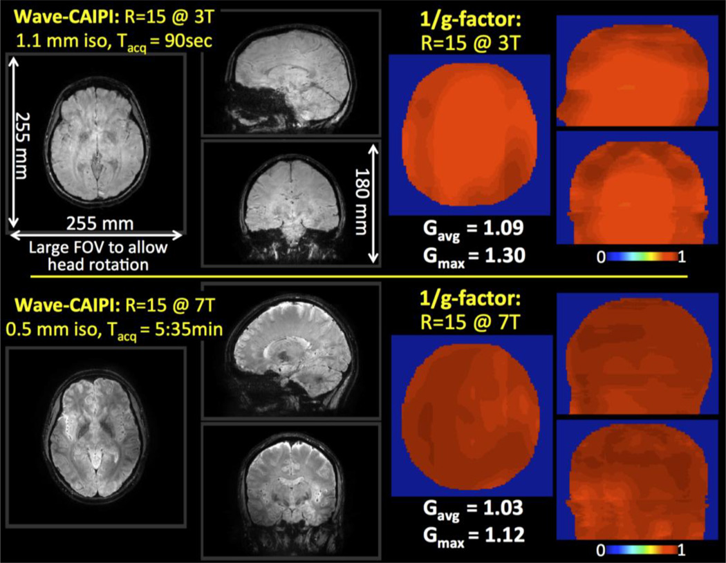
Fig. 1.
R=15 fold accelerated 3D-GRE with Wave-CAIPI at 3T and 7T. The large FOV (255×255×180 mm3) allows imaging of the entire brain across head orientations without moving the prescribed acquisition volume. G-factor analysis reveals high-quality parallel imaging with reduced noise amplification penalty at both field strengths.

