Abstract
Tuberculosis (TB) in adults can present in a large number of ways. The lung is the predominant site of TB. Primary pulmonary TB should be distinguished from postprimary pulmonary TB, which is the most frequent TB manifestation in adults (70%–80% cases). Cough is common, although the chest radiograph often raises suspicion of disease. Sputum sampling is a key step in the diagnosis of TB, and invasive procedures such as bronchoscopy may be necessary to achieve adequate samples for diagnosis. Extrapulmonary involvement, which may present many years after exposure, occurs in a variable proportion of cases (20%–45%). This reflects the country of origin of patients and also the frequency of associated human immunodeficiency virus (HIV) coinfection. In the latter case, the presentation of TB is often nonspecific, and care needs to be taken to not miss the diagnosis. Anti-TB therapy should be given in line with proven (or assumed) drug resistance. In extrapulmonary TB, adjunctive therapeutic measures may be indicated; although in all cases, support is often required to ensure that people are able to complete treatment with minimal adverse events and maximal adherence to the prescribed regimen, and so reduce risk of future disease for themselves and others.
Postprimary tuberculosis (TB) of the lungs is the prevailing TB manifestation in adults. It may spread from the lungs via the lymphatics or bloodstream to other body sites, causing various extrapulmonary TB manifestations.
In parts of the world where tuberculosis (TB) is endemic, symptomatic individuals and their health-care providers are likely to recognize and consider TB early within the diagnostic algorithm (Aït-Khaled et al. 2010). However, in countries with a declining or low prevalence, the time to diagnosis and starting treatment can be prolonged. This may be attributable to both patient and health system factors (Millen et al. 2008, Migliori et al. 2012) and can be accentuated by the stigma associated with a potential diagnosis of TB (as well as HIV coinfection) (Courtwright and Turner 2010). Hence, education for individuals, communities, and social care providers about TB symptoms, and where and how to get them investigated, is an important part of clinical management (Chowdhury et al. 2013; TB CARE 2014).
Considering TB as the cause of a person’s symptoms (or new signs in a child with a significant exposure), making a rapid and accurate diagnosis and initiating effective treatment promptly are central to good patient care and public health TB management. Not only does this alleviate symptoms and reduce mortality for the individual, but it also decreases onward transmission to others and hence impacts on the overall burden of TB.
TB in adults can present in a variety of ways. The lung is the predominant site of TB as infection with Mycobacterium tuberculosis (Mtb) or other members of the TB complex arises almost exclusively from inhaling droplets containing the bacilli. This may result in symptomatic, primary pulmonary TB disease (usually in children) and in adults, after a variable amount of time in a clinically asymptomatic state of latent TB infection (LTBI), generally as postprimary pulmonary TB. Mtb may spread directly from the lungs, via the lymphatics, or the bloodstream, to other body sites causing the various extrapulmonary TB manifestations described in this review.
The location of disease should be documented in all patients. Given that there may be multiple sites involved, it is recommended that at least two, a major and a minor site, when applicable, be recorded. Here, the definitions used are those proposed by a Working Group of the World Health Organization (WHO) and the European Region of the International Union Against Tuberculosis and Lung Disease (IUATLD) for uniform reporting of TB cases (Rieder et al. 1996).
It is important to note that TB can be diagnosed via a number of different clinical pathways and settings. In a low incidence country such as Germany, among more than 25,000 TB cases studied between 1996 and 2000, almost 80% were diagnosed through passive case finding (∼62% had symptoms suggesting TB, 16% were diagnosed during investigations for other medical causes, 1% at autopsy) and 19% by active case finding in high-risk groups, particularly in close contacts of infectious patients (Forssbohm 2004). This further reinforces the importance of thinking of TB as a possible cause of a patient’s symptoms or signs in clinical practice (Craig et al. 2009).
CLINICAL PRESENTATION AND DIAGNOSIS OF TB
Pulmonary TB
Pulmonary TB is defined as tuberculosis of the lung parenchyma and the tracheobronchial tree only. Primary pulmonary TB should be distinguished from postprimary pulmonary TB, which is the most frequent TB manifestation in adults. The classic clinical features of pulmonary TB include chronic cough, sputum production, appetite loss, weight loss, fever, night sweats, and hemoptysis (Lawn and Zumla 2011). Someone presenting with any of these symptoms should be suspected of having TB. If they are or were known to be in contact with infectious TB, they are even more likely to be suffering from TB (Ait-Khaled et al. 2010.)
Primary Pulmonary TB
In countries with a high TB prevalence, primary pulmonary disease occurs usually in childhood, but where TB is less endemic, it occurs fairly often also in adults. It is characterized by local granulomatous inflammation, usually in the periphery of the lung (Ghon focus), and may be accompanied by ipsilateral lymph node involvement, termed the Ghon complex. The infection is usually asymptomatic but can present as an acute lower respiratory tract infection. The most important clue to the diagnosis is a history of close contact with an infectious TB case. The diagnosis is suspected when a tuberculin skin test or a blood interferon-γ release assay (IGRA) converts to positive, usually 3–8 wk after infection. The chest radiograph may show the Ghon focus/complex (Fig. 1).
Figure 1.
Ghon complex. (Figure reprinted from Fuehner et al. 2007, with permission, from Springer, © 2007.)
Rare primary sites of TB are the alimentary tract caused by swallowing Mtb, usually Mycobacterium bovis, present in nonpasteurized milk products (de la Rua-Domenech 2006), or after direct cutaneous infection (usually occurring in laboratory personnel) (Menzies et al. 2003). Intravesical Bacillus Calmette–Guérin (BCG) vaccination used to treat localized bladder tumors may occasionally disseminate and present as primary BCG disease (Fig. 2) (Lamm 1992).
Figure 2.
Disseminated BCG disease. Computed tomography (CT) of the chest showing bilateral patchy ground glass shadowing and consolidation in a patient who developed fevers, cough, and positive blood cultures for BCG following treatment with intravesical BCG for bladder carcinoma. (Figure provided by Marc Lipman.)
Local complications of primary pulmonary TB may result from lymph node enlargement leading to bronchial obstruction. Tuberculous pleurisy can arise early in the course of primary pulmonary TB, either by direct spread from the pulmonary lesion or through hematogenous dissemination.
Rare, serious, early systemic complications caused by blood-borne spread of Mtb are miliary TB and meningitis. Many years later, TB of the skeletal or urogenital system and other organs may result (Wallgren 1948).
Postprimary pulmonary TB may follow primary TB. In the generally immunocompetent, there is a lifetime chance of reactivation of the dormant primary complex of 5%–10% (Horsburgh Jr 2004). These estimates were developed before the availability of molecular techniques that can distinguish reactivation from reinfection with another strain of Mtb, and it may be that the overall risk is rather less in most people with latent TB infection not exposed again to Mtb. The first 2 yr following primary infection are the period of maximal risk of progression. This can be reduced significantly by treating LTBI, which is indicated particularly in high-risk groups (Table 1) (Diel et al. 2013). It is not known why only ∼10% of individuals infected with Mtb develop active disease. Apart from diverse risk factors such as diabetes, smoking, and chronic renal failure (Hu et al. 2014), several genes have been found to be associated with increased susceptibility to, or resistance against, Mtb (Möller et al. 2010).
Table 1.
Persons at high risk of progressing from latent to active TB
| Recent contact with a TB case |
| Persons with fibrotic changes on chest radiograph consistent with old TB |
| HIV-infected persons |
| Organ transplant recipients |
| Persons immunosuppressed for other reasons (e.g., taking the equivalent of 15 mg/d of prednisone for >1 mo or taking tumor necrosis factor-α inhibitors) |
| Recent immigrants (<5 yr) from high prevalence countries |
| Persons with the following clinical conditions: |
| Diabetes mellitus |
| Chronic renal failure |
| Some hematologic disorders (e.g., leukemia and lymphomas) |
| Other specific malignancies (e.g., carcinoma of the head or neck and lung) |
| Gastrectomy and jejunoileal bypass |
| Silicosis |
| Injection drug users |
| Tobacco smokers |
| Residents and employees of high-risk congregate living facilities (e.g., correctional facilities, nursing homes, homeless shelters, hospitals, and other health-care facilities) |
| Mycobacteriology laboratory personnel |
Data modified from Diel et al. 2013.
Erythema nodosum and other skin conditions such as granulomatous panniculitis plus forms of uveitis and polyarthritis (Poncet's disease) are considered to reflect the host immune response to mycobacterial antigen. They can be seen both during primary infection and at some time distant from presumed infection (Kroot et al. 2007). The features are often nonspecific, and the key clinical issue here is to consider TB as a possible underlying cause of the presentation. This is generally based on determining whether there is a history of exposure or residence in a TB-endemic area. The increasing numbers of individuals who are immunocompromised from infections such as HIV or through medical treatments (and are at much greater risk of developing active TB) make this method of categorization less helpful than previously.
Postprimary Pulmonary TB
Postprimary TB of the lungs is the prevailing TB manifestation in adults (in 60%–80%) (Public Health England 2014). It can occur many years after exposure to an individual with infectious TB and may be provoked by temporary or permanent immunological impairment. Males are more often affected than females (Borgdorff et al. 2000).
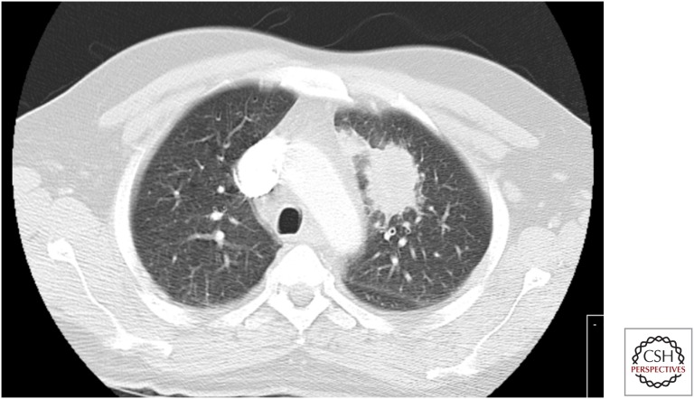
The most frequent symptoms of active disease are fever, anorexia or reduced appetite, weight loss, night sweats, anemia, and persistent cough (i.e., lasting >14 d) (TB CARE 2014) usually productive of purulent and/or blood-stained sputum. Occasionally, patients complain of localized thoracic pain attributable to accompanying pleural inflammation. In extensive and long-lasting pulmonary disease, patients may report breathlessness. Hemoptysis is usually the result of cavitating lung disease causing erosion of pulmonary blood vessels (Fig. 3).
Figure 3.
Cavitating tuberculosis presenting with hemoptysis (note arrows indicating cavities). (Figure reprinted from Fuehner et al. 2007, with permission, from Springer, © 2007.)
Hoarseness can occur if there is laryngeal involvement. Patients with laryngeal and tracheobronchial (Fig. 4) TB may have Mtb in sputum despite a normal chest X-ray, although in general, the radiograph will suggest the possible diagnosis (Bhat et al. 2009).
Figure 4.
TB in the mucosa of the left main bronchus. Histology and culture from biopsy were positive for Mtb. (Figure provided by Robert Loddenkemper.)
Tuberculoma of the lung should be considered as part of the differential diagnosis in mass lesions presenting in individuals with a history of exposure to Mtb (Fig. 5).
Figure 5.
Lung tuberculoma. Initially thought to be a primary lung cancer, bronchoscopic biopsy showed acid-fast bacilli on smear. Culture grew Mtb. (Figure provided by Marc Lipman.)
The physical signs of TB are not specific. Auscultation often does not show any pathological breath sounds or may reveal crackles, wheezing, or bronchial breathing (which is termed amphoric when related to the movement of air within a cavity). The radiographic features vary from very discrete infiltrates to extensive and bilateral changes with multiple cavities characteristic of TB. Usually, the changes are more pronounced in the upper lobes. The differential diagnosis of cavitation includes other infections as well as malignant and inflammatory lung diseases (Table 2).
Table 2.
Differential diagnosis of pulmonary cavitation
| Cause | Examples |
|---|---|
| Infection | Tuberculosis |
| Acute bacterial, e.g., Staphylococcus aureus, Klebsiella pneumoniae | |
| Subacute/chronic bacterial (e.g., Nocardia sp, melidoidosis, actinomycosis) | |
| Fungal (e.g., cryptococcosis, coccidiodomycosis) | |
| Infected pneumatocele | |
| Malignancy | Primary lung cancer (e.g., non–small cell squamous) |
| Pulmonary metastases | |
| Inflammatory | Sarcoidosis |
| Wegener's granulomatous angiitis | |
| Rheumatoid arthritis | |
| Vascular | Pulmonary infarct |
| Congenital | Sequestered lung; bronchogenic cyst |
There are no specific blood tests that aid the diagnosis of TB. Patients will have a profile that reflects their general state of health and nutrition. They may or may not be anemic and have moderately elevated inflammatory markers such as C-reactive protein. However, all of these measures are highly variable (Breen et al. 2008a). The following blood tests are recommended generally as baseline assessments rather than for any specific value in TB diagnosis. These include kidney and liver function, blood sugar, hemoglobin, white cell and differential counts, platelet count, and C-reactive protein (or ESR). It is recommended that routine HIV and hepatitis B and C testing be performed and vitamin D blood levels measured, as patients are invariably deficient (Blasi and Matteelli 2013).
The presentation of HIV-associated pulmonary TB can mimic that seen in the HIV uninfected. This is generally found in subjects with relatively well-preserved immunity (manifest as blood CD4 counts >350 cells/μL) (Cain et al. 2010). However, the associated immune dysregulation in most individuals with active TB and HIV coinfection means that although symptoms may be similar, patients can also present with very nonspecific although severe systemic features (e.g., weight loss, night sweats, and fever) and minimal specific respiratory complaints (Breen et al. 2008a).
The radiology of HIV TB is discussed in more detail elsewhere. However, an important point to note is that parenchymal cavitation is less likely in individuals with low blood CD4 counts. Extrapulmonary manifestations (including mediastinal nodal disease and pleural effusions) are also more common in HIV coinfection (Lawn and Zumla 2011).
The lack of cavities means that sputum samples in HIV-associated pulmonary TB are often smear-negative. This is despite individuals having in fact a very high mycobacterial burden (Elliott et al. 1993; Crump et al. 2012; Huang et al. 2014). Indeed, sputum cultures are generally positive as frequently in HIV-infected as -uninfected subjects. The importance of obtaining samples from other body fluids and locations cannot be overemphasized. This should include mycobacterial blood and urine cultures. Given that HIV-associated TB is often multisite, general radiological evaluation (including computed tomography [CT] chest, abdomen, and pelvis or equivalent investigations, e.g., abdominal ultrasound) may identify other areas of pathology that are amenable to diagnostic aspiration or biopsy. The high mycobacterial burden associated with HIV means that often urine antigen tests such as the cell wall component lipoarabinnoman (available in commercially produced kits) may be positive in patients with very low blood CD4 counts (<100 cells/μL). These offer, therefore, an alternative approach to TB diagnosis in advanced HIV infection (Lawn et al. 2013).
In many clinical settings, HIV is diagnosed at the same time as TB (Rice et al. 2013). This mainly follows the pattern of presentation with clinical TB, which then results in an HIV test. However, the use of antiretroviral therapy in an individual at risk of TB can lead to an unmasking paradoxical reaction (which is part of the immune reconstitution inflammatory syndrome, IRIS) (Breen et al. 2005). These are typically highly inflammatory presentations, in which individuals develop intense systemic symptoms and new onset signs and radiological changes within a matter of days.
Although the underlying mechanism has not been fully elucidated, it appears to arise as a consequence of a host immune response directed against a (generally foreign) persistent antigen. Mycobacteria fulfill this role well, although so do other chronic intracellular organisms such as fungi. Hence, a presentation of apparent unmasking TB must be investigated comprehensively. Samples should be sent for cytology, histology, and bacterial, fungal, and mycobacterial cultures to avoid diagnostic confusion and inappropriate treatment.
Sputum sampling is a key step in the diagnosis of TB. Attempts should be made to obtain at least two sputum samples from individuals with suspected TB (including extrapulmonary TB, which is often associated with positive sputum cultures despite few reported symptoms) (Parimon et al. 2008). Good-quality sputum samples should be obtained. It is standard to ask for three samples, although evidence suggests that two may strike a better balance in terms of value (Mase et al. 2007). These are generally requested to be provided first thing in the morning (i.e., at a time when individuals are most productive). However, in an ambulatory setting, this can result in delay, and often there is utility in obtaining samples in one day (e.g., in the morning and afternoon).
Should an individual be unable to produce sputum spontaneously, many centers will perform induced sputum using hypertonic saline (generally between 3% and 7% concentration). This technique is quick (in total taking <1 h), relatively inexpensive, and not as invasive as a bronchoscopy. It also has a good yield (Brown et al. 2007). However, this is operator-dependent, and the procedure requires experienced technicians to ensure that optimal samples are obtained.
Sputum induction creates a considerable aerosol of potentially infectious material (Dooley et al. 1990). It should therefore be performed in a setting where infection control is of a high standard, or there is minimal risk of transmission to others, e.g., induction performed in an open air setting, away from others.
Should sputum induction be unavailable or unsuccessful, flexible bronchoscopy with bronchoalveolar lavage is indicated. This procedure has the advantage of being able to sample specific sites (and hence in theory improve the recovery from, e.g., localized disease or a cavity) (Schoch et al. 2007). A good-quality sample, which is promptly set up for mycobacterial culture, has a high diagnostic yield (Breen et al. 2008b). This is important because the procedure is associated with significant up-front capital cost, and potential risk to both patients and health-care workers.
Bronchoscopic procedures also enable aspiration and biopsies to be performed. This includes both lung tissue (bronchial biopsies) and needle aspiration under either direct vision or using ultrasound (EBUS) (Navani et al. 2011).
Pulmonary samples can be assessed using acid-fast bacilli (AFB) smear, nucleic acid amplification techniques such as GeneXpert, mycobacterial culture, or cytological analysis. AFB sputum smear is the most long standing, although it is fairly insensitive (requiring at least 5 × 10³ bacilli per mL for detection). Molecular diagnostics are equally rapid, more sensitive, and also currently more expensive. They can also detect genetic resistance to antituberculosis drugs (Boehme et al. 2011), and with modification they might distinguish between live and dead bacilli (Honeyborne et al. 2011).
Culture techniques are still regarded as the gold standard. Liquid fluorometric systems are faster than solid media and more sensitive (Dhillon et al. 2014). However, they are also expensive and require skilled personnel to use them. At present, they are not routinely available worldwide for all patient samples that are obtained for suspected TB.
Extrapulmonary Tuberculosis
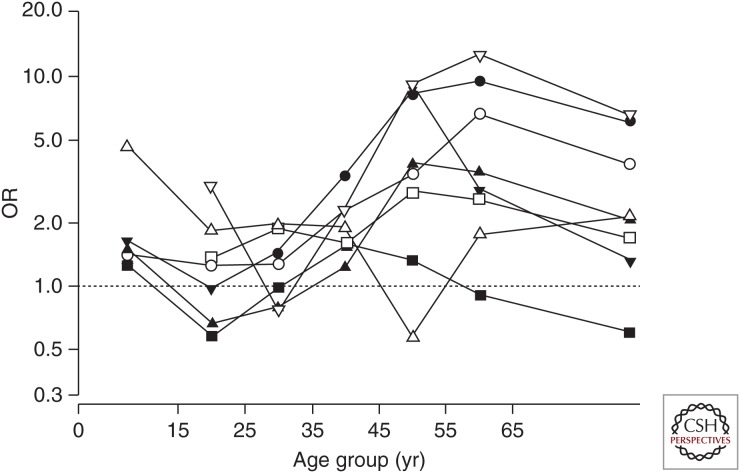
Extrapulmonary tuberculosis (EPTB) occurs in 10%–42% of adult TB patients depending on race or ethnic background, age, presence or absence of underlying disease, genotype of the Mtb strain, and immune status (Caws et al. 2008) (Table 3). EPTB is defined as TB affecting any site other than the lung. Sometimes it is accompanied by pulmonary disease (Public Health England 2014). It can affect any organ in the body and has numerous clinical manifestations, and thus requires a high index of clinical suspicion (Zumla et al. 2013). EPTB can occur many years after exposure. In addition to systemic features, patients with EPTB may complain of symptoms related to the involved site/organ. In contrast to pulmonary TB, females are more often affected (one exception to this being pleurisy) (Forssbohm et al. 2008) (Fig. 6). The differences in the proportion of EPTB by area of origin are remarkable. Patients from Africa and Asia are generally far more likely to present with EPTB than those from Germany (Forssbohm et al. 2008); genetic factors may explain some of these differences (Fernando et al. 2007). HIV and other conditions that markedly impair host immunity have led to an increase in extrapulmonary disease. This reflects an inability to contain Mtb, and hence dissemination, with positive cultures from a number of body sites, often occurs. The diagnosis can require biopsy and/or surgical removal.
Table 3.
Frequency of bacteriological culture confirmation of Mtb complex among reported tuberculosis cases by site of disease, Germany, 1996–2000
| Culture confirmation | |||
|---|---|---|---|
| Present | Absent | Total | |
| Total | 18,501 (70%) | 7801 | 26,302 |
| Pulmonary | 15,134 (73%) | 5493 | 20,627 |
| Extrapulmonary | 3367 (59%) | 2308 | 5675 |
| Lymphatic, peripheral | 859 (51%) | 826 | 1685 |
| Lymphatic, intrathoracic | 223 (39%) | 352 | 575 |
| Pleural | 579 (55%) | 470 | 1049 |
| Genitourinary | 746 (78%) | 212 | 958 |
| Osteoarticular | 326 (71%) | 131 | 457 |
| Miliary | 126 (69%) | 58 | 184 |
| Meningeal | 91 (68%) | 42 | 133 |
| Peritoneal | 65 (56%) | 51 | 116 |
| Cutaneous | 70 (61%) | 45 | 115 |
| Other | 282 (70%) | 121 | 403 |
1Data modified from Forssbohm et al. 2008.
2Data are presented as n or n (%).
Figure 6.
Age-specific female-to-male odds ratio (OR) of having extrapulmonary TB compared with pulmonary TB by site of extrapulmonary TB, Germany 1996–2000. Solid circle, lymphatic, peripheral; open circle, lymphatic intrathoracic; solid square, pleural; open square, genitourinary; solid triangle, osteoarticular; inverted solid triangle, meningeal; inverted open triangle, peritoneal. (Figure reprinted from Forssbohm et al. 2008, with permission, from the European Respiratory Society, © 2008.)
Lymph Node TB
Intrathoracic Lymph Node TB
Intrathoracic lymph node TB is relatively more frequent in children than in adults, usually in association with primary pulmonary TB. It may be noted on the chest radiograph as mediastinal enlargement, sometimes associated with calcification (Fig. 7). The use of CT scanning has shown the relative insensitivity of chest radiographs.
Figure 7.
Silicotuberculosis. (Figure reprinted from Fuehner et al. 2007, with permission, from Springer, © 2007.)
Often, enlarged lymph nodes are present only on one side of the mediastinum, although they may be bilateral. The lymph nodes may become so large that they cause bronchial obstruction and collapse of an adjacent region of the lung. If they perforate into the bronchial tract, this may result in sublobar or lobar pneumonia (Fig. 8). The differential diagnosis includes other infectious etiologies such as nontuberculosis mycobacteria (NTM) and fungal infection, malignancy (lymphoma, lung cancer), sarcoidosis, and silicosis.
Figure 8.
Pneumonia of left upper lobe (lingula) caused by lymph node perforation. (Figure provided by Robert Loddenkemper.)
The diagnosis can be established by bronchoscopy (Chung and Lee 2000) (Fig. 9), which may be used also as an adjunctive therapeutic measure by removing obstructing intrabronchial lesions or by placement of an intrabronchial stent (Lee et al. 2010). If hilar or mediastinal intrathoracic lymph node involvement is the sole TB manifestation, transbronchial needle aspiration (“blind” TBNA or under ultrasound guidance as EBUS) or mediastinoscopy may establish the etiology. Rapid molecular diagnostic methods have been successfully applied to EBUS-derived lymph node samples (Dhasmana et al. 2014). This provides the opportunity to confirm clinically suspected disease promptly and also to obtain samples for drug sensitivity testing.
Figure 9.
CT of the chest and bronchoscopy showing lymph node perforation, before and after treatment for TB. (Figure provided by Robert Loddenkemper.)
Extrathoracic Lymph Node TB
Extrathoracic lymph node TB is one of the most frequent EPTB manifestations in adults. In Germany, for example, it accounts for 30% of EPTB (Forssbohm et al. 2008). In most cases, the cervical/supraclavicular (Fig. 10), and more rarely the axillary or inguinal, lymph nodes are involved.
Figure 10.
Extrathoracic lymph node disease. (Left figure, provided by the authors; right figure, reprinted from Siemon 2007, with permission, from Springer, © 2007.)
At first, the lymph nodes are firm, painless, and not warm (Fig. 10), but they may later become fluctuant, matted together, and inflamed. If not treated, they may perforate through the skin and create nonhealing fistulas.
The differential diagnosis includes other infectious etiologies such as NTM and fungal infection, malignancy (lymphoma, [lung] cancer), and sarcoidosis. The diagnosis is established by needle aspiration, occasionally by surgical biopsy.
Tuberculous Pleurisy
Tuberculous pleurisy is one of the most frequent EPTB manifestations. In Germany, it accounts for 19% of EPTB cases (Forssbohm et al. 2008). It is more frequent (15%–90%) in HIV coinfection, being seen in patients with higher blood CD4 counts (Gopi et al. 2007). As with pulmonary TB, and contrary to other forms of EPTB, males are more often affected than females. Usually, it occurs in the young; however, in countries with a low TB incidence, pleural disease may develop at older ages as well.
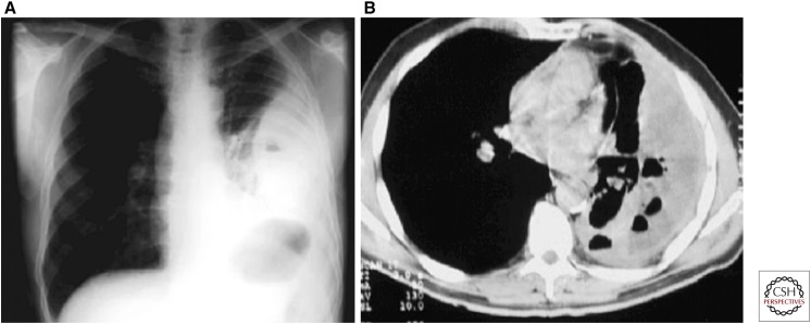
It can present acutely with high fever or as a subacute/chronic illness. Clinical features may start with pleuritic chest pain (dry pleurisy), but the predominant symptom, caused by pleural effusion, is breathlessness. Physical signs of effusion are dullness on percussion and reduced breath sounds at the affected site. The size of the effusion can vary between very small to almost complete involvement of the hemithorax; it is rarely bilateral (Fig. 11). Pulmonary TB changes are present in the minority, but studies have shown that in up to 20%, Mtb can be found in sputum (Parimon et al. 2008).
Figure 11.
Unilateral large left pleural effusion attributable to tuberculosis. (Figure reprinted from Fuehner et al. 2007 with permission, from Springer, © 2007.)
The differential diagnosis includes all causes of pleural effusion. The diagnosis is established by investigation of the pleural fluid and/or by pleural biopsies (closed-needle biopsies or thoracoscopic biopsies [Light 2010]). The diagnosis of pleural TB is hampered by its paucibacillary nature (Chegou et al. 2008). Less than 10% of pleural fluids are smear-positive, and culture is positive in only ∼30% (20%–50%) of cases. However, in HIV-positive patients, the fluid is much more likely to be smear- and culture-positive for Mtb if the CD4 count is <100 cells/μL (Heydermann et al. 1998). The exudative fluid is, in the majority of cases, predominantly lymphocytic, although in ∼10%, mainly polymorphonuclear leukocytes are found (Bielsa et al. 2013).
Several laboratory and microbiological tests have been developed during recent years, including nucleic acid amplification and detection, markers of specific and nonspecific immune response such as adenosine deaminase (ADA), and IGRA. These may help in diagnosing TB as the cause of pleural effusion and have a reasonable high sensitivity and specificity (Trajman et al. 2008; Light 2010). However, at least in countries with a low TB prevalence, the accuracy may not be sufficient in individual cases (Hooper et al. 2009, 2010). The diagnostic yield of closed-needle pleural biopsies, especially under ultrasound guidance, is up to 80% (Diacon et al. 2003; Koegelenberg et al. 2010).
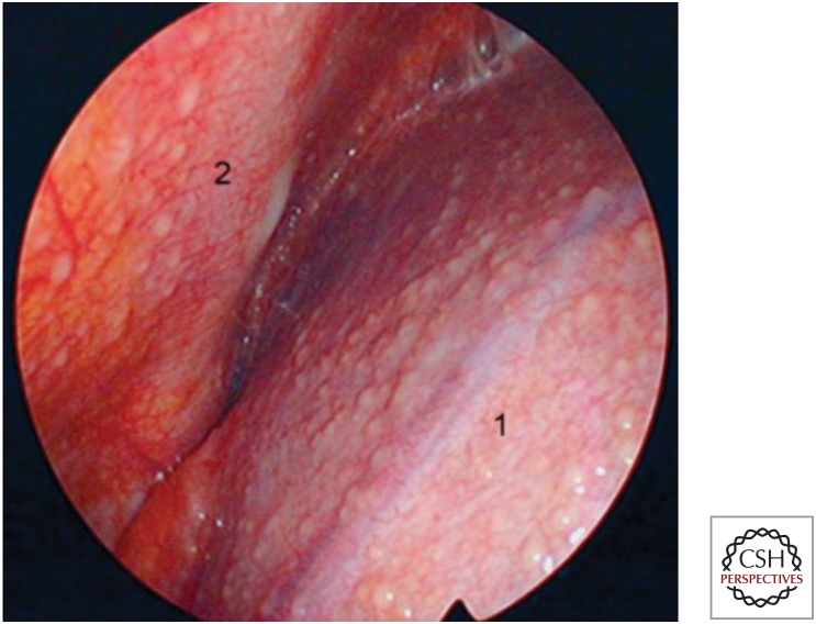
The accuracy of medical thoracoscopy (pleuroscopy) is almost 100%, as the pathologist is provided with multiple biopsies selected under direct vision (Figs. 12 and 13). Culture is therefore more often positive (78% in one study) (Fig. 14), which in turn enables drug susceptibility testing to be performed (Loddenkemper 1998; Loddenkemper et al. 2011).
Figure 12.
Thoracoscopic image of early TB pleural effusion. Following drainage of 800 mL of serous effusion, typical miliary sago-like nodules in all parts of the right parietal pleura can be seen (1) on the diaphragm and (2) the anterior chest wall. (Figure reprinted from Loddenkemper et al. 2011, with permission, from Thieme, © 2011.)
Figure 13.
Thoracoscopic image of established TB pleural effusion. Following drainage of 2000 mL of serous effusion, inflamed and hyperemic visceral and parietal pleura covered with fibrinous patches is evident. The posterior costovertebral region is shown with the diaphragm (1) and the chest wall (2). The lung cannot be visualized as it is fibrin-covered. The fibrinous adhesions pull the diaphragm cranially, producing tenting, and show vascularization at their base (arrows). (Figure reprinted from Loddenkemper et al. 2011, with permission, from Thieme, © 2011.)
Figure 14.
(A) Combined histological and bacteriological yield (%) of standard techniques in diagnosis of TB-related pleural effusion. (B) Cultural yield (%) of standard techniques in diagnosis of TB-related pleural effusion. (Figure reprinted from Loddenkemper et al. 2011, with permission, from Thieme, © 2011.)
Once again, it cannot be overemphasized that clinicians need to think of TB as a possible cause of pleural effusions—and hence perform appropriate investigations if they wish to make the diagnosis. This is particularly important given that effusions often spontaneously resolve, although the patient is at high risk of recurrence or more widespread disease if they do not receive anti-TB treatment at that stage.
Pleural thickening, trapped lung, and chronic empyema are potential sequelae of tuberculous pleural effusion. However, extensive fibrous pleural changes and tuberculous empyema (Fig. 15) are rare if an early diagnosis is made and appropriate treatment started (Sahn 2002). There are insufficient data to support evidence-based recommendations regarding the use of adjunctive corticosteroids in the treatment of tuberculous pleurisy (Engel et al. 2007; Light 2010). The early removal of pleural fluid may also reduce the long-term impact of residual pleural fibrosis (Chung et al. 2008; Light 2010).
Figure 15.
Tuberculous pleural empyema. (Figure reprinted from Siemon 2007, with permission, from Springer, © 2007.)
Bone/Joints
TB affecting the bones and/or joints is one of the most frequent EPTB manifestations in adults. In Germany, skeletal TB accounts for 8% of EPTB cases (Forssbohm et al. 2008), although in high-incidence countries, it is much more common. It presents usually several (5–7) years after the primary pulmonary infection (Wallgren 1948).
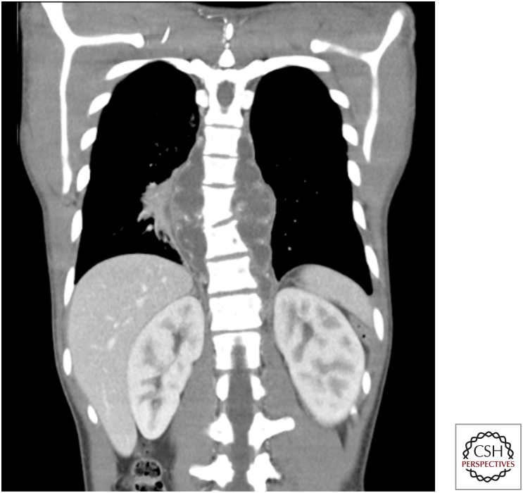
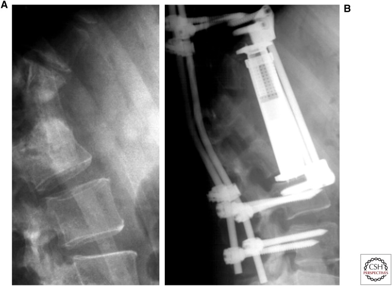
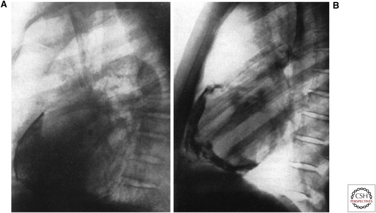
The spine is involved in almost half of skeletal TB cases (Pott’s disease). This starts as inflammation of the intervertebral disk, most often in the lower thoracic and upper lumbar vertebrae, and later involves typically two adjacent vertebrae and the intervertebral disk (Jain and Dhammi 2007). The most common first symptom is slowly progressive, often over weeks and months, back pain with occasionally associated abdominal pain. Local tenderness on palpation may be present. At later stages, kyphoscoliosis or even a gibbus may develop. Spinal cord or nerve root compression with sensory or motor symptoms, even paraplegia, can occur. A paraspinal abscess may develop within the psoas muscle and cause pain in the groin, in particular, on hip flexion. However, paraspinal collections can be seen at any site within the vertebral column (Fig. 16). Modern imaging techniques such as CT, MRI, and bone scan have considerably enhanced the diagnostic work up. Whole-spine imaging is recommended as multiple vertebral sites are involved in up to 50% of cases (Fig. 17). The differential diagnosis includes other infectious etiologies such as chronic bacterial infection, NTM, and malignancy (lymphoma, [lung] cancer). The diagnosis is established by open surgical or needle biopsy for both culture and cytology/histology. Surgical treatment may be indicated (e.g., using external fixation) if there is clear vertebral collapse or destruction or increasing pain suggesting imminent risk of spinal cord or nerve root compression (Fig. 18).
Figure 16.
Coronal CT image of young male with back pain, fever, and weight loss with tuberculous paraspinal abscess secondary to vertebral involvement. (Figure provided by Marc Lipman.)
Figure 17.
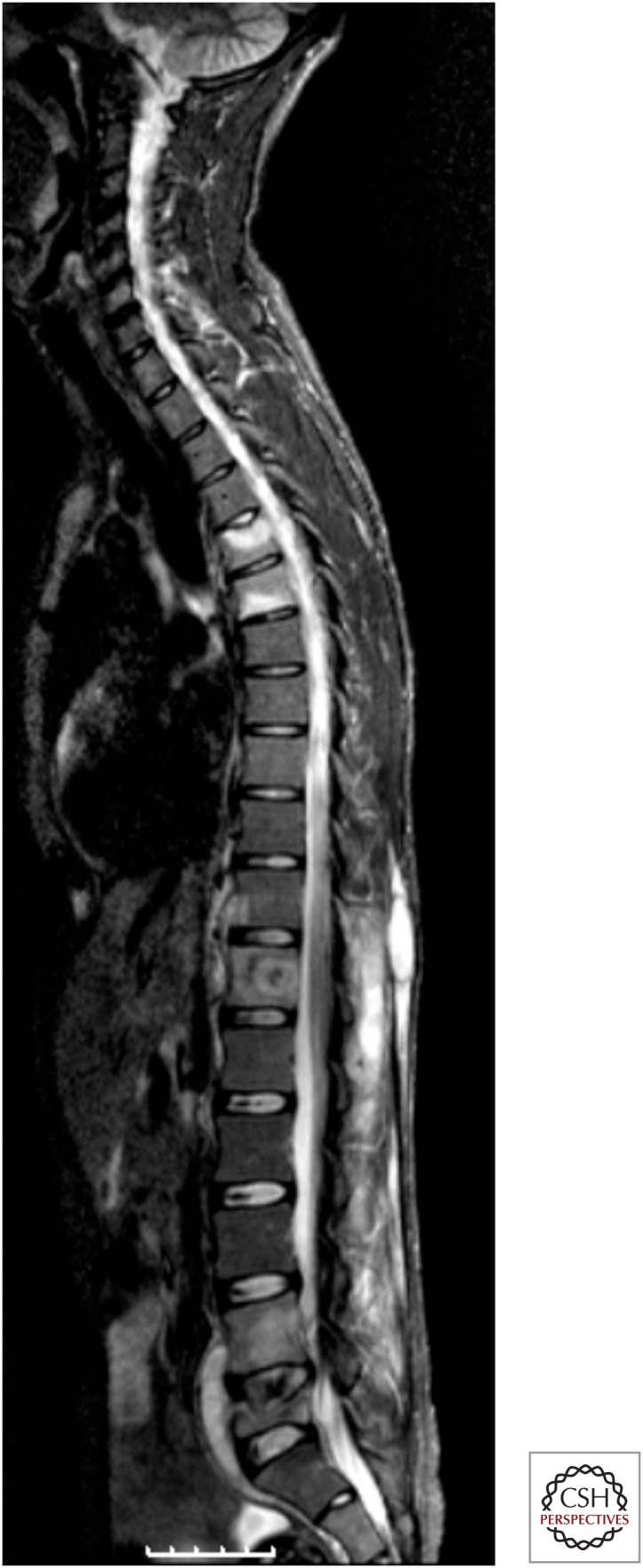
Spinal tuberculosis. Whole-spine MRI showing abnormal signal at multiple vertebral levels within cervical, thoracic, and lumbar spine, plus anterior paraspinal collections at sites of disease. (Figure provided by Marc Lipman.)
Figure 18.
Spinal tuberculosis. (A) Destruction of vertebrae after tuberculous spondylitis; (B) treatment with external fixator. (Figure reprinted from Siemon 2007, with permission, from Springer, © 2007.)
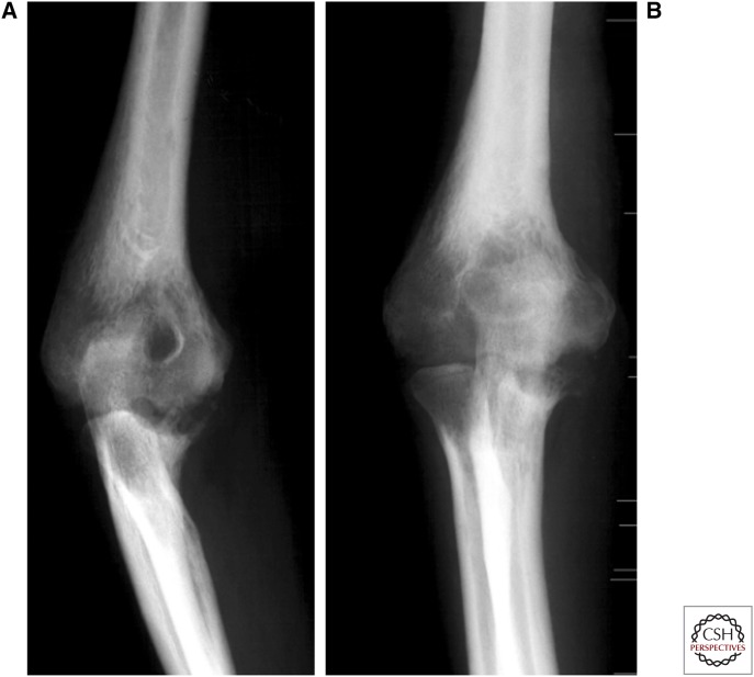
In other bones and joints, the metaphyses of long bones are the most common sites of TB, but hips, knees, shoulders, elbows (Fig. 19), ankles, wrists, or any other bone or joint may be involved (Watts and Lifeso 1996). The diagnosis is established by open surgical or needle biopsy with culture and histology.
Figure 19.
Tuberculosis of the elbow joint. Bone loss is present at the distal humerus, its epidondyles, and also the olecranon and coronoidal process of the ulna. (Figure reprinted from Siemon 2007, with permission, from Springer, © 2007.)
Genitourinary System
Genitourinary system disease includes TB of the kidney, ureter, bladder, and the male and female genital tract (Cek et al. 2005; Kulchavenya 2013). In Germany, it is a frequent extrapulmonary manifestation in adults and accounts for 17% of such cases (Forssbohm et al. 2008). This is in marked contrast to the United Kingdom, where it is responsible for ∼3% (Public Health England 2014). A variety of local symptoms can develop depending on the involved organ, usually many years after the primary infection. Systemic symptoms are less common. Many patients are asymptomatic, and TB may only be detected by urinalysis of pyuria with no apparent bacterial growth. This occurs in >90% of cases. Around 50% will have evidence of previous or active TB present on the chest radiograph (Wilberschied et al. 1999).
Urological TB can cause destruction of the involved kidney, usually unilateral, perinephric or psoas abscess, and/or may lead to stenosis of the ureter and/or involvement of the bladder with cystitis, often accompanied by hematuria.
TB of the female genital system presents as tubal or endometrial involvement (endometritis) causing pelvic pain and/or menorrhagia and may cause infertility. The ovaries (adnexitis) can be involved also, and this may mimic an ovarian cancer (Fig. 20). The diagnosis is established by image-guided needle biopsy, laparoscopy, or open surgical biopsy. TB of the vulva is uncommon.
Figure 20.
Coronal MRI showing TB of the left ovary (black arrow from caudal). Right ovary (white arrow from lateral) with cystic enlarged follicles. (Figure reprinted from Siemon 2007, with permission, from Springer, © 2007.)
TB of the male genital system presents mainly as involvement of the scrotum (epididymitis, orchitis), but the prostate may be affected as well. The diagnosis is established by image-guided needle biopsy or open surgical biopsy.
In some patients, mostly caused by late diagnosis, medical treatment may not result in the resolution of symptoms. Thus, additional surgical intervention and reconstruction may be indicated (Kulchavenya 2013).
Abdomen
Abdominal TB includes involvement of the peritoneum with ascites or any intra-abdominal organ (Tan et al. 2009). This has become rare in low-incidence countries; in Germany, it accounts for 2% of EPTB cases (Forssbohm et al. 2008). Although elsewhere, such as the Indian subcontinent and countries (e.g., the United Kingdom) that see TB relatively frequently in these populations, it is more common. Infection can result from swallowing sputum or nonpasteurized milk (Mycobacterium bovis) or by hematogenous spread. In the gut, TB may occur in any location from the mouth to the anus, although is most often in the ileum and cecum.
Abdominal pain occurs frequently, sometimes mimicking acute appendicitis. Other presentations include small bowel obstruction or gastrointestinal bleeding. The liver, spleen (Fig. 21), pancreas, and adrenal glands may all be involved. In the latter case, the presentation can be one of insidious adrenal insufficiency, in which patients report lethargy or are noted to have metabolic derangements on blood testing.
Figure 21.
Splenic tuberculomas. (Figure reprinted from Siemon 2007, with permission, from Springer, © 2007.)
The differential diagnosis (Khan et al. 2006) depends on the organs involved and includes other infectious etiologies, malignancy (Fig. 22), and inflammatory bowel disease (Almadi et al. 2009). Tuberculous peritonitis with ascites results in abdominal swelling often accompanied by acute symptoms of fever, anorexia, and weight loss. The final diagnosis is established by investigation of the ascitic fluid, which apart from cytology and mycobacterial smear, molecular diagnostics, and culture, may be tested for adenosine deaminase (Shen et al. 2013) and using IGRA (Chou et al. 2011). Laparoscopy, open surgical biopsy, or colonoscopy (Sanai and Bzeizi 2005) may need to be performed, and the diagnosis of TB of other abdominal organs is often made following surgical biopsy or tissue sampling guided by ultrasound or CT.
Figure 22.
Abdominal TB attributable to TB. Pain and weight loss in an Indian male initially considered to result from disseminated abdominal malignancy. CT abdomen: (A) Pretreatment peritoneal and mesenteric thickening leading to omental “cake” (note oral contrast given); (B) posttreatment normal peritoneum and mesentery (no oral contrast given). (Figure provided by Marc Lipman.)
Central Nervous System
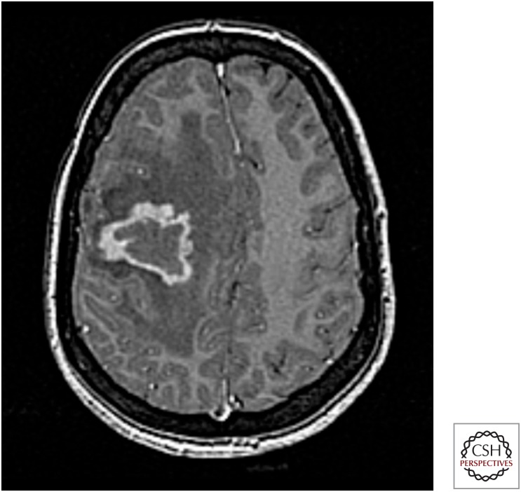
TB of the central nervous system (CNS) includes tuberculous meningitis (TBM) (the most common manifestation) and encephalitis with or without cerebral tuberculomas (Fig. 23). CNS disease is often seen as part of a disseminated illness, with radiological evidence of, for example, miliary TB on chest imaging (see below). It has become rare in low-TB-incidence countries, and in Germany, it accounts for 2% of EPTB cases in adults (Forssbohm et al. 2008). Headache is the predominant local symptom. Others include a decreased level of consciousness, seizures, neck stiffness, vomiting, and those attributable to involvement of cranial nerves. Spinal meningitis with involvement of the peripheral nerves and even paraplegia can occur (Thwaites et al. 2009). The severity of TBM at presentation is strongly associated with prognosis. Various factors and scoring models have been developed to predict the outcomes of TBM, and these are used in the evaluation of different treatment regimens (Chou et al. 2010).
Figure 23.
CNS tuberculoma. MRI brain with contrast shows enhancing lesion with associated edema and mass effect in a Somali female presenting with headache and a seizure. Biopsy-cultured Mtb (Figure provided by Marc Lipman.)
Severe complications include cerebral and spinal cord infarction from granulomatous arteritis, midbrain and the brain stem involvement with loss of automatic, involuntary function, as well as the development of hydrocephalus requiring short- and long-term intervention. The pressure effects of an abscess or granuloma in the CNS may be catastrophic. This should be considered during both investigation and subsequent treatment of patients with “high-risk” lesions in, for example, the brain substance or spinal cord (Thwaites et al. 2009).
The differential diagnosis includes other infectious etiologies of meningitis. The diagnosis is established by lumbar puncture and investigation of the spinal fluid (Thwaites et al. 2009). Highly suspicious is a cerebrospinal fluid (CSF) leucocytosis (predominantly lymphocytes) and raised protein and low-glucose values. The diagnosis is hampered by its paucibacillary nature, but it may be enhanced by simple modification of the Ziehl-Neelsen stain and an ESAT-6 intracellular stain (Feng et al. 2014). The sensitivity is improved using the Xpert MTB/RIF (Nhu et al. 2014). Rapid techniques based on nucleic acid amplification such as polymerase chain reaction (PCR) are more sensitive and specific as they detect specific DNA sequences of the organism (Thwaites et al. 2004a; Galimi 2011; Nhu et al. 2014). CT-guided needle biopsies or open surgery may be needed for the diagnosis of tuberculomas (DeLance et al. 2013). MRI will show generally more lesions than CT (Bernaerts et al. 2003), although in many parts of the world, access to MRI is restricted and, although there is a greater radiation exposure, CT may be preferable if serial imaging is required during follow up.
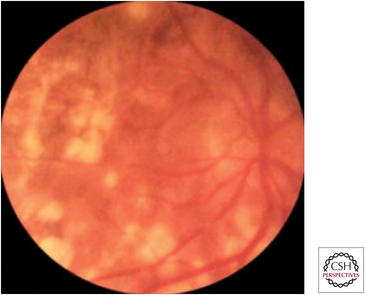
Fundoscopic examination of the eye can reveal evidence of both anterior and posterior chamber disease (Gupta et al. 2007). Apart from presumed inflammatory responses to mycobacterial antigen (e.g., phlyctenular conjunctivitis, episcleritis, and anterior uveitis), papilloedema may be present in patients with CNS involvement. Choroidal tubercles can be visualized—and are helpful in the diagnosis of disseminated TB (Fig. 24).
Figure 24.
Retinal tuberculous lesions. (Figure reprinted from Siemon 2007, with permission, from Springer, © 2007.)
Apart from specific anti-TB therapy, adjunctive corticosteroids (either dexamethasone or prednisolone) should be given to all patients with TBM, regardless of disease severity and HIV status (Thwaites et al. 2004b). Surgical shunting should be considered early in the patient with hydrocephalus and symptoms of raised intracranial pressure. Other neurosurgical operations may be indicated in patients with seizures or brain or spinal cord compression (DeLance et al. 2013).
Miliary TB
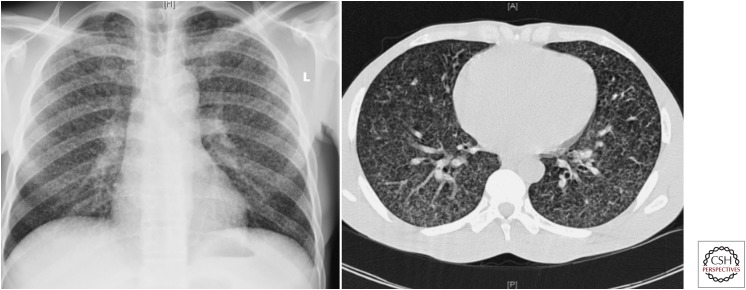
Disseminated TB can involve any number of organs (Zumla et al. 2013). Hematogenous spread, when it includes the lungs, is often characterized by the radiological feature of a “miliary” pattern on imaging. This was first described in chest radiographs (where the numerous granulomas were felt to resemble millet seed, i.e., 1–4-mm rounded opacities scattered throughout both lungs), although is also applicable to CT imaging (Fig. 25). In Germany, it accounts for 3% of EPTB cases (Forssbohm et al. 2008).
Figure 25.
Miliary TB on chest radiograph and CT. (Figure reprinted from Fuehner et al. 2007, with permission, from Springer, © 2007.)
The differential diagnosis of this radiographic appearance includes other infections (e.g., fungi, nocardia and healed varicella pneumonia) as well as malignancy (in particular, thyroid, breast, and renal cancers, plus melanoma and osteosarcoma) and conditions that result in granulomas) (e.g., sarcoidosis). Hence, clinicians must ensure that a diagnosis of miliary TB is based on as much laboratory information as possible (Ray et al. 2013) and that patients who are started on anti-TB treatment are carefully monitored for response and/or the development of new symptoms and signs. Patients may develop an acute respiratory distress syndrome (ARDS) associated with a high mortality despite intensive care (Erbes et al. 2006; Lee et al. 2011). The benefit of adjunctive corticosteroid administration in patients with miliary TB is unclear (Ray et al. 2013).
OTHER TB MANIFESTATIONS
TB can present at any body site. Therefore, clinicians should consider sending samples for mycobacterial molecular analysis, smear, and culture, as well as histo- or cytopathology. This situation occurs most frequently when patients present either with cutaneous disease (typically nonhealing lesions that have been treated for some time with standard antibacterials) (Fig. 26) or to breast, or ear, nose, and throat surgeons. In Germany, other TB manifestations account for 9% of all extrapulmonary TB cases (Forssbohm et al. 2008).
Figure 26.
Tracheo-cutaneous fistula with scrofuloderma. (Figure reprinted from Siemon 2007, with permission, from Springer, © 2007.)
TB pericarditis is a globally important condition with a distinctly poor prognosis if not diagnosed promptly (Fig. 27). It is usually seen as an active acute or subacute condition associated with systemic features plus a pericardial effusion, which may lead to tamponade (Syed and Mayosi 2007). Adjunctive treatment with corticosteroids is recommended (Evans 2008). Constrictive pericarditis secondary to pericardial calcification can also occur (Fig. 28). Here, the initial (primary) infection is thought to have occurred several years earlier and was often asymptomatic. Over time, the patient notices increasing exercise limitation, yet even then may present to health-care services with apparent acute clinical decompensation.
Figure 27.
Tuberculous pericarditis with bilateral pleural effusions. (A) CT; (B) echocardiography. (Figure reprinted from Siemon 2007, with permission, from Springer, © 2007.)
Figure 28.
Calcification after tuberculous pericarditis. (Figure reprinted from Uehlinger et al. 1959, with permission, from EMH Schweizer Ärzteverlag, © 1959.)
The calcification associated with TB is generally thick, confluent, and irregular, although these features cannot be relied on. CT or MRI are useful investigations but should not detract from efforts to isolate or detect an organism (which may be from other sites including the lung). This may be unsuccessful (particularly with the chronic presentation). The patient will often have a history of exposure to TB, usually from having spent time as a child in a TB-endemic country. Anti-TB treatment should be given, in addition to cardiac and surgical management as needed (Syed and Mayosi 2007).
TREATMENT OF TB
The diagnosis of active TB (and hence the decision to start anti-TB drug therapy without which the great majority of cases will not be cured) uses information derived from the mycobacteria (e.g., positive AFB smear, molecular diagnostic test or culture), a compatible host response (e.g., cavitation on a chest radiograph), and environmental factors (e.g., a history of contact with TB in a person with consistent clinical features, or acquisition of HIV infection leading to altered [and impaired] immunity).
Prompt initiation of treatment will reduce morbidity and mortality for the affected individual and also break the cycle of onward transmission that sustains TB. However, treatment is not without its complications. This is most evident in the management of HIV-associated TB (Khan et al. 2010; Schutz et al. 2010), in which there are considerable drug–drug interactions between antiretrovirals and anti-TB medication, and an increased risk of the intense inflammatory response provoked by concurrent use of these drugs (IRIS) (Meintjes et al. 2008). This means that in practice, the relative timing of introduction of these agents also needs to be factored (Blanc et al. 2011; Török et al. 2011).
Drug treatment is dealt with in more detail in other sections of this book. However, some of the key principles are described in brief here. Effective treatment is one that enables the individual to complete therapy and results in a very high cure rate and minimal risk of relapse owing to the original infection. Thus, drug therapy needs to be efficacious and have few adverse events (Breen et al. 2006). Both of these will maximize adherence to treatment regimens. However, factors such as duration of treatment, pill burden, cost of therapy, and even ensuring that the patient can access a sustained and secure supply of medication (Capstick et al. 2011) will impact on treatment effectiveness. Hence, at both a personal and a societal level, high-quality treatment programs must ensure that safe and effective drug supplies are available and that a TB service can provide regular monitoring with facilities that optimize the patient’s ability to adhere to the prescribed regimen.
Therapeutic combinations should, if at all possible, be based on known sensitivity of the organism to the selected drugs (World Health Organization 2011). The increasing prevalence of drug resistance (World Health Organization 2013) and the ease of global travel mean that no longer can clinicians assume that the infecting organism is likely to be drug-sensitive. This emphasizes the importance of obtaining pretreatment samples for rapid molecular diagnostics and culture, both of which can indicate likely drug sensitivity and hence guide early and ongoing treatment (Moureet al. 2011).
In presumed drug-sensitive disease, treatment should commence with a minimum of three effective drugs. In practice, this means a four-drug therapy with a rifamycin plus isoniazid as well as pyrazinamide and ethambutol for the first 2 mo (by which point, drug susceptibility should be known for any positive Mtb cultures). It can never be assumed that prescribing medication equates to the patient actually taking it, and arrangements must be in place to ensure that adequate treatment support and supervision are available for the duration of therapy.
On-treatment monitoring of individual response may use strategies such as directly observed therapy (Okanurak et al. 2007), pill counts, and urine tests, plus measures of patient characteristics that reflect disease alleviation, e.g., weight gain (Khan et al. 2006) and health status (Kruijshaar et al. 2010).
People on treatment for pulmonary TB can be followed with serial sputum cultures. However, in resource-rich environments, individuals often stop producing sputum within a few weeks of starting an effective treatment regimen (Hales et al. 2013). In cases in which there is doubt regarding adherence with medication or possible treatment failure, it is recommended that the patient be evaluated for therapeutic compliance, pharmacodynamics (e.g., is there any possibility of malabsorption?), drug resistance, or an alternative diagnosis.
The importance of close liaison with other experienced clinicians and microbiologists, as well as public health officials within the setting of a multidisciplinary team meeting or through the use of regular cohort reviews (Anderson et al. 2014), cannot be overemphasized. These provide optimal care for the individual and strengthen public health management of TB and are also an excellent method of education and training for all individuals involved in TB care (Migliori et al. 2012).
The diagnosis of TB enables health-care services to provide more than just treatment for TB. HIV testing should be routinely offered—with studies showing that “opt out” testing in TB clinics is a simple method of achieving this (Roy et al. 2013). Screening for other blood-borne viruses such as hepatitis B and C, as well as assessing nutritional status, blood pressure, blood sugar, and encouraging smoking cessation, can also be highlighted during clinical care as part of a package of person-related health management.
CONCLUSION
Given the generally excellent response to treatment if therapy is started early, there is now little excuse for “missed diagnoses” and “late presentations” of TB. It remains a condition that in many parts of the world is common and hence usually promptly diagnosed. Here, the clinical issues often center on the need to ensure that correct, effective medication is prescribed and adverse events are minimized. However, in environments where TB is less frequently seen, the recognition that it can present in multiple ways and that TB must be considered early (by both the patient and their health-care provider) within the clinical diagnostic algorithm are of paramount importance. This is made more complex by the increasing numbers of immunocompromised individuals (through, e.g., HIV infection and medical treatments) at risk of developing active TB who, by virtue of their altered host response, will often have a modified presentation of TB disease. Strategies to combat the loss of familiarity with TB in low TB burden areas include the use of clinical networks and multidisciplinary and multiprofessional team working, plus on-going, relevant education in at-risk communities, the general public, and health-care workers.
Footnotes
Editors: Stefan H.E. Kaufmann, Eric J. Rubin, and Alimuddin Zumla
Additional Perspectives on Tuberculosis available at www.perspectivesinmedicine.org
REFERENCES
- Aït-Khaled N, Alarcón E, Armengol R, Bissell K, Boillot F, Caminero JA, Chiang C-Y, Clevenbergh P, Dlodlo R, Enarson DA, et al. 2010. Management of tuberculosis: A guide to the essentials of good practice. Paris, France, International Union Against Tuberculosis and Lung Disease (The Union) http://www.theunion.org/what-we-do/publications/technical/english/pub_orange-guide_eng.pdf [Google Scholar]
- Almadi MA, Ghosh S, Aljebreen AM. 2009. Differentiating intestinal tuberculosis from Crohn’s disease: A diagnostic challenge. Am J Gastroenterol 104: 1003–1012. [DOI] [PubMed] [Google Scholar]
- Anderson C, White J, Abubakar I, Lipman M, Tamne S, Anderson SR, deKoningh J, Dart S. 2014. Raising standards in UK TB control: Introducing cohort review. Thorax 69: 187–189. [DOI] [PubMed] [Google Scholar]
- Bernaerts A, Vanhoenacker FM, Parizel PM, Van Goethem JW, Van Altena R, Laridon A, De Roeck J, Coeman V, De Schepper AM. 2003. Tuberculosis of the central nervous system: Overview of neuroradiological findings. Eur Radiol 13: 1876–1890. [DOI] [PubMed] [Google Scholar]
- Bhat VK, Latha P, Upadhya D, Hegde J. 2009. Clinicopathological review of tubercular laryngitis in 32 cases of pulmonary Kochs. Am J Otolaryngol 30: 327–330. [DOI] [PubMed] [Google Scholar]
- Bielsa S, Palma R, Pardina M, Esquerda A, Light RW, Porcel JM. 2013. Comparison of polymorphonuclear- and lymphocyte-rich tuberculous pleural effusions. Int J Tuberc Lung Dis 17: 85–89. [DOI] [PubMed] [Google Scholar]
- Blanc FX, Sok T, Laureillard D, Borand L, Rekacewicz C, Nerrienet E, Madec Y, Marcy O, Chan S, Prak N, et al. 2011. Earlier versus later start of antiretroviral therapy in HIV-infected adults with tuberculosis. N Engl J Med 365: 1471–1481. [DOI] [PMC free article] [PubMed] [Google Scholar]
- Blasi F, Matteelli A. 2013. Indicator condition-guided HIV testing in Europe: A step forward to HIV control. Eur Respir J 42: 572–575. [DOI] [PubMed] [Google Scholar]
- Boehme CC, Nicol MP, Nabeta P, Michael JS, Gotuzzo E, Tahirli R, Tarcela Gler M, Blakemore R, Worodria W, Gray C, et al. 2011. Feasibility, diagnostic accuracy, and effectiveness of decentralised use of the Xpert MTB/RIF test for diagnosis of tuberculosis and multidrug resistance: A multicentre implementation study. Lancet 377: 1495–1505. [DOI] [PMC free article] [PubMed] [Google Scholar]
- Borgdorff MW, Nagelkerke NJ, Dye C, Nunn P. 2000. Gender and tuberculosis: A comparison of prevalence surveys with notification data to explore sex differences in case detection. Int J Tuberc Lung Dis 4: 123–132. [PubMed] [Google Scholar]
- Breen RAM, Smith CJ, Cropley I, Johnson MA, Lipman MCI. 2005. Does immune reconstitution syndrome promote active tuberculosis in patients receiving HAART? AIDS 19: 1201–1206. [DOI] [PubMed] [Google Scholar]
- Breen RAM, Miller RF, Gorsuch T, Smith CJ, Schwenk A, Holmes W, Ballinger J, Swaden L, Johnson MA, Cropley I, et al. 2006. Adverse events and treatment interruption in tuberculosis patients with and without HIV co-infection. Thorax 61: 791–794. [DOI] [PMC free article] [PubMed] [Google Scholar]
- Breen RAM, Leonard O, Perrin FMR, Smith CJ, Bhagani S, Cropley I, Lipman MCI. 2008a. How good are systemic symptoms and blood inflammatory markers in detecting individuals with active tuberculosis? A prospective UK cohort study. Int J Tuberc Lung Dis 12: 44–49. [PubMed] [Google Scholar]
- Breen RA, Barry SM, Smith CJ, Shorten RJ, Dilworth JP, Cropley I, McHugh TD, Gillespie SH, Janossy G, Lipman MC. 2008b. Clinical application of a rapid lung-orientated immunoassay in individuals with possible tuberculosis. Thorax 63: 67–71. [DOI] [PubMed] [Google Scholar]
- Brown M, Varia H, Bassett P, Davidson RN, Wall R, Pasvol G. 2007. Prospective study of sputum induction, gastric washing, and bronchoalveolar lavage for the diagnosis of pulmonary tuberculosis in patients who are unable to expectorate. Clin Infect Dis 44: 1415–1420. [DOI] [PubMed] [Google Scholar]
- Cain KP, McCarthy KD, Heilig CM, Monkongdee P, Tasaneeyapan T, Kanara N, Kimerling ME, Chheng P, Thai S, Sar B, et al. 2010. An algorithm for tuberculosis screening and diagnosis in people with HIV. N Engl J Med 362: 707–716. [DOI] [PubMed] [Google Scholar]
- Capstick T, Laycock D, Lipman M. 2011. Treatment interruptions and inconsistent supply of anti-TB drugs in the UK. Int J Tuberc Lung Dis 15: 754–760. [DOI] [PubMed] [Google Scholar]
- Caws M, Thwaites G, Dunstan S, Hawn TR, Lan NT, Thuong NT, Stepniewska K, Huyen MN, Bang ND, Loc TH, et al. 2008. The influence of host and bacterial genotype on the development of disseminated disease with Mycobacterium tuberculosis. PLoS Pathog 4: e1000034. [DOI] [PMC free article] [PubMed] [Google Scholar]
- Cek M, Lenk S, Naber KG , Bishop MC, Johansen TE, Botto H, Grabe M, Lobel B, Redorta JP, Tenke P. 2005. Members of the Urinary Tract Infection (UTI) Working Group of the European Association of Urology (EAU) Guidelines Office. EAU guidelines for the management of genitourinary tuberculosis. Eur Urol 48: 353–362. [DOI] [PubMed] [Google Scholar]
- Chegou NN, Walzl G, Bolliger CT, Diacon AH, van den Heuvel MM. 2008. Evaluation of adapted whole-blood interferon-γ release assays for the diagnosis of pleural tuberculosis. Respiration 76: 131–138. [DOI] [PubMed] [Google Scholar]
- Chou CH, Lin GM, Ku CH, Chang FY. 2001. Comparison of the APACHE II, GCS and MRC scores in predicting outcomes in patients with tuberculous meningitis. Int J Tuberc Lung Dis 14: 86–92. [PubMed] [Google Scholar]
- Chou OH, Park KH, Park SJ, Kim SM, Park SY, Moon SM, Chong YP, Kim MN, Lee SO, Choi SH, et al. 2011. Rapid diagnosis of tuberculous peritonitis by T cell-based assays on peripheral blood and peritoneal fluid mononuclear cells. J Infect 62: 462–471. [DOI] [PubMed] [Google Scholar]
- Chowdhury AM, Bhuiya A, Chowdhury ME, Rasheed S, Hussain Z, Chen LC. 2013. The Bangladesh paradox: Exceptional health achievement despite economic poverty. Lancet 382: 1734–1745. [DOI] [PubMed] [Google Scholar]
- Chung HS, Lee JH. 2000. Bronchoscopic assessment of the evolution of endobronchial tuberculosis. Chest 117: 385–392. [DOI] [PubMed] [Google Scholar]
- Chung CL, Chen CH, Yeh CY, Sheu JR, Chang SC. 2008. Early effective drainage in the treatment of loculated tuberculosis pleurisy. Eur Respir J 31: 1261–1267. [DOI] [PubMed] [Google Scholar]
- Courtwright A, Turner AN. 2010. Tuberculosis and stigmatization: Pathways and interventions. Public Health Rep 125: 434–442. [DOI] [PMC free article] [PubMed] [Google Scholar]
- Craig SE, Bettinson H, Sabin CA, Gillespie SH, Lipman MCI. 2009. Think TB! Is the diagnosis of pulmonary tuberculosis delayed by antibiotics? Int J Tuberc Lung Dis 13: 208–213. [PubMed] [Google Scholar]
- Crump JA, Ramadhani HO, Morrissey AB, Saganda W, Mwako MS, Yang LY, Chow SC, Njau BN, Mushi GS, Maro VP, et al. 2012. Bacteremic disseminated tuberculosis in sub-Saharan Africa: A prospective cohort study. Clin Infect Dis 55: 242–250. [DOI] [PMC free article] [PubMed] [Google Scholar]
- DeLance AR, Safaee M, Oh MC, Clark AJ, Kaur G, Sun MZ, Bollen AW, Phillips JJ, Parsa AT. 2013. Tuberculoma of the central nervous system. J Clin Neurosci 20: 1333–1341. [DOI] [PubMed] [Google Scholar]
- de la Rua-Domenech R. 2006. Human Mycobacterium bovis infection in the United Kingdom: Incidence, risks, control measures and review of the zoonotic aspects of bovine tuberculosis. Tuberculosis (Edinb) 86: 77–109. [DOI] [PubMed] [Google Scholar]
- Dhasmana DJ, Ross C, Bradley CJ, Connell DW, George PM, Singanayagam A, Jepson A, Craig C, Wright C, Molyneaux PL, et al. 2014. Performance of Xpert MTB/RIF in the diagnosis of tuberculous mediastinal lymphadenopathy by endobronchial ultrasound. Ann Am Thorac Soc 11: 392–396. [DOI] [PMC free article] [PubMed] [Google Scholar]
- Dhillon J, Fourie PB, Mitchison DA. 2014. Persister populations of Mycobacterium tuberculosis in sputum that grow in liquid but not on solid culture media. J Antimicrob Chemother 69: 437–440. [DOI] [PubMed] [Google Scholar]
- Diacon AH, Van de Wal BW, Wyser C, Smedema JP, Bezuidenhout J, Bolliger CT, Walzl G. 2003. Diagnostic tools in tuberculous pleurisy: A direct comparative study. Eur Respir J 22: 589–591. [DOI] [PubMed] [Google Scholar]
- Diel R, Loddenkemper R, Zellweger JP, Sotgiu G, D’Ambrosio L, Centis R, van der Werf MJ, Dara M, Detjen A, Gondrie P, et al. 2013. Old ideas to innovate TB control: Preventive treatment to achieve elimination. Eur Respir J 42: 785–801. [DOI] [PubMed] [Google Scholar]
- Dooley SW Jr, Castro KG, Hutton MD, Mullan RJ, Polder JA, Snider DE Jr. 1990. Guidelines for preventing the transmission of tuberculosis in health-care settings, with special focus on HIV-related issues. MMWR Recomm Rep 39: 1–29. [PubMed] [Google Scholar]
- Elliott AM, Hayes RJ, Halwiindi B, Luo N, Tembo G, Pobee JO, Nunn PP, McAdam KP. 1993. The impact of HIV on infectiousness of pulmonary tuberculosis: A community study in Zambia. AIDS 7: 981–987. [DOI] [PubMed] [Google Scholar]
- Engel ME, Matchaba PT, Volmink J. 2007. Corticosteroids for tuberculous pleurisy. Cochrane Database Syst Rev 17: CD001876. [DOI] [PubMed] [Google Scholar]
- Erbes R, Oettel K, Raffenberg M, Mauch H, Schmidt-Ioanas M, Lode H. 2006. Characteristics and outcome of patients with active pulmonary tuberculosis requiring intensive care. Eur Respir J 27: 1223–1228. [DOI] [PubMed] [Google Scholar]
- Evans DJ. 2008. The use of adjunctive corticosteroids in the treatment of pericardial, pleural and meningeal tuberculosis: Do they improve outcome? Respir Med 102: 793–800. [DOI] [PubMed] [Google Scholar]
- Fernando SL, Saunders BM, Sluyter R, Skarratt KK, Goldberg H, Marks GB, Wiley JS, Britton WJ. 2007. A polymorphism in the P2X7 gene increases susceptibility to extrapulmonary tuberculosis. Am J Respir Crit Care Med 175: 360–366. [DOI] [PubMed] [Google Scholar]
- Feng GD, Shi M, Ma L, Chen P, Wang BJ, Zhang M, Chang XL, Su XC, Yang YN, Fan XH, et al. 2014. Diagnostic accuracy of intracellular Mycobacterium tuberculosis detection for tuberculous meningitis. Am J Respir Crit Care Med 189: 475–481. [DOI] [PMC free article] [PubMed] [Google Scholar]
- Forssbohm M. 2004. Studie des DZK zur Epidemiologie der Tuberkulose. Abschlussbericht 1996–2000 (22.333 Fälle) [Study of the German Central Committee against Tuberculosis on the epidemiology of tuberculosis. Final report 1996-2000 (22.333 cases)]. In 28. Informationsbericht des DZK (ed. Loddenkemper R), pp. 66–78. DZK, Berlin. [Google Scholar]
- Forssbohm M, Zwahlen M, Loddenkemper R, Rieder HL. 2008. Demographic characteristics of patients with extrapulmonary tuberculosis in Germany. Eur Respir J 31: 99–105. [DOI] [PubMed] [Google Scholar]
- Fuehner T, Stoll M, Bange FC, Welte T, Pletz MW. 2007. Klinik der Lungentuberkulose. Pneumologe 4: 151–162. [Google Scholar]
- Galimi R. 2011. Extrapulmonary tuberculosis: Tuberculous meningitis new developments. Eur Rev Med Pharmacol Sci 15: 365–386. [PubMed] [Google Scholar]
- Gopi A, Madhavan SM, Sharma SK, Sahn SA. 2007. Diagnosis and treatment of tuberculous pleural effusion in 2006. Chest 131: 880–889. [DOI] [PubMed] [Google Scholar]
- Gupta V, Gupta A, Rao NA. 2007. Intraocular tuberculosis—An update. Surv Ophthalmol 52: 561–587. [DOI] [PubMed] [Google Scholar]
- Hales CM, Heilig CM, Chaisson R, Leung CC, Chang KC, Goldberg SV, Gordin F, Johnson JL, Muzanyi G, Saukkonen J, et al. 2013. The association between symptoms and microbiologically defined response to tuberculosis treatment. Ann Am Thorac Soc 10: 18–25. [DOI] [PMC free article] [PubMed] [Google Scholar]
- Heyderman RS, Makunike R, Muza T, Odwee M, Kadzirange G, Manyemba J, Muchedzi C, Ndemera B, Gomo ZA, Gwanzura LK, et al. 1998. Pleural tuberculosis in Harare, Zimbabwe: The relationship between human immunodeficiency virus, CD4 lymphocyte count, granuloma formation and disseminated disease. Trop Med Int Health 3: 14–20. [DOI] [PubMed] [Google Scholar]
- Honeyborne I, McHugh TD, Phillips PJ, Bannoo S, Bateson A, Carroll N, Perrin FM, Ronacher K, Wright L, van Helden PD, et al. 2011. Molecular bacterial load assay, a culture-free biomarker for rapid and accurate quantification of sputum Mycobacterium tuberculosis bacillary load during treatment. J Clin Microbiol 49: 3905–3911. [DOI] [PMC free article] [PubMed] [Google Scholar]
- Hooper CE, Lee YC, Maskell NA. 2009. Interferon-γ release assays for the diagnosis of TB pleural effusions: Hype or real hope? Curr Opin Pulm Med 15: 358–365. [DOI] [PubMed] [Google Scholar]
- Hooper C, Lee YCG, Maskell N, on behalf of the BTS Pleural Guideline Group. 2010. Investigation of a unilateral pleural effusion in adults: British Thoracic Society pleural disease guideline 2010. Thorax 65: i4–ii17. [DOI] [PubMed] [Google Scholar]
- Horsburgh CR Jr. 2004. Priorities for the treatment of latent tuberculosis infection in the United States. N Engl J Med 350: 2060–2067. [DOI] [PubMed] [Google Scholar]
- Hu HY, Wu CY, Huang N, Chou YJ, Chang YC, Chu D. 2014. Increased risk of tuberculosis in patients with end-stage renal disease: A population-based cohort study in Taiwan, a country of high incidence of end-stage renal disease. Epidemiol Infect 142: 191–199. [DOI] [PMC free article] [PubMed] [Google Scholar]
- Huang CC, Tchetgen ET, Becerra M, Cohen T, Hughes KC, Zhang Z, Calderon R, Yataco R, Contreras C, Galea J, et al. 2014. The effect of HIV-related immunosuppression on the risk of tuberculosis transmission to household contacts. Clin Infect Dis 58: 765–774. [DOI] [PMC free article] [PubMed] [Google Scholar]
- Jain AK, Dhammi IK. 2007. Tuberculosis of the spine: A review. Clin Orthop Relat Res 460: 39–49. [DOI] [PubMed] [Google Scholar]
- Khan A, Sterling TR, Reves R, Vernon A, Horsburgh CR. 2006. Lack of weight gain and relapse risk in a large tuberculosis treatment trial. Am J Respir Crit Care Med 174: 344–348. [DOI] [PubMed] [Google Scholar]
- Khan FA, Minion J, Pai M, Royce S, Burman W, Harries AD, Menzies D. 2010. Treatment of active tuberculosis in HIV-coinfected patients: A systematic review and meta-analysis. Clin Infect Dis 50: 1288–1299. [DOI] [PubMed] [Google Scholar]
- Koegelenberg CF, Bollinger CT, Theron, Walzl G, Wright CA, Louw M, Diacon AH. 2010. A direct comparison of the diagnostic yield of ultrasound-assisted Abrams and Tru-Cut needle biopsies for pleural tuberculosis. Thorax 65: 857–862. [DOI] [PubMed] [Google Scholar]
- Kroot EJ, Hazes JM, Colin EM, Dolhain RJ. 2007. Poncet’s disease: Reactive arthritis accompanying tuberculosis. Two case reports and a review of the literature. Rheumatology (Oxford) 46: 484–489. [DOI] [PubMed] [Google Scholar]
- Kruijshaar ME, Lipman M, Essink-Bot ML, Lozewicz S, Creer D, Dart S, Sadler H, Maguire H, Abubakar I. 2010. Health status of UK patients with active tuberculosis. Int J Tuberc Lung Dis 14: 296–302. [PubMed] [Google Scholar]
- Kulchavenya E. 2013. Best practice in the diagnosis and management of urogenital tuberculosis. Ther Adv Urol 5: 143–151. [DOI] [PMC free article] [PubMed] [Google Scholar]
- Lamm DL. 1992. Complications of bacillus Calmette-Guerin immunotherapy. Urol Clin North Am 19: 565–572. [PubMed] [Google Scholar]
- Lawn SD, Zumla AI. 2011. Tuberculosis. Lancet 378: 57–72. [DOI] [PubMed] [Google Scholar]
- Lawn SD, Kerkhoff AD, Vogt M, Wood R. 2013. HIV-associated tuberculosis: Relationship between disease severity and the sensitivity of new sputum-based and urine-based diagnostic assays. BMC Med 11: 231. [DOI] [PMC free article] [PubMed] [Google Scholar]
- Lee JY, Yi CA, Kim TS, Kim H, Kim J, Han J, Kwon OJ, Lee KS, Chung MJ. 2010. CT scan features as predictors of patient outcome after bronchial intervention in endobronchial TB. Chest 138: 380–385. [DOI] [PubMed] [Google Scholar]
- Lee K, Kim JH, Lee JH, Lee WY, Park MS, Kim JY, Kim KC, Lee MG, Jung KS, Kim YS, et al. 2011. Acute respiratory distress syndrome caused by miliary tuberculosis: A multicentre survey in South Korea. Int J Tuberc Lung Dis 15: 1099–1103. [DOI] [PubMed] [Google Scholar]
- Light RW. 2010. Update on tuberculous pleural effusion. Respirology 15: 451–458. [DOI] [PubMed] [Google Scholar]
- Loddenkemper R. 1998. Thoracoscopy—State of the art. Eur Respir J 11: 213–221. [DOI] [PubMed] [Google Scholar]
- Loddenkemper, Praveen, Noppen, Pyng. 2011. Medical Thoracoscopy/Pleuroscopy: Manual and Atlas. Georg Thieme, New York. [Google Scholar]
- Mase SR, Ramsay A, Ng V, Henry M, Hopewell PC, Cunningham J, Urbanczik R, Perkins MD, Aziz MA, Pai M. 2007. Yield of serial sputum specimen examinations in the diagnosis of pulmonary tuberculosis: A systematic review. Int J Tuberc Lung Dis 11: 485–495. [PubMed] [Google Scholar]
- Meintjes G, Lawn SD, Scano F, Maartens G, French MA, Worodria W, Elliott JH, Murdoch D, Wilkinson RJ, Seyler C, et al. 2008. Tuberculosis-associated immune reconstitution inflammatory syndrome: Case definitions for use in resource-limited settings. Lancet Infect Dis 8: 516–523. [DOI] [PMC free article] [PubMed] [Google Scholar]
- Menzies D, Fanning A, Yuan L, FitzGerald JM. 2003. Canadian Collaborative Group in Nosocomial Transmission of Tuberculosis. Factors associated with tuberculin conversion in Canadian microbiologiy and pathology workers. Am J Respir Crit Care Med 167: 599–602. [DOI] [PubMed] [Google Scholar]
- Migliori GB, Zellweger JP, Abubakar I, Ibraim E, Caminero JA, De Vries G, D’Ambrosio L, Centis R, Sotgiu G, Menegale O, et al. 2012. European Union standards for tuberculosis care. Eur Respir J 39: 807–819. [DOI] [PMC free article] [PubMed] [Google Scholar]
- Millen SJ, Uys PW, Hargrove J, van Helden PD, Williams BG. 2008. The effect of diagnostic delays on the drop-out rate and the total delay to diagnosis of tuberculosis. PLoS ONE 3: e1933. [DOI] [PMC free article] [PubMed] [Google Scholar]
- Möller M, de Wit E, Hoal EG. 2010. Past, present and future directions in human genetic susceptibility to tuberculosis. FEMS Immunol Med Microbiol 58: 3–26. [DOI] [PubMed] [Google Scholar]
- Moure R, Muñoz L, Torres M, Santin M, Martín R, Alcaide F. 2011. Rapid detection of Mycobacterium tuberculosis complex and rifampin resistance in smear-negative clinical samples by use of an integrated real-time PCR method. J Clin Microbiol 49: 1137–1139. [DOI] [PMC free article] [PubMed] [Google Scholar]
- Navani N, Molyneaux PL, Breen RA, Connell DW, Jepson A, Nankivell M, Brown JM, Morris-Jones S, Ng B, Wickremasinghe M, et al. 2011. Utility of endobronchial ultrasound-guided transbronchial needle aspiration in patients with tuberculous intrathoracic lymphadenopathy: A multicentre study. Thorax 66: 889–893. [DOI] [PMC free article] [PubMed] [Google Scholar]
- Nhu NT, Heemskerk D, Thu do DA, Chau TT, Mai NT, Nghia HD, Loc PP, Ha DT, Merson L, Thinh TT, et al. 2014. Evaluation of GeneXpert MTB/RIF for diagnosis of tuberculous meningitis. J Clin Microbiol 52: 226–233. [DOI] [PMC free article] [PubMed] [Google Scholar]
- Okanurak K, Kitayaporn D, Wanarangsikul W, Koompong C. 2007. Effectiveness of DOT for tuberculosis treatment outcomes: A prospective cohort study in Bangkok, Thailand. Int J Tuberc Lung Dis 11: 762–768. [PubMed] [Google Scholar]
- Parimon T, Spitters CE, Muangman N, Euathrongchit J, Oren E, Narita M. 2008. Unexpected pulmonary involvement in extrapulmonary tuberculosis patients. Chest 134: 589–594. [DOI] [PubMed] [Google Scholar]
- Public Health England. 2014. http://www.hpa.org.uk/Topics/InfectiousDiseases/InfectionsAZ/Tuberculosis/TBUKSurveillanceData/EnhancedTuberculosisSurveillance/nTBEnhanced11site/ (accessed 18 April 2014).
- Ray S, Talukdar A, Kundu S, Khanra D, Sonthalia N. 2013. Diagnosis and management of miliary tuberculosis: Current state and future perspectives. Ther Clin Risk Manag 9: 9–26. [DOI] [PMC free article] [PubMed] [Google Scholar] [Retracted]
- Rice B, Elford J, Yin Z, Kruijshaar M, Abubakar I, Lipman M, Pozniak A, Kall M, Delpech V. 2013. Decreasing incidence of tuberculosis among HIV diagnosed heterosexuals in England and Wales. AIDS 27: 1151–1157. [DOI] [PubMed] [Google Scholar]
- Rieder HL, Watson JM, Raviglione MC, Forssbohm M, Migliori GB, Schwoebel V, Leitch AG, Zellweger JP. 1996. Surveillance of tuberculosis in Europe. Working Group of the World Health Organization (WHO) and the European Region of the International Union Against Tuberculosis and Lung Disease (IUATLD) for uniform reporting on tuberculosis cases. Eur Respir J 9: 1097–1104. [DOI] [PubMed] [Google Scholar]
- Roy A, Anaraki S, Hardelid P, Catchpole M, Rodrigues LC, Lipman M, Perkins S, Roche A, Stagg H, Figueroa J, Abubakar I. 2013. Implementing universal HIV testing in London tuberculosis clinics: A cluster randomised controlled trial. Eur Respir J 41: 627–634. [DOI] [PubMed] [Google Scholar]
- Sahn SA. 2002. Pleural thickening, trapped lung, and chronic empyema as sequelae of tuberculous pleural effusion: Don’t sweat the pleural thickening. Int J Tuberc Lung Dis 6: 461–464. [DOI] [PubMed] [Google Scholar]
- Sanai FM, Bzeizi KI. 2005. Systematic review: Tuberculous peritonitis—Presenting features, diagnostic strategies and treatment. Aliment Pharmacol Ther 22: 685–700. [DOI] [PubMed] [Google Scholar]
- Schoch OD, Rieder P, Tueller C, Altpeter E, Zellweger JP, Rieder HL, Krause M, Thurnheer R. 2007. Diagnostic yield of sputum, induced sputum, and bronchoscopy after radiologic tuberculosis screening. Am J Respir Crit Care Med 175: 80–86. [DOI] [PubMed] [Google Scholar]
- Schutz C, Meintjes G, Almajid F, Wilkinson RJ, Pozniak A. 2010. Clinical management of tuberculosis and HIV-1 co-infection. Eur Respir J 36: 1460–1481. [DOI] [PubMed] [Google Scholar]
- Shen YC, Wang T, Chen L, Yang T, Wan C, Hu QJ, Wen FQ. 2013. Diagnostic accuracy of adenosine deaminase for tuberculous peritonitis: A meta-analysis. Arch Med Sci 9: 601–607. [DOI] [PMC free article] [PubMed] [Google Scholar]
- Siemon G. 2007. Extrapulmonale tuberkulose. Pneumolog 4: 163–174. [Google Scholar]
- Syed FF, Mayosi BM. 2007. A modern approach to tuberculous pericarditis. Prog Cardiovasc Dis 50: 218–236. [DOI] [PubMed] [Google Scholar]
- Tan KK, Chen K, Sim R. 2009. The spectrum of abdominal tuberculosis in a developed country: A single institution’s experience over 7 years. J Gastrointest Surg 13: 142–147. [DOI] [PubMed] [Google Scholar]
- TB CARE I. 2014. International standards for tuberculosis care. 3rd ed TB CARE I, The Hague, Netherlands. [Google Scholar]
- Thwaites GE, Caws M, Chau TT, Dung NT, Campbell JI, Phu NH, Hien TT, White NJ, Farrar JJ. 2004a. Comparison of conventional bacteriology with nucleic acid amplification (amplified mycobacterium direct test) for diagnosis of tuberculous meningitis before and after inception of antituberculosis chemotherapy. J Clin Microbiol 42: 996–1002. [DOI] [PMC free article] [PubMed] [Google Scholar]
- Thwaites GE, Nguyen DB, Nguyen HD, Hoang TQ, Do TT, Nguyen TC, Nguyen QH, Nguyen TT, Nguyen NH, Nguyen TN, et al. 2004b. Dexamethasone for the treatment of tuberculous meningitis in adolescents and adults. N Engl J Med 351: 1741–1751. [DOI] [PubMed] [Google Scholar]
- Thwaites G, Fisher M, Hemingway C, Scott G, Solomon T, Innes J, British Infection Society. 2009. British Infection Society guidelines for the diagnosis and treatment of tuberculosis of the central nervous system in adults and children. J Infect 59: 167–187. [DOI] [PubMed] [Google Scholar]
- Török ME, Yen NT, Chau TT, Mai NT, Phu NH, Mai PP, Dung NT, Chau NV, Bang ND, Tien NA, et al. 2011. Timing of initiation of antiretroviral therapy in human immunodeficiency virus (HIV)—Associated tuberculous meningitis. Clin Infect Dis 52: 1374–1383. [DOI] [PMC free article] [PubMed] [Google Scholar]
- Trajman A, Pai M, Dheda K, van Zyl Smit R, Zwerling AA, Joshi R, Kalantri S, Daley P, Menzies D. 2008. Novel tests for diagnosing tuberculous pleural effusion: What works and what does not? Eur Respir J 31: 1098–1106 [DOI] [PubMed] [Google Scholar]
- Uehlinger A, Schaub F, Bühlmann A. 1959. Zur klinik und pathophysiologie der pericarditis constrictiva. Schweiz Med Wochenschr 89: 853–862. [PubMed] [Google Scholar]
- Wallgren A. 1948. The time-table of tuberculosis. Tubercle 29: 245–251. [DOI] [PubMed] [Google Scholar]
- Watts HG, Lifeso RM. 1996. Tuberculosis of bones and joints. J Bone Joint Surg Am 78: 288–298. [DOI] [PubMed] [Google Scholar]
- Wilberschied LA, Kaye K, Fujiwara PI, Frieden TR. 1999. Extrapulmonary tuberculosis among foreign-born patients, New York City, 1995 to 1996. J Immigr Health 1: 65–75. [DOI] [PubMed] [Google Scholar]
- World Health Organization. 2011. Guidelines for the programmatic management of drug-resistant tuberculosis—2011 update, 6th ed. WHO/HTM/TB/2011 WHO, Geneva. [PubMed] [Google Scholar]
- World Health Organization. 2013. Global tuberculosis report [Google Scholar]
- Zumla A, Raviglione M, Hafner R, von Reyn CF. 2013. Tuberculosis. N Engl J Med 368: 745–755. [DOI] [PubMed] [Google Scholar]


