



Abstract
Cortisol production by hepatocellular carcinoma (HCC) has not been previously reported and dehydroepiandrosterone (DHEA) secretion by HCC is rare. We report a case of a 53-year-old woman admitted with dyspnoea and headache. Serum cortisol by immunoassay (IA) was 42.3 μg/dL, urine free cortisol (UFC) by liquid chromatography mass spectrometry (LC/MS/MS) was 106.1 μg/24 h, serum DHEA by LC/MS/MS was 4886 ng/mL, serum DHEA-S by LC/MS/MS was 4477 ng/mL and plasma adrenocorticotrophic hormone (ACTH) by IA was 10 pg/mL. CT showed likely HCC metastatic to the left adrenal gland, brain and lungs. Liver and adrenal gland biopsies confirmed HCC. ACTH tumour staining was negative. High serum and UFC levels and high serum DHEA and DHEA-S with low-normal plasma ACTH and negative tumour ACTH staining suggested ACTH-independent ectopic Cushing's syndrome (CS); cortisol and DHEA being likely secreted by the HCC. To the best of our knowledge, this is the first reported case of HCC associated with CS.
Background
Cushing's syndrome (CS) is a group of disorders defined by glucocorticoid excess. When not iatrogenic, it is most frequently due to a corticotroph adenoma (Cushing's disease) or, rarely, to an adrenal or ectopic neuroendocrine neoplasm. CS often results in considerable morbidity/mortality. Clinical presentation varies. Characteristics of CS include central, dorsal, supraclavicular and temporal fat distribution, proximal muscle weakness, wide (sometimes) purple striae, thinned skin, acne, hirsutism, irregular menses, affective lability, sleep apnoea, hypertension, abnormal carbohydrate metabolism and decreased linear growth with ongoing weight gain in children. CS may result from excessive ACTH synthesis (80–85%), typically by pituitary adenomas (Cushing's disease), ectopic ACTH secretion, or, very rarely, from ectopic corticotrophin-releasing hormone (CRH) secretion. CS can also be ACTH independent, resulting from hypersecretion of cortisol by primary adrenocortical tumours, or bilateral autonomous nodular adrenal hyperplasia, sometimes in association with the Carney complex.1 Neuroendocrine tumours may cause ectopic CS (ECS). Neuroendocrine tumours causing ECS include small cell lung carcinoma, bronchial carcinoid and medullary thyroid cancer (MCT). ECS results from primary increased secretion of cortisol, ACTH or CRH.2
An association between cancer and CS was first reported in 1928 in a patient with oat cell carcinoma.3 Hepatocellular carcinoma (HCC) is the fifth most common cancer in men and the seventh most common in women. Risk factors for HCC include chronic hepatitis B and C, alcoholic liver disease and non-alcoholic fatty liver disease. Rarer causes are hereditary haemochromatosis, α1 antitrypsin deficiency, autoimmune hepatitis, the porphyrias and Wilson’s disease. Common findings are anorexia, malaise, abdominal discomfort and distension.4 Huh et al5 found a paraneoplastic incidence of 43.6% in patients with HCC during the course of their disease. Frequent paraneoplastic syndromes (PNS) in HCC are erythrocytosis, hypoglycaemia, hypercalcaemia, high cholesterol, porphyria cutanea tarda and myositis. Less common PNS are thrombocytosis and hypereosinophilia. The prevalence of PNS was 11.4–12.1% for hypercholesterolaemia, 2.8–5.3% for hypoglycaemia, 1.8–4.1% for hypercalcaemia and 2.5–3.1% for erythrocytosis.6–11 Our extensive literature search did not reveal any reported cases of CS associated with HCC. This case is critical to report because it is the first case, to the best of our knowledge, of ACTH-independent, cortisol hypersecretion by HCC accompanied by a very rare instance of DHEA hypersecretion by the same neoplasm.
Case presentation
Our patient was a 53-year-old married woman who was admitted to our hospital, with dyspnoea and headache. Her medical history included chronic hepatitis B, diet-controlled type 2 diabetes, hypertension, morbid obesity, vitamin D deficiency, malignant mast cell tumour and mood disorders. She had no history of steroid use. On physical examination, she had moon facies, hirsutism, plethora, truncal obesity and proximal muscle weakness. Her blood pressure was 100/58 mm Hg, blood glucose 118 (65–115) mg/dL, serum sodium 149 (135–147) mmoL/dL and serum potassium was 4.0 (3.5–5.0) mmoL/dL. Her body mass index was 51.4 kg/m2. Her leucocyte count was 14 900 (neutrophils 87%, lymphocytes 9%, and monocytes 4%), aspartate transaminase 234 (0–40) U/L, alanine transaminase 111 (0–45) U/L, alkaline phosphatase 344 (30–120) U/L, total bilirubin 12.1 (0.0–1.5) mg/dL, direct bilirubin 8.7 (0.0–0.5) mg/dL, creatinine 2.5 (0.8–2.0) mg/dL and blood urea nitrogen 49 (8–26) mg/dL.
We could not perform formal dexamethasone suppression testing as the patient was already on dexamethasone for cerebral oedema from metastases. Prior to starting dexamethasone, her serum cortisol had been 39.4 (4.6–20.6) μg/dL and urine free cortisol had been 106.1 (4.0–50.0) μg/24 h with creatinine 0.9 (0.63–2.50) g/24 h. After five doses of 4 mg intravenous dexamethasone given every 8 h by intravenous piggyback (IVPB), her serum cortisol was 42.3 (4.6–20.6) μg/dL. The above value of cortisol is at least equivalent to what would be obtained with a high-dose dexamethasone suppression test, as cortisol was measured after five doses of therapeutically administered dexamethasone and serum cortisol did not suppress. Prior to initiating dexamethasone therapy for her cerebral oedema, the following assays had been performed: serum DHEA by liquid chromatography tandem mass spectrometry (LC/MS/MS) was 4886 (102–1185) ng/dL, DHEA-S by LC/MS/MS was 4477 (15–170) ng/dL, plasma ACTH was 10 (6–50) pg/mL and plasma renin activity by LC/MS/MS was 10.7 (0.25–5.82) ng/mL/h. Urinary metanephrine was 38 (90–135) μg/24 h, normetanephrine by LC/MS/MS was 224 (122–676) μg/24 h, total metanephrine by LC/MS/MS 262 (224–832) μg/24 h, urine vanillylmandelic acid (VMA) was 6.1 mg/L and 24 h urine VMA was 3.4 (<6.0) mg/24 h. Her catecholamine metabolites were normal, excluding phaeochromocytoma and the serum aldosterone of 3 (3–16) ng/dL ruled out primary hyperaldosteronism. 17-OH progesterone, androstenedione and testosterone levels were not measured. Chest CT showed innumerable bilateral pulmonary nodules. CT of the abdomen showed a necrotic, heterogeneously enhancing 9.1×11.9 cm hepatic mass and an 11.7 cm left adrenal mass, without any evidence of a concomitant functional adrenal adenoma, such as smooth outline, homogeneous texture or low Hounsfield unit reading. In addition, the CT of the adrenal was not consistent with autonomous, macronodular adrenal hyperplasia (figure 1). Furthermore, a functioning adrenal adenoma would generally secrete only a single steroid, for example, cortisol or aldosterone. Brain MRI showed a mass in the frontal horn of the left lateral ventricle. Adrenal scintigraphy (NP59) was not performed, as this isotope was not available and we already had pathological confirmation that the adrenal mass was HCC. Pathological tissue analyses of CT-guided liver and adrenal biopsy specimen (H&E stain) were consistent with HCC with metastasis to the left adrenal (figure 2). Masses in the lungs and brain were read as metastases from the HCC. Immunoperoxidase staining for ACTH of the biopsy specimen (figure 3) was negative. We could not carry out a CRH stain as this was not available from our suppliers nor were special reagents for cortisol, DHEA, steroidogenic enzymes, or their messenger RNA (mRNA) as this is a clinical setting. Taking all these findings into consideration, this was likely to be a case of ectopic, ACTH independent, CS, resulting from the HCC metastasis to the left adrenal gland. This was also unlikely to be ectopic CRH syndrome as the ACTH level is generally high in such cases.
Figure 1.
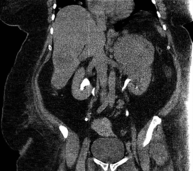
Abdominal CT image showing hepatocellular carcinoma with metastasis to left adrenal gland.
Figure 2.
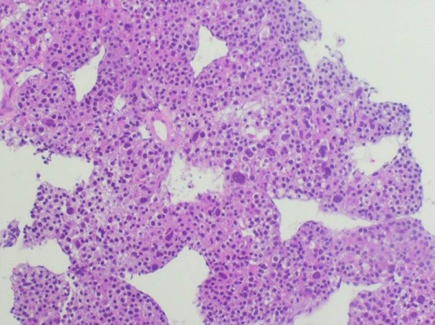
H&E tumour stain showing hepatocellular carcinoma.
Figure 3.
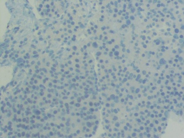
Negative immunoperoxidase tumour stain for ACTH.
Investigations
Serum cortisol levels, baseline and on dexamethasone treatment, were determined by immunoassay (IA). Urine free cortisol, serum DHEA and serum DHEA-S, all before dexamethasone treatment, were performed by LC/MS/MS. Plasma ACTH was measured by IA before initiation of dexamethasone therapy. Plasma renin activity and urinary metanephrines were determined by LC/MS/MS. Urine VMA was measured by high-performance liquid chromatography. Serum aldosterone was measured by LC/MS/MS. Imaging studies included CT scan of the chest and abdomen, and brain MRI. Pathology studies included H&E stain of the sample from CT-guided biopsy of the left adrenal gland and ACTH Immunoperoxidase staining of the tumour tissue sample.
Differential diagnosis
Ectopic CRH syndrome, ectopic ACTH syndrome, functioning adrenal adenoma, Cushing's disease, ACTH-independent autonomous adrenocortical macronodular hyperplasia, functioning adrenal carcinoma and functioning ovarian tumour.
Treatment
Cerebral oedema from metastases was treated with dexamethasone 4 mg IVPB every 8 h, after samples for the initial set of analyses were collected for ACTH level, DHEA, DHEA-S, serum cortisol and 24 h urine cortisol.
The endocrine consultants ordered aminoglutethimide 250 mg every 6 h. When the hospital pharmacy was unable to obtain this medication, the order was changed to mitotane 2 g every 8 h. The patient expired before the pharmacy was able to obtain the second drug.
Outcome and follow-up
Cerebral oedema from metastases was treated with dexamethasone. The patient's prognosis was poor and she was placed on palliative care. She expired 20 days after hospital admission.
Discussion
ECS is most often due to ACTH or CRH-secreting tumours. Most ACTH-dependent CS is due to Cushing's disease (90%). Ectopic ACTH comprises <10% of cases.2 Patients with ectopic ACTH syndrome have high plasma ACTH levels (>20 ng/L), non-suppressible cortisol on both the 2 and 8 mg dexamethasone suppression tests, and no ACTH response to CRH. Many ACTH-secreting tumours remain hidden, even after extensive imaging. Tumours secreting ACTH ectopically include: small cell lung carcinomas (27%), bronchial carcinoids (21%), islet cell tumours (16%) and thymus carcinoids (10%). Others are chromaffinomas and MCT. Imaging helps detect lung tumours, while elevated gut hormones, calcitonin and catecholamine metabolites help in diagnosing conditions such as pancreatic tumours, MCT and phaeochromocytoma. Confirmation of ectopic ACTH production requires demonstration of immunostaining positivity for ACTH in the resected tumour. Recently, extraction of appropriate mRNA by real-time PCR (RT-PCR) has proven to be a highly specific means of confirming that these tumours actually synthesise ACTH. Management of patients with ectopic ACTH syndrome requires rapid control of hypercortisolaemia.12 Ketoconazole and metyrapone appear to be reasonably safe and effective.13 14 Patients with localisable, operable sources of ectopic ACTH should have their tumours resected.15 Shah et al,16 in reviewing reported cases of ECS from 1969–2005, stated that 38% were bronchial carcinoids, 26% thymus carcinoids, 16% pancreatic tumours, 11% phaeochromocytomas and 9% were other neoplasia.
Ectopic CRH syndrome is much rarer than ectopic ACTH syndrome; only a handful of reports of ACTH producing tumours that co-secrete CRH and even fewer describing isolated ectopic CRH production exist. An extensive literature search found only 20 cases of isolated ectopic CRH secretion. MCT (33%), phaeochromocytoma (19%) and prostate cancer are among the most common sources of isolated ectopic CRH.17–23 Although the clinical picture overlaps with ectopic ACTH syndrome, it is important to keep ectopic CRH syndrome in mind. Another distinct subtype of CS is ACTH-independent macronodular hyperplasia (AIMAH). Using the clinical findings and serum cortisol level, Su and colleagues classified AIMAH into three types; subclinical, clinical and high risk. Salient features of AIMAH are male dominance, higher mean age, autonomous adrenal cortisol secretion with a low plasma ACTH level, dexamethasone non-suppressibility, normal pituitary imaging, marked, bilateral adrenal enlargement and increased isotope uptake on adrenal nuclear scan.24
The principal tests used to screen for CS are: measurement of 24 h urine free cortisol, late night salivary cortisol levels and the dexamethasone-suppression test.1 ECS confirmation requires a combination of tests, which include inferior petrosal sinus sampling for ACTH and imaging. The diagnosis is based on elevated levels of cortisol with normal or elevated levels of ACTH, usually not suppressible by high-dose dexamethasone, and lack of other possible causes of CS. Immunohistochemistry with in situ detection of ACTH and/or CRH mRNA is helpful in some instances.19
In a study of 43 patients with Cushing's disease, serum DHEA was reported to be elevated in all, with a 19.6% reduction of mean DHEA values after trans-sphenoidal hypophysectomy. High-serum adrenal androgens, for example, DHEA-S and increased urinary 17-ketosteroids, have been suggested by several authors to indicate the presence of functional adrenocortical carcinoma.25 26 Midorikawa et al reported an in vitro steroidogenesis experiment comparing adrenal carcinoma (right side) and adrenal adenoma (left side) in a single patient. 21-Hydroxylase, 17α-hydroxylase and 18-hydroxylase were found in both tumours, while 11-hydroxylase activity was found only in cancer. 21-Hydroxylase activity was more abundant in the adenoma than in the carcinoma, but 17α-hydroxylase activity was more abundant in the cancer than in the adenoma. In the adrenal steroidogenic cascade 17α-hydroxylase converts pregnenolone to 17α-hydroxy-pregnenolone, explaining the higher levels of the subsequent metabolites, DHEA and its metabolite, DHEA-S, in cancer. Immunohistochemistry of steroidogenic enzymes in pathological specimens is consistent with these in vitro studies. In vitro studies of steroidogenic enzymes showed that 21-hydroxylase activity is lower in cancers than in adenomas. Tissue culture studies showed that adrenal androgens including androstenedione, DHEA and DHEA-S were synthesised more in cancers than in adenomas.27
Typically, patients with ECS, except those with indolent bronchial carcinoids, present with cachexia, hyperglycaemia and hypokalaemia; characteristic clinical findings of CS, for example, facial mooning, abdominal striae, truncal obesity, hirsutism and increased supraclavicular fat, as seen in our patient, are not encountered. Furthermore, our patient's diabetes was easily controlled.
Lacroix and colleagues28 reported the presence of aberrant receptors in primary adrenal neoplasia that stimulate steroidogenesis in response to atypical ligands, for example, vasopressin. Our patient did not have a primary adrenal gland tumour, but had metastasis of primary HCC to the left adrenal gland. Significantly, there was no history of an adrenal adenoma or Cushing-associated symptoms preceding this admission for HCC. There was no radiographic or histopathological evidence for adrenal gland hyperplasia. While it is possible she had aberrant receptors in the metastatic or primary lesion, this has not been previously reported.
Recently, Isidori et al29 reported extraordinarily high sensitivity (100%) of 68Ga-SSTR-PET/CT in the localisation of covert tumours causing ECS. Use of this imaging modality was not widely reported before 2014, and its use in the USA is currently limited to pilot studies at a very few centres. It was not available in 2013, when our patient was evaluated. Since our patient's primary and metastatic tumour masses were readily seen on conventional CT, and tumour histology was confirmed for the hepatic and adrenal gland masses, the only additional information that might have been gleaned from 68Ga-SSTR-PET/CT would have been further identification of the apparently metastatic lung and brain lesions, and the possible identification of additional tiny metastatic foci.
A tumour may metastasise to an adrenal gland and lodge adjacent to or embed itself within a pre-existing benign adenoma. Only one such adrenal ‘collision tumour’ has been reported30 and the primary malignancy was an adenocarcinoma of the lung, not a HCC, as was the case in our patient. In the aforementioned single case, the adrenal gland adenoma had been identified several years before the adrenal gland metastasis had occurred. Our patient had no history of—or biochemical, radiographic or pathological evidence for—a pre-existing benign adrenal adenoma.
A tumour may secrete an ACTH-like substance, which could then stimulate the secretion of cortisol, aldosterone and DHEA by the adrenal gland cortices. For example, pro-opiomelanocortin (the precursor of ACTH) secretion resulting in CS has been reported in breast cancer,31 small cell lung cancer, bronchial and thymic carcinoid, and islet cell tumours,32 but not in HCC. A placental/amniotic ACTH-like peptide, dubbed chorionic corticotrophin, has also been identified.33 In all such reported cases, these peptides are measureable as ACTH by IA, and result in high-normal to high measured plasma ACTH and generalised adrenocortical hyperplasia, while in our patient, plasma ACTH was low and there was no radiographic evidence of generalised adrenocortical hyperplasia. One could postulate a compound that stimulates adrenal glandular steroidogenesis and growth via the ACTH receptor, yet is not recognised immunologically as ACTH, however, this is yet to be reported. As mentioned earlier, aberrant receptors in primary adrenal neoplasia that stimulate steroidogenesis in response to atypical ligands, for example, vasopressin, have been reported,28 but would be expected to produce a radiographically identifiable adenoma, or nodular or generalised adrenal glandular hyperplasia producing a single steroid hormone in excess. In addition, constitutive activation of the human ACTH receptor due to an interaction between two missense mutations in the MC2R gene has been reported,34 but, again, such a constitutive activation should result in a biochemically/radiographically/histologically identifiable adenoma or hyperplasia.
Finally, one could postulate the existence of an agonist, autoimmune antibody against the ACTH receptor, similar to the thyroid-stimulating immunoglobulin in Graves’ disease, however, such an antibody has yet to be reported.
The strengths of this case report include:
The very high serum levels of DHEA, DHEA-S (which exclude adrenal adenoma) and of serum and urine free cortisol associated with a metastatic HCC, confirmed by biopsy.
The absence of high-dose dexamethasone suppressibility, excluding Cushing's disease.
The suppressed low-normal plasma ACTH level-pre-dexamethasone treatment excluding Cushing's disease and ECS.
To the best of our knowledge, this is the first reported case of CS associated with this tumour type.
The case has an unusual combination of an aggressive tumour type with classical CS findings.
The systematic, methodical, virtual exclusion of other possible causes of ACTH-independent Cushing's syndrome in this setting by the exhaustively demonstrated lack of historical, biochemical, histopathological and immunopathological, as well as radiographic features consistent with these other causes, should be noted.
The limitations of this report are non-availability of stains for CRH, cortisol, DHEA and steroidogenic enzymes, and their respective mRNAs, of the pathological specimen. Also, androstenedione, 17-OH progesterone and testosterone levels were not performed. While culturing of the HCC cells with direct confirmation of their production of cortisol and DHEA would be desirable in a research setting, these techniques were not available to us and would not be realistic to obtain in a terminal patient. Thus, our ‘proof’ that the HCC produced cortisol and DHEA partially remains one of relentless deductive logic (based on painstaking exclusion of all other potential causes by the available historical, biochemical, radiographic, and pathologic data and review of the related literature) rather than direct logic.
Learning points.
Hepatocellular carcinoma can be associated with paraneoplastic Cushing's syndrome, which is very rare, and to the best of our knowledge, this is the first reported case.
This patient also manifested rare DHEA-secreting hepatocellular carcinoma.
Tumoural production of cortisol and DHEA may be inferred by demonstration of high, non-suppressible serum levels of these metabolites coupled with systematic exclusion of all other possible causes, without the need to directly demonstrate synthesis within the tumour. Occam's razor dictates that the simpler single diagnosis compatible with all the findings is to be preferred to a more complex multidiagnosis explanation due to its potentially more straightforward testability.
Functional adrenal carcinoma may be associated with high DHEA and DHEA-S levels, which may aid in diagnosis.
High DHEA and DHEA-S levels in adrenal gland malignancy are explained by lower 21-hydroxylase activity and higher 17α-hydroxylase, while adrenal adenomas have lower activity of 17α-hydroxylase and higher activity of 21-hydroxylase.
Acknowledgments
The authors wish to thank Dr Oskar Salamon of our radiology department for the CT image and Dr Jose Ruiz of our pathology department for the photomicrographs.
Footnotes
Competing interests: None declared.
Patient consent: Obtained.
Provenance and peer review: Not commissioned; externally peer reviewed.
References
- 1.Castro MD, Moreira AC. Screening and diagnosis of Cushing's syndrome. Arq Bras Endocrinol Metab 2007;51:1191–8. [DOI] [PubMed] [Google Scholar]
- 2.Shahani S, Nudelman RJ, Nalini R et al. Ectopic corticotrophin-releasing hormone (CRH) syndrome from metastatic small cell carcinoma: a case report and review of the literature. Diagn Pathol 2010;5:56.. 10.1186/1746-1596-5-56 [DOI] [PMC free article] [PubMed] [Google Scholar]
- 3.Isdiori AM, Lenzi A. Ectopic ACTH syndrome. Arq Bras Endocrinol Metab 2007;51:1217–25. 10.1590/S0004-27302007000800007 [DOI] [PubMed] [Google Scholar]
- 4.El-Serag HB. Hepatocellular carcinoma. N Engl J Med 2011;365:1118–27. 10.1056/NEJMra1001683 [DOI] [PubMed] [Google Scholar]
- 5.Huh UY, Kim JH, Kim BH et al. The incidence and clinical significance of paraneoplastic syndromes in patients with hepatocellular carcinoma. Korean J Hepatol 2005;11:275–6. [PubMed] [Google Scholar]
- 6.Kishore D, Khurana V, Raj A et al. Hepatocellular carcinoma presenting as polymyositis: a paraneoplastic syndrome. Ann Saudi Med 2011;31:533–5. 10.4103/0256-4947.84636 [DOI] [PMC free article] [PubMed] [Google Scholar]
- 7.Sultan-e-Rome, Sharif S, Shah AA. Parathyroid hormone related peptide causing hypercalcaemia in a patient with hepatocellular carcinoma. J Pak Med Assoc 2013;63:263–4. [PubMed] [Google Scholar]
- 8.Tekaya R, Abdelghni K, Abdelmoula L et al. Hepatocellular carcinoma with polymyositis as an initial symptom: a case report. Acta Clin Belq 2011;66:53–4. 10.1179/ACB.66.1.2062515 [DOI] [PubMed] [Google Scholar]
- 9.Kamińska-Winciorek G, Brzezińska-Wcisło L, Lis-Swiety A et al. Paraneoplastic type of acanthosis nigricans in patient with hepatocellular carcinoma. Adv Med Sci 2007;52:254–6. [PubMed] [Google Scholar]
- 10.Vautravers C, Rat P, Cercueil JP et al. Hepatocellular carcinoma presenting as paraneoplastic myasthenia gravis. Eur J Intern Med 2008;19:e86–7. 10.1016/j.ejim.2008.04.014 [DOI] [PubMed] [Google Scholar]
- 11.Chang PE, Tan CK. Paraneoplastic erythrocytosis as a primary presentation of hepatocellular carcinoma. Indian J Med Sci 2009;63:202–3. 10.4103/0019-5359.53167 [DOI] [PubMed] [Google Scholar]
- 12.Wajchenberg BL, Mendonca B, Liberman B et al. Ectopic ACTH syndrome. J Steroid Biochem Mol Biol 1995;53:139–51. 10.1016/0960-0760(95)00044-Z [DOI] [PubMed] [Google Scholar]
- 13.Bayraktar F, Kebapcilar L, Kocdor MA et al. Cushing's syndrome due to ectopic CRH secretion by adrenal pheochromocytoma accompanied by renal infarction. Exp Clin Endocrinol Diabetes 2006;114:444–7. 10.1055/s-2006-924154 [DOI] [PubMed] [Google Scholar]
- 14.Saeger W, Reincke M, Scholz GH et al. Ectopic ACTH- or CRH- secreting tumors in Cushing's syndrome. Zentralbl Pathol 1993;139:157–63. [PubMed] [Google Scholar]
- 15.Kenchaiah M, Hyer S. Cushing's Syndrome due to Ectopic ACTH from bronchial carcinoid: a case report and review. Case Rep Endocrinol 2012;2012:215038 10.1155/2012/215038 [DOI] [PMC free article] [PubMed] [Google Scholar]
- 16.Shah NA, Urusova IA, D'agnolo A et al. Primary hepatic carcinoid presenting as Cushing's syndrome. J Endocrinol Invest 2007;30:327–33. 10.1007/BF03346308 [DOI] [PubMed] [Google Scholar]
- 17.Oates SK, Roth SI, Molitch ME. Corticotropin-releasing hormone-producing medullary thyroid carcinoma causing Cushing's syndrome: clinical and pathological findings. Endocr Pathol 2000;11:277–85. 10.1385/EP:11:3:277 [DOI] [Google Scholar]
- 18.Eng PH, Tan LH, Wong KS et al. Cushing's syndrome in a patient with a corticotropin-releasing hormone producing pheochromocytoma. Endocr Pract 1999;5:84–7. 10.4158/EP.5.2.84 [DOI] [PubMed] [Google Scholar]
- 19.Ruggeri RM, Ferrau F, Campenni A et al. Immunohistochemical localization and functional characterization of somatostatin receptor subtypes in a corticotropin releasing hormone secreting adrenal phaeochromocytoma: review of the literature and report of a case. Eur J Histochem 2009;53:1–6. 10.4081/ejh.2009.e1 [DOI] [PubMed] [Google Scholar]
- 20.Chrisoulidou A, Pazaitou-Panayiotou K, Georgiou E et al. Ectopic Cushing's syndrome due to CRH secreting liver metastasis in a patient with medullary thyroid carcinoma. Hormones (Athens) 2008;7:259–62. [DOI] [PubMed] [Google Scholar]
- 21.Parenti G, Nassi R, Silvestri S et al. Multi-step approach in a complex case of Cushing's syndrome and medullary thyroid carcinoma. J Endocrinol Invest 2006;29:177–81. 10.1007/BF03344094 [DOI] [PubMed] [Google Scholar]
- 22.Jessop DS, Cunnah D, Millar JG et al. A pheochromocytoma presenting with Cushing's syndrome associated with increased concentrations of circulating corticotrophin-releasing factor. J Endocrinol 2008;7:259–62. [DOI] [PubMed] [Google Scholar]
- 23.Auchus RJ, Mastorakos G, Friedman TC et al. Corticotropin-releasing hormone production by a small cell carcinoma in a patient with ACTH-dependent Cushing's syndrome. J Endocrinol Invest 1994;17:447–52. 10.1007/BF03347737 [DOI] [PubMed] [Google Scholar]
- 24.Su HC, Dai J, Huang X et al. Classification, diagnosis and treatment of ACTH-independent macronodular adrenal hyperplasia. Can Urol Assoc J 2013;7:E594–7. 10.5489/cuaj.420 [DOI] [PMC free article] [PubMed] [Google Scholar]
- 25.Yoshida A, Nishikawa T, Tamura Y et al. Evidence for decreased activity of guanine nucleotide binding protein in adenylate cyclase of cell membrane in human ACTH-unresponsive adrenocortical carcinoma. Endocrinol Jpn 1986;33:891–9. 10.1507/endocrj1954.33.891 [DOI] [PubMed] [Google Scholar]
- 26.Margioris AN, Chrousos GP. Cushing's syndrome evaluation. In: Biglieri EG, Merby JC, eds. Endocrine hypertension. New York: Raven Press, 1990:99–111. [Google Scholar]
- 27.Midorikawa S, Hashimoto S, Kuriki M et al. A patient with preclinical Cushing's DHEA-S secretion having unilateral adrenal carcinoma and contralateral adenoma. Endocr J 1999;46:59–66. 10.1507/endocrj.46.59 [DOI] [PubMed] [Google Scholar]
- 28.Christopoulos S, Bourdeau I, Lacroix A. Aberrant expression of hormone receptors in adrenal Cushing's syndrome. Pituitary 2004;7:225–35. 10.1007/s11102-005-1083-7 [DOI] [PubMed] [Google Scholar]
- 29.Isidori AM, Sbardella E, Zatelli MC et al. , ABC Study Group. Conventional and nuclear medicine imaging in ectopic Cushing's syndrome: a systematic review. J Clin Endocrinol Metab 2015;100:3231–44. 10.1210/JC.2015-1589 [DOI] [PMC free article] [PubMed] [Google Scholar]
- 30.Anderson SB, Webb MD, Banks KP. Adrenal collision tumor diagnosed by F-18 fluorodeoxyglucose PET/CT. Clin Nuc Med 2010;35:414–17. 10.1097/RLU.0b013e3181db4df1 [DOI] [PubMed] [Google Scholar]
- 31.Pelte MF, Schwaller J, Cerrato C et al. Pro-opiomelanocortin expression in a metastatic breast carcinoma with ectopic ACTH expression. Breast J 2004;10:350–4. 10.1111/j.1075-122X.2004.21467.x [DOI] [PubMed] [Google Scholar]
- 32.Beuschlein F, Hammer GD. Ectopic pro-opiomelanocortin syndrome. Endocrinol Metab Clin North Am 2002;31:191–234. 10.1016/S0889-8529(01)00025-1 [DOI] [PubMed] [Google Scholar]
- 33.Genazzani AR, Hürlimann J, Fioretti P et al. In vitro synthesis of an ACTH-like hormone and chorionic somatomammotrophin by placental and amniotic cells. Experientia 1974;30:430–2. 10.1007/BF01921705 [DOI] [PubMed] [Google Scholar]
- 34.Swords FM, Noon LA, King PJ et al. Constitutive activation of the human ACTH receptor resulting from a synergistic interaction between two naturally occurring missense mutations in the MC2R gene. Mol Cell Endocrinol 2004;213:149–54. 10.1016/j.mce.2003.10.052 [DOI] [PubMed] [Google Scholar]