Abstract
Hydatid cyst disease, which is endemically observed and an important health problem in our country, involves the spleen at a frequency ranking third following the liver and the lungs. In this study, we aimed to evaluate the efficacy and results of management in splenic hydatid cysts. The demographic data, localization, diagnosis, treatment methods, and the length of postoperative hospital stay of patients with splenic hydatid cysts in a 12-year period were evaluated retrospectively. Seventeen cases were evaluated. Among these, 13 were females and four were males. Seven had solitary splenic involvement, eight had involvement of both the spleen and the liver, and two had multiple organ involvement. Ten had undergone splenectomy, one had undergone distal splenectomy, and the remaining cases had undergone different surgical procedures. The patients had received albendazole treatment in the pre- and postoperative period. One patient had died secondary to hypernatremia on the first postoperative day. The clinical picture in splenic hydatid cysts, which is seen rarely, is usually asymptomatic. The diagnosis is established by ultrasonography and abdominal CT. Although splenectomy is the standard mode of treatment, spleen-preserving methods may be used.
Keywords: Splenic, Hydatid cysts, Splenectomy
Introduction
Hydatid cyst disease maintains its importance as a common health problem in our country. This disease is endemic in many regions of the world. It is caused by Echinococcus granulosus. It most frequently involves the liver (50−70 %) and the lungs (11−17 %). Primary splenic involvement (2.5−5.8 %) is in the third rank for frequency following the liver and the lungs [1, 2]. Besides, soft tissues (2.4−5.3 %), the heart (0.5−3 %), the pericardium (5 %), muscle, and subcutaneous tissues (0.5−4.7 %) have also been reported to be involved [2–4]. While splenic involvement may be solitary, it may also accompany other organ involvement, commonly the liver. The treatment protocols of hydatid cysts have generally been established, but cases with splenic involvement are still controversial. We hereby present 17 splenic hydatid cysts.
Material and Method
The data of 17 patients, who had undergone surgery for splenic hydatid cysts at the Atatürk University Medical Faculty Department of Surgery between January 1998 and August 2010, were collected retrospectively. The demographic data, symptoms, clinical findings, radiological findings, intraoperative findings, surgical interventions, postoperative course, complications, length of hospital stay, and mortality were recorded.
Findings
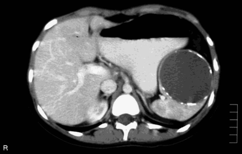
Seventeen patients had undergone surgery for splenic hydatid cyst at the Atatürk University Medical Faculty Department of Surgery between January 1998 and August 2010 and were followed up. The gender distribution was 13 females and four males. The mean age was 40.8 years (17−70 years). All the patients had presented with complaints of pain, fullness, and discomfort in the left upper quadrant. On physical examination, there was tenderness in the left upper quadrant and epigastrium. Six patients had dullness to percussion over the Traube’s space. There were no patients with abnormal laboratory findings. Serological tests were not used for the diagnosis. Thirteen patients underwent ultrasonography and abdominal tomography, while the remaining four underwent abdominal tomography only (Fig. 1). On radiology, seven were found to have solitary splenic involvement, eight had accompanying liver involvement, and two had multiple organ involvement. The data including organ involvement, cyst diameters, numbers, and localizations have been presented in Table 1. All the cases received albendazole (10 mg/kg/day) for at least 2 weeks preoperatively. Following this medication, ten patients underwent splenectomy, one underwent distal splenectomy, two underwent partial cystectomy + omentoplasty, and four underwent cystotomy + omentoplasty.
Fig. 1.
Computed tomography shows splenic hydatid cyst
Table 1.
The data including tissue involvement, cyst diameters, numbers, and localizations
| Cases | Splenic localization | Diameter (cm) | Type (WHO) | Others tissue | Surgery |
|---|---|---|---|---|---|
| 1 | Upper pole | 15 × 12 | 3 | – | Splenectomy |
| 2 | Lower pole | 6.5 × 8 | 3 | – | Distal splenectomy |
| 3 | Upper pole | 16 × 17 | 3 | – | Splenectomy |
| 4 | Lower pole | 20 × 15 | 3 | – | Cystotomy + omentoplasty |
| 5 | Lower pole | 12 × 14 | 3 | – | Splenectomy |
| 6 | Hilus | 15 × 10 | 3 | – | Splenectomy |
| 7 | Upper pole | 12 × 11 | 3 | – | Splenectomy |
| 8 | Middle pole | 15 × 12 | 3 | Liver | Partial cystectomy + omentoplasty |
| 9 | Upper pole | 8 × 10 | 3 | Liver | Splenectomy |
| 10 | Upper pole | 10 × 9 | 3 | Liver | Partial cystectomy + omentoplasty |
| 11 | Middle pole | 8 × 10 | 3 | Liver | Cystotomy + omentoplasty |
| 12 | Lower pole | 4 × 6 | 3 | Liver | Cystotomy + omentoplasty |
| 13 | Upper pole | 10 × 11 | 3 | Liver | Splenectomy |
| 14 | Upper pole | 3 × 2 | 3 | Liver | Splenectomy |
| 15 | Middle pole | 7 × 8 | 3 | Liver, kidney | Cystotomy + omentoplasty |
| 16 | Lower pole | 10 × 8 | 3 | Liver | Splenectomy |
| 17 | Upper pole | 10 × 8 | 3 | Liver, pelvis, intestine mezoperitoneum | Splenectomy |
Ten of the 17 cases had not undergone previous surgery for hydatid cysts. Five of these ten cases had solitary splenic involvement, while only two of the seven cases previously having undergone surgery for hydatid cysts had solitary splenic involvement.
The length of postoperative hospital stay has a mean of 9.2 (1−17) days. During this period, three patients had developed surgical site infection. One patient that had accompanying liver and omentum involvement died on the first postoperative day. That patient had 11 different cyst foci on the omentum and was thought to have died secondary to hypernatremia developing on the night of surgery. According to our clinical protocol, all the cases received postoperative albendazole treatment for 6 weeks at a dose of 10 mg/kg/day.
Discussion
Hydatid cyst disease is most frequently observed in the liver at a rate of 50−70 %, and in the lungs at a rate of 10−30 % as the second most frequent site. The third frequent site is the spleen with a rate of 2.5−5.8 % [2–4]. In our study, splenic involvement was found at a rate of 6.3 %. Solitary splenic involvement was found at a rate of 2.6 %. In 20−50 % of the cases, there is an accompanying organ involvement. Seven of our 17 cases had solitary splenic involvement, and the rate of accompanying organ involvement was 58 %.
In splenic hydatid cysts, the clinical picture is usually asymptomatic. Although the patients’ complaints are not so great as to interrupt their daily activities, hydatid disease is generally identified incidentally during radiological investigations for some other reasons [4–6]. The most frequent complaints are left upper quadrant pain, fullness, and discomfort [4, 6, 7]. Left upper quadrant fullness and discomfort are secondary to splenomegaly, which were also observed in six cases in our series (35.3 %). The size of the hydatid cysts was at a minimum of 4 × 6 cm and a maximum of 20 × 15 cm.
The diagnosis is established by radiological imaging and serological tests [6]. Özdoğan et al. [8] reported that serological tests were not necessary. In general, in the diagnosis of hydatid cyst, the Casoni test has been found to be positive at a rate of 50−80 % and the Weinberg test at a rate of 70 %. Both tests have a high false-positivity rate [2–4]. There are different views in the literature. In this respect, the use and the diagnostic value of serology have decreased. Furthermore, factors such as some of the cases having previously undergone hydatid cyst surgery, our region being endemic for the disease, and the radiological findings providing valuable information for the diagnosis, have decreased the value of serological tests. Therefore, serological tests are not used for routine evaluation; however, they are used when it is difficult to differentiate type I hydatid cysts from simple cysts. In our opinion, serology should be used in such controversial cases and in patients living in non-endemic regions.
Moreover, in the same study, it was stated that ultrasonography and CT findings were not specific for hydatid cysts, but that they were the most valuable diagnostic methods [8]. We generally agree on this issue. Ultrasonography and CT were used together in 13 cases in our series. CT was used in four cases. The diagnostic accuracy rate of radiological tools was 100 % in our series. Abdominal CT does not only provide information about the diagnosis but also provides information regarding the cyst localization, neighborhood, type, and contents. Once the diagnosis is made, systemic examination should be performed and other organs should be investigated for involvement [9].
The choice of treatment to be preferred is preoperative medical treatment and intraoperative scolocidal injection together with splenectomy. Cases in which the cyst has a low degree of adhesion to the surrounding tissue and in which it is thought that splenectomy can be performed with low complications, splenectomy is the most appropriate choice [4, 8]. Although splenectomy has been a preferred surgical procedure in the literature and in our series, six patients had undergone spleen-preserving surgical interventions. Different from liver hydatid cysts, cystotomy is an adequate surgical procedure in cases with splenic hydatid cysts as it carries no risk such as bile leakage. It provides preservation of the spleen. Furthermore, enucleation and partial splenectomy have a higher risk of hemorrhage than cystotomy. Therefore, partial cystectomy or cystotomy that protects the fibrous tissue is more rational. In our series, ten cases had undergone splenectomy and one had undergone distal splenectomy. In six cases, the spleen was preserved. Other surgical methods include PAIR, partial splenectomy, laparoscopic splenectomy, partial cystectomy + omentoplasty, spleen-preserving enucleation in children and adolescents, and marsupialization [4, 7, 8]. Cases undergoing splenectomy also received pneumococcus vaccine postoperatively.
In splenic hydatid cyst cases, the use of antiparasitic medication following an uneventful splenectomy is controversial. Özdoğan et al. [8] approved of continuing the medical treatment for a few months. Despite this recommendation by Özdoğan, there has been no controlled study on whether to administer albendazole following splenectomy or not, and this information depends on experience. Albendazole treatment may not be recommended following unproblematic splenectomy (without rupturing the cyst content) in cases with solitary splenic involvement, but we think that withholding albendazole treatment in those with multiple organ involvement would not be wise. We think that short-term medical treatment after surgery has a low complication rate and may decrease the recurrence. Unfortunately, there is no controlled study to support this opinion.
Conclusion
Splenic hydatid cyst is a rather rare condition. In recent years, successful results have been obtained by ultrasonography and CT-guided percutaneous cyst drainage and cavity irrigation using scolicidal agents. PAIR has not been completely established yet. The aim of surgical treatment should be to preserve the spleen. Medical treatment should begin in the preoperative period and continue following surgery.
References
- 1.Amman RW, Eckert J. Cestodes. Echinococcus. Gastroenterol Clin N Am. 1996;25:655–689. doi: 10.1016/S0889-8553(05)70268-5. [DOI] [PubMed] [Google Scholar]
- 2.Arikanoglu Z, Taskesen F, Gumus H, Onder A, Aliosmanoglu I, Gul M, et al. Selecting a surgical modality to treat a splenic hydatid cyst: total splenectomy or spleen-saving surgery? J Gastrointest Surg. 2012;16:1189–1193. doi: 10.1007/s11605-012-1837-2. [DOI] [PubMed] [Google Scholar]
- 3.Malik AA, ul Bari S, Younis M, Wani KA, Rather AA. Primary splenic hydatidosis. Indian J Gastroenterol. 2011;30:175–177. doi: 10.1007/s12664-011-0104-x. [DOI] [PubMed] [Google Scholar]
- 4.Ramia-Angel JM, Gasz A, de la Plaza-Llamas R, Quinones-Sampedro J, Sancho E, Garcia Parreno J. Hidatidosis the spleen. Pol Przegl Chir. 2011;83:271–275. doi: 10.2478/v10035-011-0042-4. [DOI] [PubMed] [Google Scholar]
- 5.Durgun V, Kapan S, Kapan M, Karabıçak I, Aydoğan F, Goksoy E. Primary splenic hydatidosis. Dig Surg. 2003;20:38–41. doi: 10.1159/000068864. [DOI] [PubMed] [Google Scholar]
- 6.Dar MA, Shah OJ, Wani NA, Kahn FA, Shah P. Surgical management of splenic hydatidosis. Surg Today. 2002;32:224–229. doi: 10.1007/s005950200025. [DOI] [PubMed] [Google Scholar]
- 7.Berrada S, Ridai M, Mokhtari M. Hydatid cysts of the spleen: splenectomy or conservative surgery? Ann Chir. 1991;45:434–464. [PubMed] [Google Scholar]
- 8.Özdoğan M, Baykal A, Keşkek M, Yorgancı K, Hamaloğlu E, Sayek I. Hydatid cyst of the spleen: treatment options. Int Surg. 2001;86:122–126. [PubMed] [Google Scholar]
- 9.Uriarte C, Pomares N, Martin M, Conde A, Alonso N, Bueno MG. Splenic hydatidosis. Am J Trop Med Hyg. 1991;44:420–423. doi: 10.4269/ajtmh.1991.44.420. [DOI] [PubMed] [Google Scholar]


