Abstract
Focused assessment with sonography for trauma (FAST) is a limited ultrasound examination, primarily aimed at the identification of the presence of free intraperitoneal or pericardial fluid. In the context of blunt trauma abdomen (BTA), free fluid is usually due to hemorrhage, bowel contents, or both; contributes towards the timely diagnosis of potentially life-threatening hemorrhage; and is a decision-making tool to help determine the need for further evaluation or operative intervention. Fifty patients with blunt trauma abdomen were evaluated prospectively with FAST. The findings of FAST were compared with contrast-enhanced computed tomography (CECT), laparotomy, and autopsy. Any free fluid in the abdomen was presumed to be hemoperitoneum. Sonographic findings of intra-abdominal free fluid were confirmed by CECT, laparotomy, or autopsy wherever indicated. In comparing with CECT scan, FAST had a sensitivity, specificity, and accuracy of 77.27, 100, and 79.16 %, respectively, in the detection of free fluid. When compared with surgical findings, it had a sensitivity, specificity, and accuracy of 94.44, 50, and 90 %, respectively. The sensitivity of FAST was 75 % in determining free fluid in patients who died when compared with autopsy findings. Overall sensitivity, specificity, and accuracy of FAST were 80.43, 75 and 80 %, respectively, for the detection of free fluid in the abdomen. From this study, we can safely conclude that FAST is a rapid, reliable, and feasible investigation in patients with BTA, and it can be performed easily, safely, and quickly in the emergency room with a reasonable sensitivity, specificity, and accuracy. It helps in the initial triage of patients for assessing the need for urgent surgery.
Keywords: FAST, BTA, CECT scan, Hemoperitoneum
Introduction
Trauma is a major cause of death during the first four decades of life and is often associated with permanent disability, resulting in the loss of productive years in young individuals [1]. Trauma commonly affects the age group of 15–44 years, which is economically the most productive age group [1]. The incidence of abdominal trauma is 20 % of all trauma cases, and the relative incidence of blunt/penetrating abdominal trauma differs according to the geographic area. In urban areas, the incidence of gunshot and stab wounds (penetrating wounds) is higher than blunt trauma, and the reverse is the case in rural areas [1].
Blunt injuries to the abdomen are most common following road traffic crashes and fall from heights [1, 2] and often pose a diagnostic and management challenge. The rapid diagnosis and appropriate and timely intervention of these patients are essential to avoid significant morbidity and mortality associated with delay in treatment [3].
Because there are inadequacies in physical examination with low sensitivity for the detection of intra-abdominal injuries, particularly in patients with blunt multisystem trauma [2–4], focused assessment with sonography for trauma (FAST) has become a safe, reliable, and common modality worldwide in the evaluation of blunt abdominal trauma. The most important factor in the management of blunt abdominal trauma is triage of the patients who require immediate laparotomy or observation only. For this, we need a rapid, reliable, cost-effective, and reproducible investigation. FAST is used for this purpose in most trauma centers in primary screening for the diagnosis of intraperitoneal hemorrhage or intra-abdominal injury [2, 3].
FAST is a rapid bedside examination that has now become an extension of the physical examination of the patient with blunt trauma abdomen (BTA) and can be performed during the resuscitation of such patients [5]. There are various studies that show the good sensitivity of ultrasound in the detection of hemoperitoneum, but its role in the detection of solid organ injuries is still debatable [6].
Materials and Methods
Fifty consecutive patients with history of BTA presenting in our emergency department (ED) were included in this study from 1 April 2004 to 31 May 2006. The patients who presented with unrecordable blood pressure or in shock, with an indication for an immediate laparotomy, were excluded from the study. The mechanism of injury and physical examination findings were recorded. An FAST examination was done during initial resuscitation in the ED.
Hemodynamically stable patients with a positive FAST for free fluid underwent an abdominal CT scan with intravenous and oral contrast. If the patients were hemodynamically unstable and not responding to resuscitation or if there was any other indication for laparotomy, the patients underwent immediate surgery without any further investigation. If FAST did not detect any fluid, serial physical examination was performed. The patients were followed up until the time of discharge. Serial ultrasounds were obtained to monitor the progress of the patients while under observation.
FAST was performed by the resident radiologist in the ED before contrast-enhanced computed tomography (CECT), laparotomy, or autopsy. The ultrasound machine installed in the ED was used for this purpose (Sonoline Versa Pro, Siemens, Germany). A curvilinear/sector probe of 3.5 Hz was used. Any free fluid in the abdomen was presumed to be due to hemoperitoneum. FAST findings were confirmed by CECT, laparotomy, or autopsy wherever indicated. For patients in whom nonoperative management was decided, a CECT scan of the abdomen and pelvis including lower chest was performed to confirm the findings of FAST, and the patients were kept under observation. The progress was recorded and followed up until the time of discharge or until the termination of nonoperative management. CECT scan was done in hemodynamically stable patients or those who responded to resuscitation and for whom a nonoperative management was planned with no immediate indication for laparotomy. By statistical methods, sensitivity, specificity, positive predictive value, and negative predictive values were calculated. A p < 0.05 was considered significant.
Results
Age of the patients ranged from 3 to 65 years, and mean age was 28.62 years. Most patients were males (male to female ratio, 42:8). History was available in 48 patients regarding the time lag between trauma and arrival in hospital. Of these, almost one third of the patients reached within the first hour of trauma. All of them, except one patient, reached within 24 h of trauma. All 50 patients underwent FAST examination at presentation.
Of 50 patients, 28 patients presented with shock at initial presentation (Table 1). The resuscitative efforts failed in eight patients, and they died in the emergency room itself within 30 min of their arrival before they could be shifted to the operation theater for emergency surgery. Twelve patients underwent immediate surgery for hemodynamic instability. Eight of these patients responded to resuscitation with crystalloids and blood products. After adequate stabilization, they underwent CECT scan and were planned for nonoperative management.
Table 1.
FAST compared to CECT findings for free fluid
| Parameter | Free fluid (n = 24) |
|---|---|
| True positive | 17 |
| True negative | 2 |
| False positive | 0 |
| False negative | 5 |
| Sensitivity | 77.27 % |
| Specificity | 100 % |
| Positive predictive value | 100 % |
| Negative predictive value | 28.57 % |
Twenty-two patients were hemodynamically stable at presentation. Of these, three patients had frank signs of peritonitis and underwent laparotomy. Another 3 patients had suspicion of bowel injury, and the remaining 16, who were stable, underwent CECT abdomen.
After initial resuscitation, 24 patients were planned for nonoperative management on the basis of hemodynamic stability and with no other immediate indication for laparotomy. Eighteen patients were taken up for immediate laparotomy based on their clinical findings and imaging studies, and the remaining eight died during the initial resuscitation.
Of 50 patients, FAST was positive for free fluid in 38 patients (37 true positive and 1 false positive) [nonoperative management (NOM) Group (Gp), 17/24 + immediate death Gp, 6/8 + immediate laparotomy Gp, 15/18 (including one false positive) = 38/50]. FAST detected one case of intrapericardial fluid in a case of blunt trauma to the lower anterior chest and upper abdomen, which was confirmed by echocardiography and surgery.
Nonoperative Management
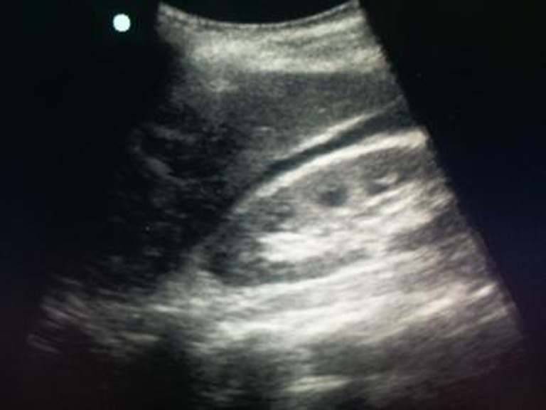
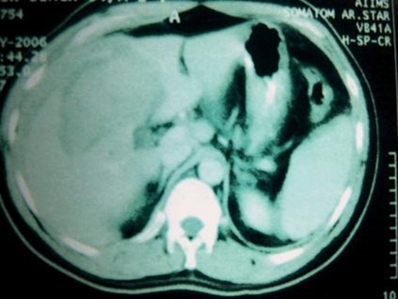
A total of 24 patients underwent nonoperative management. Of these, eight patients were in hypotension at presentation, and they responded to fluid resuscitation and packed red blood cell transfusion. Sixteen patients were hemodynamically stable at presentation. All these 24 patients underwent FAST as well as CECT scan of the lower chest, abdomen, and pelvis with oral and intravenous contrast. Of these, FAST showed 17 patients with free intraperitoneal fluid and 7 patients with no free fluid in abdomen, whereas CECT was positive in 22 and negative in 2 patients for free intraperitoneal fluid (Figs. 1 and 2). CECT findings were compared with FAST findings for free fluid.
Fig. 1.

FAST examination showing free fluid in the hepatorenal fossa
Fig. 2.
CECT scan of the abdomen showing perihepatic fluid collection
FAST had a sensitivity, specificity, and accuracy of 77.27, 100, and 79.2 %, respectively, for the detection of free fluid when compared to CECT scan (Table 1).
There were two failures of nonoperative management: one underwent splenorrhaphy on day 5 and the other patient with grade IV liver injury needed laparotomy after 4 days of nonoperative management.
Follow-up of the patients was carried out at 1 and 4 weeks following discharge. Ultrasound was done at 1 week. No significant complications occurred in any of these patients needing hospitalization or any intervention.
Operative Management
Twenty patients required laparotomy. Of these, 18 patients required immediate laparotomy after initial resuscitation for various indications as described earlier. Two patients were operated upon due to the failure of conservative management.
Of 20 patients, including 2 FAST-positive patients with failure of NOM, who underwent laparotomy, FAST detected free fluid in 17 patients, whereas laparotomy confirmed the presence of free fluid in 18 patients.
The FAST had a sensitivity, specificity, and accuracy of 94.44, 50, and 90 %, respectively, in the detection of free fluid in the operative group (Table 2).
Table 2.
FAST compared to surgical findings for free fluid
| Parameter | Free fluid |
|---|---|
| True positive | 17 |
| True negative | 1 |
| False positive | 1 |
| False negative | 1 |
| Sensitivity | 94.44 % |
| Specificity | 50 % |
| Positive predictive value | 94.44 % |
| Negative predictive value | 50 % |
The overall sensitivity, specificity, and accuracy of FAST were 80.4, 75, and 80 %, respectively, for the detection of free fluid in the abdomen after BTA (Table 3).
Table 3.
FAST compared to CECT/surgery/autopsy for free fluid
| Parameter | Free fluid (n = 50) |
|---|---|
| True positive | 37 |
| True negative | 3 |
| False positive | 1 |
| False negative | 9 |
| Sensitivity | 80.43 % |
| Specificity | 75 % |
| Positive predictive value | 97.36 % |
| Negative predictive value | 27.27 % |
Mortality
A total of 15 (30 %) patients died. Eight patients (16 %) died during resuscitation or during shifting to the operation theater for laparotomy. Five patients (10 %) died following operation, and two patients (4 %) died while on nonoperative management. All patients who died underwent autopsy.
Of the eight patients who died during resuscitation, FAST detected six patients with free fluid and two were false negatives for free fluid. Of all 15 patients (30 %) who died in the study, FAST detected 10 patients with free fluid, and autopsy showed all 15 patients with free fluid. The sensitivity and positive predictive value of FAST were 75 and 100 %, respectively, in the detection of free fluid as compared to autopsy.
Discussion
BTA is often a great diagnostic and management challenge to trauma surgeons. The rapid diagnosis and appropriate and timely intervention in patients of intra-abdominal injury are essential to avoid significant morbidity and mortality that, in the case of delayed diagnosis and treatment, are very high, while the outcome of early diagnosis and optimal intervention is extremely rewarding [7]. A number of studies have been done for accurate diagnosis of intra-abdominal bleeding and its management [2–4, 6, 8–12].
The most important factor in the management of blunt abdominal trauma is triaging the patients who require immediate laparotomy or observation only. The history and physical examination may be unreliable because of various factors. No single investigation has been found to accurately identify patients who require immediate laparotomy. For this, we need a rapid, reliable, safe, cost-effective, and repeatable investigation. In this clinical scenario, FAST is used frequently in most trauma centers in primary screening for the diagnosis of intraperitoneal hemorrhage [2, 3].
In a study by Bode et al. [8], 1,671 patients underwent FAST. Four hundred seventy patients were with negative sonographic findings and were discharged approximately 12 h after admission, without confirmation by a gold standard test. This gave a sensitivity of 88 %, a specificity of 100 %, and an accuracy of 99 %.
In a study by Healy et al. [13], 800 patients were screened by ultrasound in blunt abdominal trauma. They determined that sonography had a sensitivity of 88 % and a specificity of 98 %.
In a study by Richards et al. [14], 3,264 patients were taken and all underwent FAST, and the findings were compared with CECT/surgery/clinical outcome. In this study, the sensitivity, specificity, and accuracy were 60, 98, and 80 %, respectively, for free intraperitoneal fluid. In a large review, Adams et al. [15] concluded that FAST examination has 82 % sensitivity and 99 % specificity for detecting intra-abdominal injuries in adults with blunt abdominal trauma. Fleming et al. [16], in a study, concluded that FAST had a specificity of 94.7 % [95 % confidence interval (CI), 0.75–0.99], sensitivity of 46.2 % (95 % CI, 0.33–0.60), positive predictive value of 0.96 (0.81–0.99), and negative predictive value of 0.39 (0.26–0.54).
In our study, we have used the best available gold standards for comparison of the results of FAST, and the sensitivity of FAST has ranged from 63 to 100 %. The overall sensitivity, specificity, and accuracy of FAST were 80.4, 75, and 80 %, respectively, for free intraperitoneal fluid. When FAST findings were compared with only the CECT findings, the sensitivity, specificity, and accuracy were 77.3, 100, and 79.2 %, respectively, for free intraperitoneal fluid. A total of 20 patients underwent surgery (18 immediately and 2 out of failure in nonoperative management). When FAST findings were compared with the surgical findings, the sensitivity, specificity, and accuracy were 94.4, 50, and 90 %, respectively, for free intraperitoneal fluid.
Conclusion
From this study, it can be reliably concluded that FAST is a feasible investigation in patients with BTA, and it can be performed easily and quickly in the emergency room with a reasonable sensitivity, specificity, and accuracy. It helps in the initial triage of patients for conservative management or immediate operation. FAST can be used safely in patients with blunt abdominal and chest trauma for the diagnosis of intraperitoneal bleeding and traumatic pericardial tamponade, without any added complications. CECT scan can be used in BTA for the diagnosis of hemoperitoneum and hollow viscus injuries. CECT scan is more sensitive and specific in detecting free intraperitoneal fluid following BTA than FAST, but it is time consuming and expensive. Though the present study was a small pilot study, we need to perform a larger study to reach a definite conclusion.
Acknowledgments
Acknowledgments
No financial grant has been received for this study.
Contributor Information
Subodh Kumar, Email: Subodh6@gmail.com.
Virinder Kumar Bansal, Phone: +91-11-26593686, FAX: +91-11-26588324, Email: drvkbansal@gmail.com.
References
- 1.Timothy CF, Martin AC. Abdominal trauma. In: Mattox KL, Feliciano DV, Moore EE, editors. Trauma. 4. USA: McGraw-Hill; 2000. p. 583. [Google Scholar]
- 2.Rozycki GS, Ochsner MG, Schmidt JA, Frankel HL, Davis TP, Wang D, et al. A prospective study of surgeon-performed ultrasound as the primary adjuvant modality for injured patient assessment. J Trauma. 1995;39:492–8. doi: 10.1097/00005373-199509000-00016. [DOI] [PubMed] [Google Scholar]
- 3.Ingeman JE, Plewa MC, Okasinski RE, King RW, Knotts FB. Emergency physician use of ultrasonography in blunt abdominal trauma. Acad Emerg Med. 1996;3:931–7. doi: 10.1111/j.1553-2712.1996.tb03322.x. [DOI] [PubMed] [Google Scholar]
- 4.Kristensen JK, Buemann B, Kühl E. Ultrasonic scanning in the diagnosis of splenic haematomas. Acta Chir Scand. 1971;137(7):653–657. [PubMed] [Google Scholar]
- 5.Ascher WM, Parvin S, Virgilio RW. Echographic evaluation of splenic injury after blunt trauma. Radiology. 1976;118:411–415. doi: 10.1148/118.2.411. [DOI] [PubMed] [Google Scholar]
- 6.Raum MR, Bouillon B, Eypasch E, Tiling T. Technology assessment of ultrasound in acute diagnosis of blunt abdominal trauma. Langenbecks Arch Chir Suppl Kongressbd. 1997;114:461–464. [PubMed] [Google Scholar]
- 7.Chiu WC, Cushing BM, Rodriguez A, Ho SM, Mirvis SE, Shanmuganathan K, et al. Abdominal injuries without hemoperitoneum: a potential limitation of focused abdominal sonography for trauma (FAST) J Trauma. 1997;42:617–622. doi: 10.1097/00005373-199704000-00006. [DOI] [PubMed] [Google Scholar]
- 8.Bode PJ, Niezen RA, van Vugt AB, Schipper J. Abdominal ultrasound as a reliable indicator for conclusive laparotomy in blunt abdominal trauma. J Trauma. 1993;34:27–31. doi: 10.1097/00005373-199301000-00005. [DOI] [PubMed] [Google Scholar]
- 9.Liu M, Lee CH, P’eng FK. Prospective comparison of diagnostic peritoneal lavage, computed tomographic scanning, and ultrasonography for the diagnosis of blunt abdominal trauma. J Trauma. 1993;35:267–270. doi: 10.1097/00005373-199308000-00016. [DOI] [PubMed] [Google Scholar]
- 10.Branney SW, Wolfe RE, Moore EE, Albert NP, Heinig M, Mestek M, et al. Quantitative sensitivity of ultrasound in detecting free intraperitoneal fluid. J Trauma. 1995;39:375–380. doi: 10.1097/00005373-199508000-00032. [DOI] [PubMed] [Google Scholar]
- 11.Chambers JA, Pilbrow WJ. Ultrasound in abdominal trauma: an alternative to peritoneal lavage. Arch Emerg Med. 1988;5:26–33. doi: 10.1136/emj.5.1.26. [DOI] [PMC free article] [PubMed] [Google Scholar]
- 12.Jehle D, Guarino J, Karamanoukian H. Emergency department ultrasound in the evaluation of blunt abdominal trauma. Am J Emerg Med. 1993;11:342–346. doi: 10.1016/0735-6757(93)90164-7. [DOI] [PubMed] [Google Scholar]
- 13.Healy MA, Simons RK, Winchell RJ, et al. A prospective evaluation of abdominal ultrasound in blunt trauma: is it useful? J Trauma. 1996;40:875. doi: 10.1097/00005373-199606000-00004. [DOI] [PubMed] [Google Scholar]
- 14.Richards JR, Schleper NH, Woo BD, Bohnen PA, McGahan JP. Sonographic assessment of blunt abdominal trauma: a 4-year prospective study. J Clin Ultrasound. 2002;30(2):59–67. doi: 10.1002/jcu.10033. [DOI] [PubMed] [Google Scholar]
- 15.Adams B, Sisson C. Review: bedside ultrasonography has 82% sensitivity and 99% specificity for blunt intraabdominal injury. Ann Intern Med. 2012;157(4):2–12. doi: 10.7326/0003-4819-157-4-201208210-02012. [DOI] [PubMed] [Google Scholar]
- 16.Fleming S, Bird R, Ratnasingham K, Sarker SJ, Walsh M, Patel B. Accuracy of FAST scan in blunt abdominal trauma in a major London trauma centre. Int J Surg. 2012;10(9):470–4. doi: 10.1016/j.ijsu.2012.05.011. [DOI] [PubMed] [Google Scholar]


