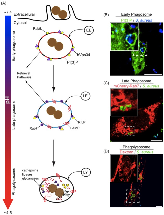
Figure 2.
Maturation of phagosomes in macrophages. In (A) the maturation pathway that a newly formed phagosome typically follows is depicted. Nascent phagosomes interact sequentially with compartments of the endo/lysosomal network, giving rise to phagosomes that, at each maturation stage, possess biochemically distinct properties. Phagosomes can be classified as early phagosomes, late phagosomes, or phagolysosomes and the presence of specific markers aid in defining the state of phagosome maturation. Early phagosomes are marked with the small GTPase Rab5, the phosphatidylinositol 3 kinase Vps34, and the phosphoinositide PI(3)P. Late phagosomes are decorated with LAMP-1/2, Rab7, RILP and are devoid of PI(3)P and Rab5. Finally, phagolysosomes that are formed by fusion between late phagosomes and lysosomes, are enriched with hydrolytic enzymes (e.g., proteases, glycanases, and lipases) and are markedly acidic; In (B–D) representative laser scanning confocal micrographs depicting RAW macrophages containing S. aureus USA300 that reside in early phagosomes, late phagosomes, or in phagolysosomes, respectively, are shown. S. aureus in green are expressing GFP and S. aureus in blue were stained with eFluor670; In (B) accumulation of PI(3)P is detected by expression of the lipid biosensor 2xFYVE-GFP; In (C) the distribution of Rab7 expressed as a mCherry-Rab7 fusion protein is shown; In (D) dextran pulse chase experiments were performed to label lysosomes with TMR-dextran (10 kDa) prior to phagocytosis of S. aureus. For each micrograph the hashed box demarcates the region of the micrograph presented in the inset; Note in (D) the GFP channel was omitted from the inset to clearly show the accumulation of dextran around each coccus that appears as a void in the fluorescence. Bars equal ~10 μm. Abbreviations: PI(3)P, phosphatidylinositol-3-phosphate; LAMP, lysosome-associated membrane proteins; RILP, Rab7 lysosomal interacting protein; GFP, green fluorescent protein; TMR, tetramethylrhodamine.

