Abstract
Voltage-gated Na+ channels (VGSCs) are heteromeric protein complexes containing pore-forming α subunits together with non-pore-forming β subunits. There are nine α subunits, Nav1.1-Nav1.9, and four β subunits, β1-β4. The β subunits are multifunctional, modulating channel activity, cell surface expression, and are members of the immunoglobulin superfamily of cell adhesion molecules. VGSCs are classically responsible for action potential initiation and conduction in electrically excitable cells, including neurons and muscle cells. In addition, through the β1 subunit, VGSCs regulate neurite outgrowth and pathfinding in the developing central nervous system. Reciprocal signalling through Nav1.6 and β1 collectively regulates Na+ current, electrical excitability and neurite outgrowth in cerebellar granule neurons. Thus, α and β subunits may have diverse interacting roles dependent on cell/tissue type. VGSCs are also expressed in non-excitable cells, including cells derived from a number of types of cancer. In cancer cells, VGSC α and β subunits regulate cellular morphology, migration, invasion and metastasis. VGSC expression associates with poor prognosis in several studies. It is hypothesised that VGSCs are up-regulated in metastatic tumours, favouring an invasive phenotype. Thus, VGSCs may have utility as prognostic markers, and/or as novel therapeutic targets for reducing/preventing metastatic disease burden. VGSCs appear to regulate a number of key cellular processes, both during normal postnatal development of the CNS and during cancer metastasis, by a combination of conducting (i.e. via Na+ current) and non-conducting mechanisms.
Keywords: Cancer, Development, Migration, Metastasis, Voltage-gated Na+ channel
Introduction
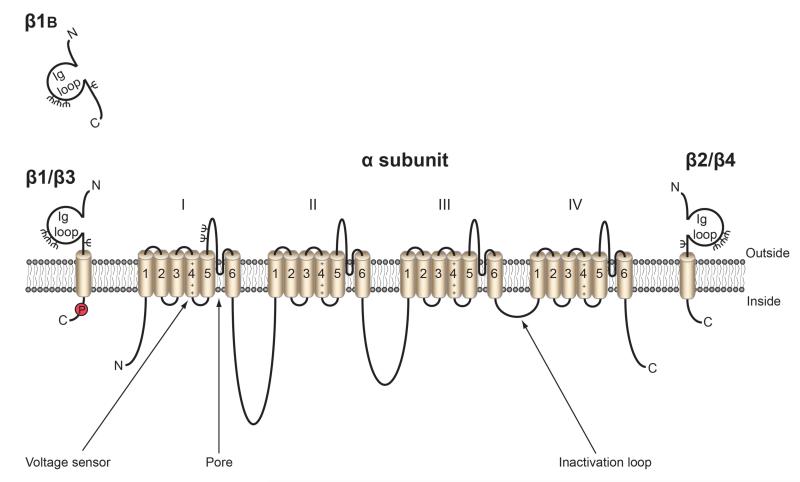
Voltage-gated Na+ channels (VGSCs) are heteromeric membrane protein complexes containing pore-forming α subunits in association with non-pore-forming β subunits (Figure 1) (Catterall, 2000). The β subunits regulate channel gating and are also cell adhesion molecules (CAMs) (Brackenbury and Isom, 2011). The classical role of VGSCs is the initiation and conduction of action potentials in electrically excitable cells, e.g. neurons (Hille, 1992). However, VGSCs are also expressed in a number of “non-excitable” cells, including fibroblasts, glia, immune cells, and cancer cells, where their role is less well understood (Brackenbury et al., 2008b). Clearly, in both excitable and non-excitable cells, VGSCs regulate a number of key cellular processes, by a combination of conducting (i.e. via Na+ current) and non-conducting mechanisms. The purpose of this article is to provide an up-to-date review of the current evidence suggesting a dual role for VGSCs in regulating cellular migration during central nervous system development and cancer progression.
Figure 1.
Topology of VGSCs. VGSCs contain a pore-forming α subunit that has four homologous domains, each containing six transmembrane segments. The voltage sensor is in segment 4 (Catterall, 2000). The β subunits contain an extracellular immunoglobulin (Ig) loop, transmembrane domain, and an intracellular C-terminal domain, with the exception of β1B, which lacks a transmembrane domain, and is thus a soluble protein (Patino et al., 2011). Red P, tyrosine phosphorylation site in β1 C-terminus (Malhotra et al., 2004); ψ, glycosylation sites. Figure as originally published in Brackenbury WJ and Isom LL (2011) Na+ Channel β Subunits: Overachievers of the Ion Channel Family. Front. Pharmacol. 2:53. doi: 10.3389/fphar.2011.00053.
Structure and function of VGSCs
The pore-forming α subunit consists of four homologous domains, each with six transmembrane segments. The pore is formed from the membrane dipping loop between the 5th and 6th transmembrane segments of each domain (Figure 1) (Catterall, 2000). There are nine α subunits, Nav1.1-Nav1.9, encoded by SCN1A-SCN11A (Catterall, 2000). The different α subunits have unique, but often overlapping, tissue-specific expression patterns (Table 1A) (Goldin et al., 2000). There is considerable electrophysiological and pharmacological diversity between α subunits, which may, in part explain their tissue specificity (Catterall, 2000). Alternative splicing of α subunits provides additional functional, developmental, and tissue-specific variability (Diss et al., 2004). Four genes (SCN1B-SCN4B) encode five different β subunits, β1, and its splice variant β1B, and β2-4 (Table 1B) (Brackenbury and Isom, 2011). With the exception of β1B, the β subunits are type 1 topology transmembrane proteins, with a small intracellular C-terminus, and an extracellular N-terminus containing an immunoglobulin loop (Figure 2) (Gilchrist et al., 2013, Isom et al., 1992, Namadurai et al., 2014). β1B is a splice variant of β1, which, through the retention of exon 3A, transcribes an early stop codon and does not contain the transmembrane region of β1 (Kazen-Gillespie et al., 2000, Qin et al., 2003). β1 and β3 are non-covalently linked to α subunits, whereas β2 and β4 are covalently linked (Isom et al., 1992, Isom et al., 1995, Morgan et al., 2000, Yu et al., 2003).
Table 1.
Tissue and cancer expression of VGSCs.
| (A) α subunits (Brackenbury, 2012, Goldin et al., 2000). | |||
|---|---|---|---|
| Protein | Gene | Tissue location | Cancer type |
| Nav1.1 | SCN1A | CNS, PNS, heart | Ovarian |
| Nav1.2 | SCN2A | CNS, PNS | Cervical, mesothelioma, ovarian, prostate |
| Nav1.3 | SCN3A | CNS, PNS | Ovarian, prostate, small cell lung cancer |
| Nav1.4 | SCN4A | Skeletal muscle | Cervical, ovarian, prostate |
| Nav1.5 | SCN5A | Uninnervated skeletal muscle, heart, brain | Breast, colon, lymphoma, neuroblastoma, non-small cell lung cancer, ovarian, small cell lung cancer |
| Nav1.6 | SCN8A | CNS, PNS, heart | Breast, cervical, lymphoma, melanoma, mesothelioma, non-small cell lung cancer, prostate, small cell lung cancer |
| Nav1.7 | SCN9A | PNS, neuroendocrine cells, sensory neurons | Breast, cervical, lymphoma, mesothelioma, non-small cell lung cancer, ovarian, prostate |
| Nav1.8 | SCN10A | sensory neurons | Prostate |
| Nav1.9 | SCN11A | sensory neurons | Lymphoma, small-cell lung cancer |
| (B) β subunits (Brackenbury, 2012, Brackenbury and Isom, 2011). | |||
|---|---|---|---|
| Protein | Gene | Tissue location | Cancer type |
| β1 | SCN1B | Heart, skeletal muscle, adrenal gland, CNS, glia, PNS | Breast, cervical, non-small cell lung cancer, prostate |
| β2 | SCN2B | CNS, PNS, heart, glia | Breast, cervical, non-small cell lung cancer, prostate |
| β3 | SCN3B | CNS, adrenal gland, kidney, PNS | Non-small cell lung cancer, prostate |
| β4 | SCN4B | Heart, skeletal muscle, CNS, PNS | Breast, cervical, non-small cell lung cancer, prostate |
Figure 2.
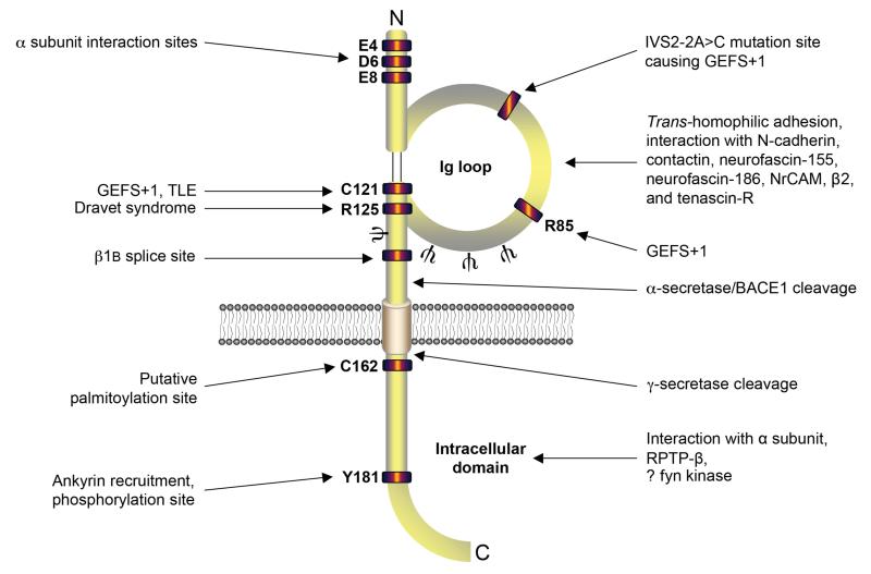
Functional map of β1. The extracellular immunoglobulin loop interacts with other cell adhesion molecules and extracellular matrix proteins (Kazarinova-Noyes et al., 2001, Malhotra et al., 2000, McEwen and Isom, 2004, Ratcliffe et al., 2001, Xiao et al., 1999). A number of mutations at the indicated sites in and adjacent to the immunoglobulin loop have been identified as responsible for causing temporal lobe epilepsy (TLE) and genetic epilepsy with febrile seizures plus (GEFS+) (Patino and Isom, 2010). Other sites indicated: alternative splice site, putative palmitoylation site, secretase cleavage sites, glycosylation sites (ψ), sites for interaction with ankyrin, receptor tyrosine phosphatase β (RPTPβ), and fyn kinase (Brackenbury et al., 2008a, Malhotra et al., 2002, Malhotra et al., 2004, Patino et al., 2011, Wong et al., 2005). Figure as originally published in Brackenbury WJ and Isom LL (2011) Na+ Channel β Subunits: Overachievers of the Ion Channel Family. Front. Pharmacol. 2:53. doi: 10.3389/fphar.2011.00053.
Classically, the β subunits modulate the biophysical properties of the α subunit. For example, β1 and β2 increase current density, accelerate inactivation, and hyperpolarize the voltage dependence of inactivation in heterologous cells (Isom et al., 1992, Isom et al., 1995). In contrast, β3 depolarizes the voltage dependence of activation and inactivation of Nav1.3 in HEK-293 cells (Cusdin et al., 2010), and increases Na+ current density by enhancing trafficking of Nav1.5 to the plasma membrane (Ishikawa et al., 2013). β4 hyperpolarizes the voltage-dependence of activation of Nav1.2 in tsA-201 cells (Qu et al., 2001, Yu et al., 2003). In addition, the intracellular domain of β4 has been proposed to act as an open-channel blocker in cerebellar Purkinje neurones (Grieco et al., 2005). However, there have been inconsistent reports on the type and magnitude of alteration of the Na+ current by individual β subunits, which may be dependent on the cell line/type used (Meadows and Isom, 2005, Moran et al., 2003). This variability may be due to differences in the endogenous levels of α subunits and β subunits and different glycosylation states. For example, β1 and β3 have recently been shown to alter glycosylation of Nav1.7 in HEK293 cells (Laedermann et al., 2013). Interestingly, the β subunits may also regulate other classes of ion channels. For example, β1 regulates A-type K+ currents in isolated cortical neurons (Marionneau et al., 2012), and modifies KV4.3 gating in cardiomyocytes (Deschenes et al., 2008, Deschenes and Tomaselli, 2002). β1 has also been shown to modulate the biophysical properties of KV1.1, KV1.2, KV1.3, KV1.6, and KV7.2 (Nguyen et al., 2012).
In addition to regulating Na+/K+ current, the presence of the immunoglobulin loop means that the β subunits are also CAMs. β1 can interact both homophilically and heterophilically with a number of extracellular proteins and other CAMs, including β2, contactin, neurofascin-186, NrCAM, N-cadherin, and tenascin-R (Figure 2) (Kazarinova-Noyes et al., 2001, Malhotra et al., 2000, McEwen and Isom, 2004, Ratcliffe et al., 2001, Xiao et al., 1999). β2 also interacts with tenascin-C and tenascin-R (Srinivasan et al., 1998). In Chinese hamster lung cells, phosphorylation of the intracellular Y181 residue on β1 abolishes recruitment of ankyrinG and ankyrinB (Malhotra et al., 2002). Further, phosphorylation of Y181 regulates subcellular localization of β1 to the intercalated disks in cardiomyocytes (Malhotra et al., 2004). β3 shows significant homology to β1 however, when expressed in Drosophila S2 cells, β3 does not participate in trans-homophilic adhesion, nor does it interact with β1 or contactin in Chinese hamster lung cells, but does interact with neurofascin-186 (McEwen et al., 2009, McEwen and Isom, 2004). In contrast, a recent study has shown that in HEK-293 cells, the immunoglobulin domain of β3 can indeed participate in trans-homophilic binding, and can interact heterophilically with β1 (Yereddi et al., 2013). Clearly, further work is required to resolve these conflicting observations, and β3-mediated adhesive interactions may be dependent on species/cell type.
VGSCs in central nervous system development
Electrical activity is required for axonal and dendritic development and synaptogenesis in the retinogeniculate pathway and visual cortex (Casagrande and Condo, 1988, Riccio and Matthews, 1985). Similarly, deletion of Nav1.1, Nav1.2, or Nav1.6 in mice results in central nervous system (CNS) defects and premature lethality (Harris and Pollard, 1986, Planells-Cases et al., 2000, Yu et al., 2006). Thus, α subunit expression and activity appear to be critical for normal CNS development. Fine-tuning of electrical activity via VGSC α subunit expression is tightly regulated during development. For example, Nav1.3 is expressed during foetal development and is replaced by Nav1.1, Nav1.2, and Nav1.6 postnatally (Beckh et al., 1989, Schaller and Caldwell, 2000). Later in postnatal development, Nav1.6 replaces Nav1.2 at the axon initial segment, and nodes of Ranvier following myelination (Boiko et al., 2001, Boiko et al., 2003, Kaplan et al., 2001). Interestingly however, α subunits may also play a non-conducting role (independent of Na+ current) in regulating tissue development. For example, Nav1.5 expression is required for normal heart development in zebrafish (Chopra et al., 2010). Further developmental regulation of VGSCs is achieved by alternative splicing. Alternative splicing in domain I segment 3 (DI:S3) occurs in a number of the α subunits, and is developmentally regulated for Nav1.2, Nav1.3 and Nav1.5 (Diss et al., 2004).
β subunit expression is also developmentally regulated. During CNS development, the SCN1B splice variant β1B is predominantly expressed embryonically (Kazen-Gillespie et al., 2000, Patino et al., 2011). In contrast, β1 expression increases from birth, peaking at postnatal day 14 in mice (Kazen-Gillespie et al., 2000). Finally, the developmentally regulated expression profile of VGSCs is disrupted in CNS diseases. For example, in multiple sclerosis (MS), Nav1.2, Nav1.6 and Nav1.8 are up-regulated in CNS neurons in response to demyelination (Black et al., 2000, Craner et al., 2004). Interestingly, Scn2b deletion is neuroprotective in the experimental allergic encephalomyelitis MS model in mice, possibly by reducing α subunit up-regulation (O'Malley et al., 2009).
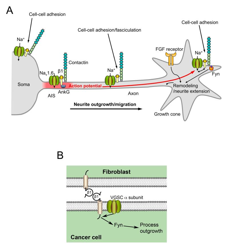
The β subunits also play critical roles in CNS development. Scn1b null mice are ataxic and display spontaneous generalized seizures (Chen et al., 2004). Mutations in SCN1B result in genetic epilepsy with febrile seizures plus (GEFS+; OMIM 604233; reviewed in (Patino and Isom, 2010). In cerebellar granule neurons (CGNs), β1 promotes neurite outgrowth via trans-homophilic adhesion (Davis et al., 2004). β1-mediated neurite outgrowth also requires fyn kinase and contactin (Brackenbury et al., 2008a). In addition, β1 is required for neuronal pathfinding and fasciculation in the postnatally developing CNS (Brackenbury et al., 2008a, Brackenbury et al., 2013). β1B can also promote neurite outgrowth (Patino et al., 2011). β1 is required for normal localization of Nav1.6 to the axon initial segment in CGNs and the resultant inward Na+ current is required for β1 mediated neurite outgrowth suggesting a specific reciprocal relationship between these two subunits (Figure 3A) (Brackenbury et al., 2010).
Figure 3.
Functional reciprocity between α and β subunits regulating neurite outgrowth and migration during CNS development and metastasis. (A) β1 is required for localization of Nav1.6 to the axon initial segment and high frequency action potential firing. The electrical activity and resultant membrane depolarisation promotes β1-mediated neurite outgrowth towards the growth cone. (B) A similar mechanism is proposed for β1-mediated process outgrowth in breast cancer cells. β1 from an adjacent fibroblast or cancer cell interacts with β1 on the cancer cell, initiating a signalling cascade that requires Na+ current and fyn kinase. Figure panels reproduced with permission (Brackenbury et al., 2010, Nelson et al., 2014).
Scn2b null mice appear normal in neurological tests, although they display increased seizure susceptibility, and altered sensitivity to pain stimuli (Chen et al., 2002, Lopez-Santiago et al., 2006). Electrical activity is reduced in the optic nerve of Scn2b null mice, and Na+ current is reduced in hippocampal and dorsal root ganglion neurons, compared to wildtype animals (Chen et al., 2002, Lopez-Santiago et al., 2006). Scn3b null mice have altered cardiac function but show no abnormalities in the CNS (Hakim et al., 2008). It is possible that β1 may compensate for the lack of β3 allowing for an apparently normal neurological phenotype. Overexpression of β4 in Neuro2a cells increases neurite outgrowth, dendrite formation, and filopodia-like protrusions (Oyama et al., 2006), suggesting that, like β1, β4 may regulate migration and pathfinding in vivo.
The β subunits may play a role in downstream signalling pathways and gene transcription. The β subunits are substrates for proteolytic processing by α, β and γ-secretases (Kim et al., 2005, Wong et al., 2005). Sequential cleavage of β2 by β-secretase (BACE1) and γ-secretase release the β2 intracellular domain, which is proposed to translocate to the nucleus and regulate expression of Nav1.1 (Kim et al., 2007, Kim et al., 2005). Secretase-mediated cleavage of β1 regulates neurite outgrowth, suggesting that proteolytic processing of β subunits may be an essential step in transducing the adhesion signal to promote migration (Brackenbury and Isom, 2011).
In summary, VGSC α and β subunit expression is temporally regulated during CNS development. Regulated expression of specific subtypes is critical for maintaining electrical excitability and activity-dependent synaptic connections on the one hand, and adhesive interactions, neurite outgrowth, fasciculation and migration on the other. Several studies point towards a potential causal relationship between altered VGSC expression, developmental aberrations, and CNS pathophysiologies, which requires further investigation.
VGSCs and cancer
α subunits
VGSC α subunits are widely expressed in a range of different types of cancer, including breast cancer, cervical cancer, colon cancer, glioma, leukaemia, lung cancer, lymphoma, melanoma, mesothelioma, neuroblastoma, ovarian cancer, prostate cancer (Table 1A) (Brackenbury, 2012). Although the majority of evidence is based on studies using cell lines cultured in vitro, a number of reports have now confirmed that α subunit expression occurs in tumours in vivo, e.g. (Fraser et al., 2005, Gao et al., 2010, Hernandez-Plata et al., 2012, House et al., 2010). In several cancers where multiple α subunits have been detected, one α subunit has been identified as most highly expressed, e.g. Nav1.5 is predominant in breast cancer (Fraser et al., 2005), whereas Nav1.7 is predominant in prostate cancer (Diss et al., 2004). Interestingly, Nav1.5 and Nav1.7 have been shown to be mainly expressed in their neonatal DI:S3 splice forms in several cancers (Brackenbury, 2012). However, this splicing pattern is not conserved across all the tumour types studied, e.g., the neonatal DI:S3 splice form is absent in colon cancer cells, and the adult variant is expressed instead (House et al., 2010). There also appears to be a cancer type-specific relationship between α subunit expression and metastatic propensity. For example, Nav1.5 is more highly expressed in strongly metastatic MDA-MB-231 breast cancer cells than weakly metastatic MCF-7 cells, and elevated Nav1.5 expression in tumours correlates with increased risk of recurrence, metastasis and reduced overall survival (Fraser et al., 2005, Yang et al., 2012). A similar pattern has been shown for α subunit expression in colon, prostate and ovarian cancers. However, there is an inverse correlation between α subunit expression and clinical grade in glioma, and no relationship has been found in lung cancer cell lines (reviewed in (Brackenbury, 2012).
The mechanisms by which VGSCs are up-regulated in cancer cells are not well understood. Several studies suggest that growth factors may play a role (Fraser et al., 2014). Epidermal growth factor (EGF) and nerve growth factor (NGF) both increase Na+ current in prostate cancer cells, the latter via activation of protein kinase A (PKA) (Brackenbury and Djamgoz, 2007, Ding et al., 2008). Similarly, EGF signalling via the extracellular signal-regulated kinase (ERK)1/2 pathway increases expression of Nav1.7 and Na+ current (Campbell et al., 2013). In breast cancer cells, oestrogen increases Na+ current, suggesting that steroid hormones may also regulate VGSC expression/activity (Fraser et al., 2010). Further fine-tuning of VGSC expression in cancer cells is achieved through positive feedback auto-regulation. In both metastatic breast and prostate cancer cells, Na+ current activates PKA, which in turn, promotes functional expression of Nav1.5 and Nav1.7, respectively (Brackenbury and Djamgoz, 2006, Chioni et al., 2010).
In vitro, the α subunits have been shown to enhance various cellular behaviours associated with metastasis, including endocytosis (Mycielska et al., 2003), galvanotaxis (Djamgoz et al., 2001), gene expression (Mycielska et al., 2005), invasion (Grimes et al., 1995), migration (Fraser et al., 2003), and process outgrowth (Fraser et al., 1999) (Table 2). Conflicting reports suggest that α subunits may, or may not, also regulate proliferation (Abdul and Hoosein, 2002, Fraser et al., 2000, Roger et al., 2003). These discrepancies may be due to the differing specificity of the various pharmacological approaches used in different studies. Several studies have indicated that specific α subunits contribute to the invasive capacity of different cancer cell types. For example, the neonatal DI:S3 splice variant of Nav1.5 enhances migration and invasion of metastatic breast cancer cells (Brackenbury et al., 2007). In contrast, Nav1.6 enhances invasion of cervical cancer cells (Hernandez-Plata et al., 2012), and Nav1.6 and Nav1.7 enhance invasion and endocytosis in prostate cancer cells (Nakajima et al., 2009). Nonetheless, expression of any subtype may be sufficient to promote invasion. For example, overexpression of Nav1.4 increases the invasiveness of LNCaP prostate cancer cells (Bennett et al., 2004).
Table 2.
Metastatic cell behaviours regulated by VGSCs (Brackenbury, 2012).
| Cellular activity | Cancer | Subunit(s) implicated |
|---|---|---|
| Process outgrowth | Breast, prostate | Nav1.5, Nav1.7, β1 |
| Galvanotaxis | Breast, prostate | Nav1.5, Nav1.7 |
| Lateral motility (wound healing) | Breast, mesothelioma, prostate | Nav1.5, Nav1.7, β1, β2 |
| Transwell migration | Breast, prostate | Nav1.5, Nav1.7 |
| Endocytic membrane activity | Breast, prostate, small cell lung cancer | Nav1.5, Nav1.7 |
| Vesicular patterning | Breast, prostate | Nav1.7 |
| Adhesion | Breast, prostate | Nav1.5, Nav1.7, β1, β2 |
| Gene expression | Breast, colon, prostate | Nav1.5, Nav1.7, β1 |
| Invasion | Breast, cervical, colon, lymphoma, melanoma, non-small cell lung cancer, prostate | Nav1.5, Nav1.6, Nav1.7, β1, β2 |
The fact that α subunits appear to be up-regulated in cancer cells and promote metastasis-like behaviour suggests that they may be useful therapeutic targets. Indeed, the VGSC-inhibiting drugs phenytoin and ranolazine have both recently been shown to inhibit metastasis in xenograft mouse models of breast cancer (Driffort et al., 2014, Nelson et al., 2015). In support of this, several other VGSC-targeting antiepileptic drugs, including phenytoin, carbamazepine and riluzole, have been shown to inhibit secretory activity, cellular migration, proliferation and invasion in cell lines from several different cancers (Abdul and Hoosein, 2001, Abdul and Hoosein, 2002, Fraser et al., 2003, Yang et al., 2012). Given that the membrane potential (Vm) of cancer cells is relatively depolarised compared with terminally differentiated cells (Yang and Brackenbury, 2013), it is likely that the majority of VGSCs are in the inactivated state. Therefore, the persistent Na+ current, which is typically a few per cent of the transient current, is likely to be predominant and may prove to be an important therapeutic target (Yang et al., 2012).
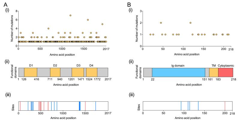
Several theories have been proposed to explain how Na+ flux through VGSCs contributes to invasion and metastasis. In breast cancer cells, Nav1.5-mediated Na+ influx has been shown to increase H+ efflux through the Na+/H+ exchanger (NHE1), causing intracellular alkalinisation and extracellular perimembrane acidification, thus enhancing the activity of pH-dependent cathepsin proteases and invadopodia formation (Brisson et al., 2013, Gillet et al., 2009). An additional possibility is that VGSCs may regulate gene expression (Brackenbury and Djamgoz, 2006). In colon cancer cells, Nav1.5 has been proposed to be a key regulator of a network of invasion-promoting genes (House et al., 2010). However, the intermediate steps between Na+ current and gene transcription remain to be elucidated. A third possibility is that VGSCs may regulate the intracellular Ca2+ level. For example, activation of VGSCs present on intracellular membranes in macrophages and melanoma cells causes Na+ release from cationic stores, followed by Na+ uptake by mitochondria, and Ca2+ release, which then increases podosome and invadopodia formation, and enhanced invasiveness (Carrithers et al., 2009). Finally, a significant number of somatic mutations have been identified in SCN5A in tumours (Figure 4A), which span all functional domains (Figure 1). Further work is required to establish whether and how these mutations may confer a functional advantage on the VGSC to promote invasive behaviour.
Figure 4.
Nav1.5 and β1 mutations in cancer. (A) Nav1.5: (i) Number of mutations reported in the COSMIC database (http://cancer.sanger.ac.uk/cancergenome/projects/cosmic/) for each amino acid position on x-axis. (ii) Location of Domains 1-4 (yellow) (Catterall, 2000). (iii) Putative phosphorylation (red) and glycosylation sites (blue). (B) β1: (i) Number of mutations reported in the COSMIC database for each amino acid position. (ii) Location of immunoglobulin (Ig) domain (blue), transmembrane (TM) domain (yellow) and cytoplasmic domain (red) (Brackenbury and Isom, 2011). (iii) Phosphorylation (red) and glycosylation sites (blue).
β subunits
VGSC β subunits have been detected in prostate, breast, lung, and cervical cancers (Table 1B) (Brackenbury, 2012). Subtype-specific expression varies across cancer types: β3 is present in prostate and lung cancer cells, but is absent in breast and cervical cancer cells. In contrast, β1 is predominant in breast, prostate, and cervical cancer cells (Chioni et al., 2009, Diss et al., 2008, Hernandez-Plata et al., 2012). Similar to SCN5A, a number of somatic mutations have been identified in SCN1B in tumours (Figure 4B), in both the immunoglobulin and cytoplasmic domains (Figure 2). β1 and β2 expression levels correlate with metastatic potential in prostate cancer (Diss et al., 2008, Jansson et al., 2012). However, this pattern is not reflected in breast cancer (Chioni et al., 2009, Nelson et al., 2014). Thus, β subunit expression may vary across cancer type and grade, dependent on specific functional specialisations and heterotypic interactions.
In breast cancer cell lines cultured in vitro, β1 enhances cell-cell and cell-substrate adhesion, and retards migration in wound healing and transwell assays (Chioni et al., 2009). In an orthotopic mouse model of breast cancer, β1 overexpression increases tumour growth and metastasis (Nelson et al., 2014). β1 overexpression also increases vascular endothelial growth factor (VEGF) secretion and angiogenesis, and reduces apoptosis. Interestingly, β1 promotes neurite-like process outgrowth from breast cancer cells via trans-homophilic adhesion, thus recapitulating its functional role in neurons (Figure 3A) (Davis et al., 2004, Nelson et al., 2014). As in neurons, β1-mediate process outgrowth in breast cancer cells requires fyn kinase activity and Na+ current (Figure 3B) (Brackenbury et al., 2010, Brackenbury et al., 2008a). Thus, it appears that β1 plays parallel roles in regulating neuronal migration during CNS development, on the one hand, and cancer cell invasion during metastasis, on the other. Therefore, targeting the adhesive function of β1 may provide a novel approach to anti-cancer therapy (Brackenbury and Isom, 2008).
In LNCaP prostate cancer cells, over-expression of β2 induces a bipolar morphology and increases overall length with a concurrent reduction in volume (Jansson et al., 2012). These changes could allow for greater invasion and motility. In agreement with this, β2 over-expressing cells have increased migratory capability compared to control cells in a wound healing assay (Jansson et al., 2012). β2 over-expressing cells plated on various substrates preferentially adhere to vitronectin and Matrigel over fibronectin, suggesting that β2 may selectively increase adhesion dependent on the surrounding tissue/substrate (Jansson et al., 2012). In contrast, β2 over-expression reduces tumour take and growth following subcutaneous implantation of LNCaP cells into nude mice (Jansson et al., 2012). Thus, β2 may enhance invasion and metastasis whilst also reducing the ability of tumours to form localized masses. In support of this notion, β2 over-expression increases invasion and growth on laminin, and enhances association between prostate cancer cells and nerve axons in organotypic cultures (Jansson et al., 2014). Therefore, β2 may permit association between prostate cancer cells and neural matrices, enhancing perineural invasion, thus enabling glandular egress and subsequent metastatic dissemination.
In contrast to β1 and β2, β3 may function as a tumour suppressor. SCN3B (encoding β3) contains two functional p53 response elements, suggesting that it may be directly regulated by the tumour suppressor p53 (Adachi et al., 2004). In addition, Scn3b is up-regulated in wildtype mouse embryo fibroblasts (MEFs), but not p53 null MEFs following treatment with adriamycin (Adachi et al., 2004). Furthermore, β3 suppresses colony formation, and in association with various anticancer agents, β3 promotes apoptosis in a p53-dependent manner (Adachi et al., 2004).
Less is known about the expression/function of β4 in cancers. Interestingly, a strong down-regulation of β4 has been reported in primary cultures of cervical cancer cells relative to cells from noncancerous cervix (Hernandez-Plata et al., 2012). A similar pattern of expression has also been reported in prostate cancer cell lines (Diss et al., 2008). However, β4 expression is increased in cervical cancer biopsies compared to noncancerous cervix. This difference in relative expression levels between biopsies and primary cell cultures may be due to the adhesive function differing in vivo as opposed to in vitro (Hernandez-Plata et al., 2012). Further work is required to investigate this possibility.
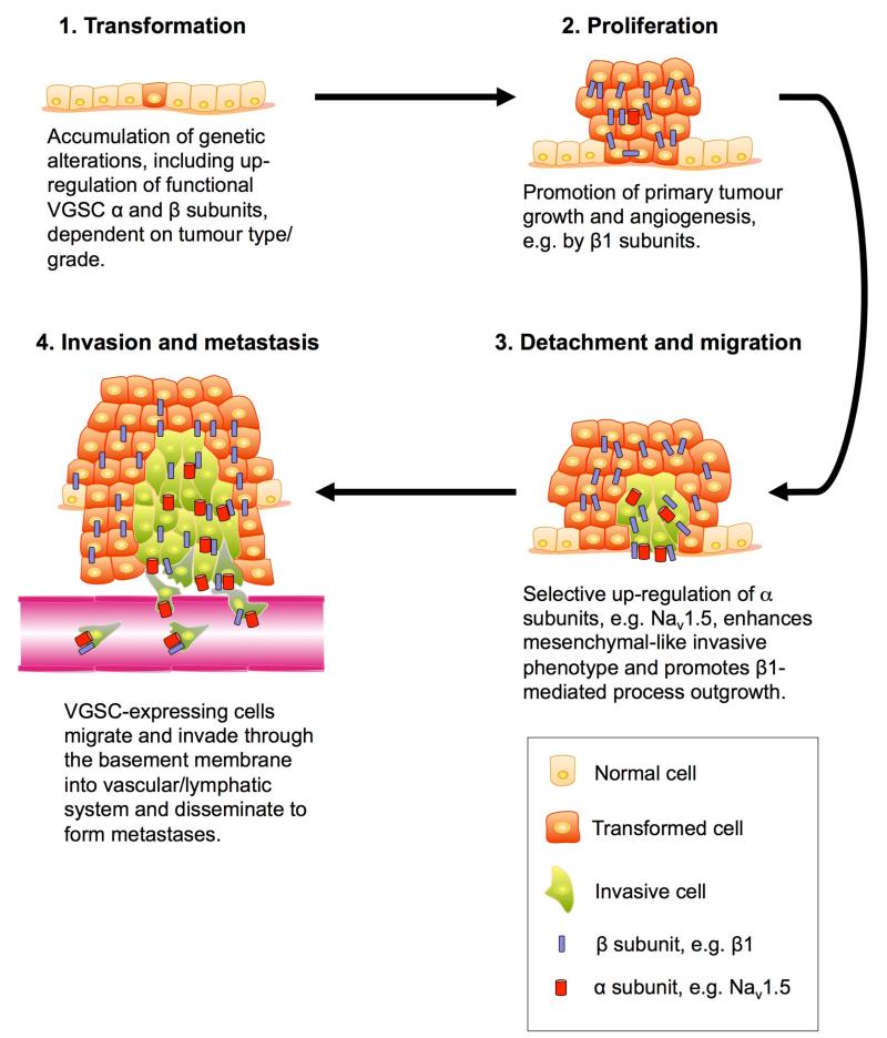
In summary, VGSCs are up-regulated in a number of different types of cancer. Increasing evidence suggests that α and β subunits both play an important role in promoting various aspects of cancer progression and metastasis (Figure 5). The role(s) played by specific subtypes appears to be complex, and may be dependent on tumour type. A common theme is that α subunits regulate invasion via Na+ current, whereas β subunits regulate adhesion interactions. The next step is to establish the extent and distribution of expression of VGSCs across tumour types, and the precise involvement of different α and β subunits, with the goal of harnessing their therapeutic potential.
Figure 5.
A model for VGSC involvement in cancer progression. β subunits are expressed in proliferating primary tumours, contributing to adhesion (Chioni et al., 2009), and in the case of β1, promoting angiogenesis and resistance to apoptosis (Nelson et al., 2014). Up-regulation of α subunits, e.g. Nav1.5, promotes a mesenchymal-like phenotype (Brisson et al., 2013, Nelson et al., 2014), activation of proteases (Gillet et al., 2009) and local invasion from the primary tumour (Brackenbury et al., 2007, Fraser et al., 2005, Roger et al., 2003). VGSC-expressing cells subsequently intravasate and metastasise to distant sites (Fraser et al., 2005, Jansson et al., 2012, Nelson et al., 2014). Figure adapted with permission (Brackenbury et al., 2008b)
Conclusions
VGSCs function as macromolecular signalling complexes in which Na+ current through the α subunit pore is coupled with non-conducting signalling via the β subunits. For example, in migrating neurons, complexes of Nav1.6, β1, fyn and contactin are proposed to localise to the growth come, regulating neurite outgrowth and migration (Brackenbury et al., 2010, Brackenbury et al., 2008a). Similarly, complexes of Nav1.5 and β1 may occur in breast cancer cells, regulating morphological changes and metastasis (Nelson et al., 2014, Yang et al., 2012). Additional complexity may be provided by further interactions with the cytoskeleton, e.g. via ankyrin, and secretases (Brackenbury and Isom, 2011, Kim et al., 2005, Malhotra et al., 2000). The challenge now is to understand how signalling through these complexes gives rise to morphological changes and cellular motility, and how variations in the composition of the complex might relate to cell/tissue type, functional specialisation and subcellular domain. An important observation is that VGSC α and β subunits appear to play multifunctional and parallel roles (1) in excitable cells, e.g. CNS neurons, and (2) in metastatic cancer cells, regulating Na+ current, migration and invasion in both systems. It is therefore essential to better understand the identity, function and composition of these complexes during development and in pathophysiological situations. An intriguing possibility is that VGSCs may be useful prognostic markers, and/or novel therapeutic targets for reducing/preventing metastasis.
Acknowledgements
This work was supported by the Medical Research Council [grant numbers MR/K016296/1 and G1000508].
Abbreviations
- BACE1
β-secretase
- CAM
cell adhesion molecule
- CGN
cerebellar granule neuron
- CNS
central nervous system
- DI:S3
domain I segment 3
- EGF
epidermal growth factor
- ERK
extracellular signal-regulated kinase
- GEFS+
genetic epilepsy with febrile seizures plus
- MEF
mouse embryonic fibroblast
- MS
multiple sclerosis
- NGF
nerve growth factor
- NHE1
Na+/H+ exchanger 1
- PKA
protein kinase A
- PNS
peripheral nervous system
- RPTPβ
receptor tyrosine phosphatase β
- TLE
temporal lobe epilepsy
- VEGF
vascular endothelial growth factor
- VGSC
voltage-gated Na+ channel
- Vm
membrane potential
References
- ABDUL M, HOOSEIN N. Inhibition by anticonvulsants of prostate-specific antigen and interleukin-6 secretion by human prostate cancer cells. Anticancer Res. 2001;21:2045–8. [PubMed] [Google Scholar]
- ABDUL M, HOOSEIN N. Voltage-gated sodium ion channels in prostate cancer: expression and activity. Anticancer Res. 2002;22:1727–30. [PubMed] [Google Scholar]
- ADACHI K, TOYOTA M, SASAKI Y, YAMASHITA T, ISHIDA S, OHE-TOYOTA M, MARUYAMA R, HINODA Y, SAITO T, IMAI K, et al. Identification of SCN3B as a novel p53-inducible proapoptotic gene. Oncogene. 2004;23:7791–8. doi: 10.1038/sj.onc.1208067. [DOI] [PubMed] [Google Scholar]
- BECKH S, NODA M, LUBBERT H, NUMA S. Differential regulation of three sodium channel messenger RNAs in the rat central nervous system during development. EMBO J. 1989;8:3611–6. doi: 10.1002/j.1460-2075.1989.tb08534.x. [DOI] [PMC free article] [PubMed] [Google Scholar]
- BENNETT ES, SMITH BA, HARPER JM. Voltage-gated Na+ channels confer invasive properties on human prostate cancer cells. Pflugers Arch. 2004;447:908–14. doi: 10.1007/s00424-003-1205-x. [DOI] [PubMed] [Google Scholar]
- BLACK JA, DIB-HAJJ S, BAKER D, NEWCOMBE J, CUZNER ML, WAXMAN SG. Sensory neuron-specific sodium channel SNS is abnormally expressed in the brains of mice with experimental allergic encephalomyelitis and humans with multiple sclerosis. Proc. Natl. Acad. Sci. U. S. A. 2000;97:11598–602. doi: 10.1073/pnas.97.21.11598. [DOI] [PMC free article] [PubMed] [Google Scholar]
- BOIKO T, RASBAND MN, LEVINSON SR, CALDWELL JH, MANDEL G, TRIMMER JS, MATTHEWS G. Compact myelin dictates the differential targeting of two sodium channel isoforms in the same axon. Neuron. 2001;30:91–104. doi: 10.1016/s0896-6273(01)00265-3. [DOI] [PubMed] [Google Scholar]
- BOIKO T, VAN WART A, CALDWELL JH, LEVINSON SR, TRIMMER JS, MATTHEWS G. Functional specialization of the axon initial segment by isoform-specific sodium channel targeting. J. Neurosci. 2003;23:2306–13. doi: 10.1523/JNEUROSCI.23-06-02306.2003. [DOI] [PMC free article] [PubMed] [Google Scholar]
- BRACKENBURY WJ. Voltage-gated sodium channels and metastatic disease. Channels (Austin) 2012;6:352–61. doi: 10.4161/chan.21910. [DOI] [PMC free article] [PubMed] [Google Scholar]
- BRACKENBURY WJ, CALHOUN JD, CHEN C, MIYAZAKI H, NUKINA N, OYAMA F, RANSCHT B, ISOM LL. Functional reciprocity between Na+ channel Nav1.6 and β1 subunits in the coordinated regulation of excitability and neurite outgrowth. Proc. Natl. Acad. Sci. U. S. A. 2010;107:2283–2288. doi: 10.1073/pnas.0909434107. [DOI] [PMC free article] [PubMed] [Google Scholar]
- BRACKENBURY WJ, CHIONI AM, DISS JK, DJAMGOZ MB. The neonatal splice variant of Nav1.5 potentiates in vitro metastatic behaviour of MDA-MB-231 human breast cancer cells. Breast Cancer Res. Treat. 2007;101:149–60. doi: 10.1007/s10549-006-9281-1. [DOI] [PMC free article] [PubMed] [Google Scholar]
- BRACKENBURY WJ, DAVIS TH, CHEN C, SLAT EA, DETROW MJ, DICKENDESHER TL, RANSCHT B, ISOM LL. Voltage-gated Na+ channel β1 subunit-mediated neurite outgrowth requires fyn kinase and contributes to central nervous system development in vivo. J. Neurosci. 2008a;28:3246–3256. doi: 10.1523/JNEUROSCI.5446-07.2008. [DOI] [PMC free article] [PubMed] [Google Scholar]
- BRACKENBURY WJ, DJAMGOZ MB. Activity-dependent regulation of voltage-gated Na+ channel expression in Mat-LyLu rat prostate cancer cell line. J. Physiol. 2006;573:343–56. doi: 10.1113/jphysiol.2006.106906. [DOI] [PMC free article] [PubMed] [Google Scholar]
- BRACKENBURY WJ, DJAMGOZ MB. Nerve growth factor enhances voltage-gated Na+ channel activity and Transwell migration in Mat-LyLu rat prostate cancer cell line. J. Cell. Physiol. 2007;210:602–8. doi: 10.1002/jcp.20846. [DOI] [PMC free article] [PubMed] [Google Scholar]
- BRACKENBURY WJ, DJAMGOZ MB, ISOM LL. An emerging role for voltage-gated Na+ channels in cellular migration: regulation of central nervous system development and potentiation of invasive cancers. Neuroscientist. 2008b;14:571–83. doi: 10.1177/1073858408320293. [DOI] [PMC free article] [PubMed] [Google Scholar]
- BRACKENBURY WJ, ISOM LL. Voltage-gated Na+ channels: potential for beta subunits as therapeutic targets. Expert Opin. Ther. Targets. 2008;12:1191–203. doi: 10.1517/14728222.12.9.1191. [DOI] [PMC free article] [PubMed] [Google Scholar]
- BRACKENBURY WJ, ISOM LL. Na Channel beta Subunits: Overachievers of the Ion Channel Family. Front. Pharmacol. 2011;2:53. doi: 10.3389/fphar.2011.00053. [DOI] [PMC free article] [PubMed] [Google Scholar]
- BRACKENBURY WJ, YUAN Y, O'MALLEY HA, PARENT JM, ISOM LL. Abnormal neuronal patterning occurs during early postnatal brain development of Scn1b-null mice and precedes hyperexcitability. Proc. Natl. Acad. Sci. U. S. A. 2013;110:1089–94. doi: 10.1073/pnas.1208767110. [DOI] [PMC free article] [PubMed] [Google Scholar]
- BRISSON L, DRIFFORT V, BENOIST L, POET M, COUNILLON L, ANTELMI E, RUBINO R, BESSON P, LABBAL F, CHEVALIER S, et al. NaV1.5 Na(+) channels allosterically regulate the NHE-1 exchanger and promote the activity of breast cancer cell invadopodia. J. Cell Sci. 2013;126:4835–42. doi: 10.1242/jcs.123901. [DOI] [PubMed] [Google Scholar]
- CAMPBELL TM, MAIN MJ, FITZGERALD EM. Functional expression of the voltage-gated Na(+)-channel Nav1.7 is necessary for EGF-mediated invasion in human non-small cell lung cancer cells. J. Cell Sci. 2013;126:4939–49. doi: 10.1242/jcs.130013. [DOI] [PubMed] [Google Scholar]
- CARRITHERS MD, CHATTERJEE G, CARRITHERS LM, OFFOHA R, IHEAGWARA U, RAHNER C, GRAHAM M, WAXMAN SG. Regulation of podosome formation in macrophages by a novel splice variant of the sodium channel SCN8A. J. Biol. Chem. 2009;284:8114–8126. doi: 10.1074/jbc.M801892200. [DOI] [PMC free article] [PubMed] [Google Scholar]
- CASAGRANDE VA, CONDO GJ. The effect of altered neuronal activity on the development of layers in the lateral geniculate nucleus. J. Neurosci. 1988;8:395–416. doi: 10.1523/JNEUROSCI.08-02-00395.1988. [DOI] [PMC free article] [PubMed] [Google Scholar]
- CATTERALL WA. From ionic currents to molecular mechanisms: the structure and function of voltage-gated sodium channels. Neuron. 2000;26:13–25. doi: 10.1016/s0896-6273(00)81133-2. [DOI] [PubMed] [Google Scholar]
- CHEN C, BHARUCHA V, CHEN Y, WESTENBROEK RE, BROWN A, MALHOTRA JD, JONES D, AVERY C, GILLESPIE PJ, KAZEN-GILLESPIE KA, et al. Reduced sodium channel density, altered voltage dependence of inactivation, and increased susceptibility to seizures in mice lacking sodium channel beta 2-subunits. Proc. Natl. Acad. Sci. U. S. A. (3RD) 2002;99:17072–7. doi: 10.1073/pnas.212638099. [DOI] [PMC free article] [PubMed] [Google Scholar]
- CHEN C, WESTENBROEK RE, XU X, EDWARDS CA, SORENSON DR, CHEN Y, MCEWEN DP, O'MALLEY HA, BHARUCHA V, MEADOWS LS, et al. Mice lacking sodium channel beta1 subunits display defects in neuronal excitability, sodium channel expression, and nodal architecture. J. Neurosci. 2004;24:4030–42. doi: 10.1523/JNEUROSCI.4139-03.2004. [DOI] [PMC free article] [PubMed] [Google Scholar]
- CHIONI AM, BRACKENBURY WJ, CALHOUN JD, ISOM LL, DJAMGOZ MB. A novel adhesion molecule in human breast cancer cells: voltage-gated Na+ channel beta1 subunit. Int. J. Biochem. Cell Biol. 2009;41:1216–27. doi: 10.1016/j.biocel.2008.11.001. [DOI] [PMC free article] [PubMed] [Google Scholar]
- CHIONI AM, SHAO D, GROSE R, DJAMGOZ MB. Protein kinase A and regulation of neonatal Nav1.5 expression in human breast cancer cells: activity-dependent positive feedback and cellular migration. Int. J. Biochem. Cell Biol. 2010;42:346–58. doi: 10.1016/j.biocel.2009.11.021. [DOI] [PubMed] [Google Scholar]
- CHOPRA SS, STROUD DM, WATANABE H, BENNETT JS, BURNS CG, WELLS KS, YANG T, ZHONG TP, RODEN DM. Voltage-gated sodium channels are required for heart development in zebrafish. Circ. Res. 2010;106:1342–50. doi: 10.1161/CIRCRESAHA.109.213132. [DOI] [PMC free article] [PubMed] [Google Scholar]
- CRANER MJ, NEWCOMBE J, BLACK JA, HARTLE C, CUZNER ML, WAXMAN SG. Molecular changes in neurons in multiple sclerosis: altered axonal expression of Nav1.2 and Nav1.6 sodium channels and Na+/Ca2+ exchanger. Proc. Natl. Acad. Sci. U. S. A. 2004;101:8168–73. doi: 10.1073/pnas.0402765101. [DOI] [PMC free article] [PubMed] [Google Scholar]
- CUSDIN FS, NIETLISPACH D, MAMAN J, DALE TJ, POWELL AJ, CLARE JJ, JACKSON AP. The sodium channel {beta}3-subunit induces multiphasic gating in NaV1.3 and affects fast inactivation via distinct intracellular regions. J. Biol. Chem. 2010;285:33404–12. doi: 10.1074/jbc.M110.114058. [DOI] [PMC free article] [PubMed] [Google Scholar]
- DAVIS TH, CHEN C, ISOM LL. Sodium channel beta1 subunits promote neurite outgrowth in cerebellar granule neurons. J. Biol. Chem. 2004;279:51424–32. doi: 10.1074/jbc.M410830200. [DOI] [PubMed] [Google Scholar]
- DESCHENES I, ARMOUNDAS AA, JONES SP, TOMASELLI GF. Post-transcriptional gene silencing of KChIP2 and Navbeta1 in neonatal rat cardiac myocytes reveals a functional association between Na and Ito currents. J. Mol. Cell. Cardiol. 2008;45:336–46. doi: 10.1016/j.yjmcc.2008.05.001. [DOI] [PMC free article] [PubMed] [Google Scholar]
- DESCHENES I, TOMASELLI GF. Modulation of Kv4.3 current by accessory subunits. FEBS Lett. 2002;528:183–8. doi: 10.1016/s0014-5793(02)03296-9. [DOI] [PubMed] [Google Scholar]
- DING Y, BRACKENBURY WJ, ONGANER PU, MONTANO X, PORTER LM, BATES LF, DJAMGOZ MB. Epidermal growth factor upregulates motility of Mat-LyLu rat prostate cancer cells partially via voltage-gated Na+ channel activity. J. Cell. Physiol. 2008;215:77–81. doi: 10.1002/jcp.21289. [DOI] [PMC free article] [PubMed] [Google Scholar]
- DISS JK, FRASER SP, DJAMGOZ MB. Voltage-gated Na+ channels: multiplicity of expression, plasticity, functional implications and pathophysiological aspects. Eur. Biophys. J. 2004;33:180–93. doi: 10.1007/s00249-004-0389-0. [DOI] [PubMed] [Google Scholar]
- DISS JK, FRASER SP, WALKER MM, PATEL A, LATCHMAN DS, DJAMGOZ MB. Beta-subunits of voltage-gated sodium channels in human prostate cancer: quantitative in vitro and in vivo analyses of mRNA expression. Prostate Cancer Prostatic Dis. 2008;11:325–33. doi: 10.1038/sj.pcan.4501012. [DOI] [PubMed] [Google Scholar]
- DJAMGOZ MBA, MYCIELSKA M, MADEJA Z, FRASER SP, KOROHODA W. Directional movement of rat prostate cancer cells in direct-current electric field: involvement of voltage gated Na+ channel activity. J. Cell Sci. 2001;114:2697–705. doi: 10.1242/jcs.114.14.2697. [DOI] [PubMed] [Google Scholar]
- DRIFFORT V, GILLET L, BON E, MARIONNEAU-LAMBOT S, OULLIER T, JOULIN V, COLLIN C, PAGES JC, JOURDAN ML, CHEVALIER S, et al. Ranolazine inhibits NaV1.5-mediated breast cancer cell invasiveness and lung colonization. Mol. Cancer. 2014;13:264. doi: 10.1186/1476-4598-13-264. [DOI] [PMC free article] [PubMed] [Google Scholar]
- FRASER SP, DING Y, LIU A, FOSTER CS, DJAMGOZ MB. Tetrodotoxin suppresses morphological enhancement of the metastatic MAT-LyLu rat prostate cancer cell line. Cell Tissue Res. 1999;295:505–12. doi: 10.1007/s004410051256. [DOI] [PubMed] [Google Scholar]
- FRASER SP, DISS JK, CHIONI AM, MYCIELSKA ME, PAN H, YAMACI RF, PANI F, SIWY Z, KRASOWSKA M, GRZYWNA Z, et al. Voltage-gated sodium channel expression and potentiation of human breast cancer metastasis. Clin. Cancer. Res. 2005;11:5381–9. doi: 10.1158/1078-0432.CCR-05-0327. [DOI] [PubMed] [Google Scholar]
- FRASER SP, GRIMES JA, DJAMGOZ MB. Effects of voltage-gated ion channel modulators on rat prostatic cancer cell proliferation: comparison of strongly and weakly metastatic cell lines. Prostate. 2000;44:61–76. doi: 10.1002/1097-0045(20000615)44:1<61::aid-pros9>3.0.co;2-3. [DOI] [PubMed] [Google Scholar]
- FRASER SP, OZERLAT-GUNDUZ I, BRACKENBURY WJ, FITZGERALD EM, CAMPBELL TM, COOMBES RC, DJAMGOZ MB. Regulation of voltage-gated sodium channel expression in cancer: hormones, growth factors and auto-regulation. Philos. Trans. R. Soc. Lond. B Biol. Sci. 2014;369:20130105. doi: 10.1098/rstb.2013.0105. [DOI] [PMC free article] [PubMed] [Google Scholar]
- FRASER SP, OZERLAT-GUNDUZ I, ONKAL R, DISS JK, LATCHMAN DS, DJAMGOZ MB. Estrogen and non-genomic upregulation of voltage-gated Na(+) channel activity in MDA-MB-231 human breast cancer cells: role in adhesion. J. Cell. Physiol. 2010;224:527–39. doi: 10.1002/jcp.22154. [DOI] [PubMed] [Google Scholar]
- FRASER SP, SALVADOR V, MANNING EA, MIZAL J, ALTUN S, RAZA M, BERRIDGE RJ, DJAMGOZ MB. Contribution of functional voltage-gated Na+ channel expression to cell behaviors involved in the metastatic cascade in rat prostate cancer: I. lateral motility. J. Cell. Physiol. 2003;195:479–487. doi: 10.1002/jcp.10312. [DOI] [PubMed] [Google Scholar]
- GAO R, SHEN Y, CAI J, LEI M, WANG Z. Expression of voltage-gated sodium channel alpha subunit in human ovarian cancer. Oncol. Rep. 2010;23:1293–9. doi: 10.3892/or_00000763. [DOI] [PubMed] [Google Scholar]
- GILCHRIST J, DAS S, VAN PETEGEM F, BOSMANS F. Crystallographic insights into sodium-channel modulation by the beta4 subunit. Proc. Natl. Acad. Sci. U. S. A. 2013;110:E5016–24. doi: 10.1073/pnas.1314557110. [DOI] [PMC free article] [PubMed] [Google Scholar]
- GILLET L, ROGER S, BESSON P, LECAILLE F, GORE J, BOUGNOUX P, LALMANACH G, LE GUENNEC JY. Voltage-gated Sodium Channel Activity Promotes Cysteine Cathepsin-dependent Invasiveness and Colony Growth of Human Cancer Cells. J. Biol. Chem. 2009;284:8680–91. doi: 10.1074/jbc.M806891200. [DOI] [PMC free article] [PubMed] [Google Scholar]
- GOLDIN AL, BARCHI RL, CALDWELL JH, HOFMANN F, HOWE JR, HUNTER JC, KALLEN RG, MANDEL G, MEISLER MH, NETTER YB, et al. Nomenclature of voltage-gated sodium channels. Neuron. 2000;28:365–368. doi: 10.1016/s0896-6273(00)00116-1. [DOI] [PubMed] [Google Scholar]
- GRIECO TM, MALHOTRA JD, CHEN C, ISOM LL, RAMAN IM. Open-channel block by the cytoplasmic tail of sodium channel b4 as a mechanism for resurgent sodium current. Neuron. 2005;45:233–244. doi: 10.1016/j.neuron.2004.12.035. [DOI] [PubMed] [Google Scholar]
- GRIMES JA, FRASER SP, STEPHENS GJ, DOWNING JE, LANIADO ME, FOSTER CS, ABEL PD, DJAMGOZ MB. Differential expression of voltage-activated Na+ currents in two prostatic tumour cell lines: contribution to invasiveness in vitro. FEBS Lett. 1995;369:290–4. doi: 10.1016/0014-5793(95)00772-2. [DOI] [PubMed] [Google Scholar]
- HAKIM P, GURUNG IS, PEDERSEN TH, THRESHER R, BRICE N, LAWRENCE J, GRACE AA, HUANG CL. Scn3b knockout mice exhibit abnormal ventricular electrophysiological properties. Prog. Biophys. Mol. Biol. 2008;98:251–66. doi: 10.1016/j.pbiomolbio.2009.01.005. [DOI] [PMC free article] [PubMed] [Google Scholar]
- HARRIS JB, POLLARD SL. Neuromuscular transmission in the murine mutants “motor end-plate disease” and “jolting”. J. Neurol. Sci. 1986;76:239–53. doi: 10.1016/0022-510x(86)90172-3. [DOI] [PubMed] [Google Scholar]
- HERNANDEZ-PLATA E, ORTIZ CS, MARQUINA-CASTILLO B, MEDINA-MARTINEZ I, ALFARO A, BERUMEN J, RIVERA M, GOMORA JC. Overexpression of Na(V) 1.6 channels is associated with the invasion capacity of human cervical cancer. Int. J. Cancer. 2012;130:2013–2023. doi: 10.1002/ijc.26210. [DOI] [PubMed] [Google Scholar]
- HILLE B. Ionic channels of excitable membranes. Sinauer Associates Inc.; Sunderland (Massachusetts): 1992. [Google Scholar]
- HOUSE CD, VASKE CJ, SCHWARTZ A, OBIAS V, FRANK B, LUU T, SARVAZYAN N, IRBY RB, STRAUSBERG RL, HALES T, et al. Voltage-gated Na+ channel SCN5A is a key regulator of a gene transcriptional network that controls colon cancer invasion. Cancer Res. 2010;70:6957–67. doi: 10.1158/0008-5472.CAN-10-1169. [DOI] [PMC free article] [PubMed] [Google Scholar]
- ISHIKAWA T, TAKAHASHI N, OHNO S, SAKURADA H, NAKAMURA K, ON YK, PARK JE, MAKIYAMA T, HORIE M, ARIMURA T, et al. Novel SCN3B mutation associated with brugada syndrome affects intracellular trafficking and function of Nav1.5. Circ. J. 2013;77:959–67. doi: 10.1253/circj.cj-12-0995. [DOI] [PubMed] [Google Scholar]
- ISOM LL, DE JONGH KS, PATTON DE, REBER BF, OFFORD J, CHARBONNEAU H, WALSH K, GOLDIN AL, CATTERALL WA. Primary structure and functional expression of the beta1 subunit of the rat brain sodium channel. Science. 1992;256:839–842. doi: 10.1126/science.1375395. [DOI] [PubMed] [Google Scholar]
- ISOM LL, RAGSDALE DS, DE JONGH KS, WESTENBROEK RE, REBER BF, SCHEUER T, CATTERALL WA. Structure and function of the b2 subunit of brain sodium channels, a transmembrane glycoprotein with a CAM motif. Cell. 1995;83:433–42. doi: 10.1016/0092-8674(95)90121-3. [DOI] [PubMed] [Google Scholar]
- JANSSON KH, CASTILLO DG, MORRIS JW, BOGGS ME, CZYMMEK KJ, ADAMS EL, SCHRAMM LP, SIKES RA. Identification of beta-2 as a key cell adhesion molecule in PCa cell neurotropic behavior: a novel ex vivo and biophysical approach. PLoS One. 2014;9:e98408. doi: 10.1371/journal.pone.0098408. [DOI] [PMC free article] [PubMed] [Google Scholar]
- JANSSON KH, LYNCH JE, LEPORI-BUI N, CZYMMEK KJ, DUNCAN RL, SIKES RA. Overexpression of the VSSC-associated CAM, beta-2, enhances LNCaP cell metastasis associated behavior. Prostate. 2012;72:1080–92. doi: 10.1002/pros.21512. [DOI] [PubMed] [Google Scholar]
- KAPLAN MR, CHO MH, ULLIAN EM, ISOM LL, LEVINSON SR, BARRES BA. Differential control of clustering of the sodium channels Na(v)1.2 and Na(v)1.6 at developing CNS nodes of Ranvier. Neuron. 2001;30:105–19. doi: 10.1016/s0896-6273(01)00266-5. [DOI] [PubMed] [Google Scholar]
- KAZARINOVA-NOYES K, MALHOTRA JD, MCEWEN DP, MATTEI LN, BERGLUND EO, RANSCHT B, LEVINSON SR, SCHACHNER M, SHRAGER P, ISOM LL, et al. Contactin associates with Na+ channels and increases their functional expression. J. Neurosci. 2001;21:7517–7525. doi: 10.1523/JNEUROSCI.21-19-07517.2001. [DOI] [PMC free article] [PubMed] [Google Scholar]
- KAZEN-GILLESPIE KA, RAGSDALE DS, D’ANDREA MR, MATTEI LN, ROGERS KE, ISOM LL. Cloning, localization, and functional expression of sodium channel b1A subunits. J. Biol. Chem. 2000;275:1079–1088. doi: 10.1074/jbc.275.2.1079. [DOI] [PubMed] [Google Scholar]
- KIM DY, CAREY BW, WANG H, INGANO LA, BINSHTOK AM, WERTZ MH, PETTINGELL WH, HE P, LEE VM, WOOLF CJ, et al. BACE1 regulates voltage-gated sodium channels and neuronal activity. Nat. Cell Biol. 2007;9:755–64. doi: 10.1038/ncb1602. [DOI] [PMC free article] [PubMed] [Google Scholar]
- KIM DY, INGANO LA, CAREY BW, PETTINGELL WH, KOVACS DM. Presenilin/gamma-secretase-mediated cleavage of the voltage-gated sodium channel beta2-subunit regulates cell adhesion and migration. J. Biol. Chem. 2005;280:23251–61. doi: 10.1074/jbc.M412938200. [DOI] [PubMed] [Google Scholar]
- LAEDERMANN CJ, SYAM N, PERTIN M, DECOSTERD I, ABRIEL H. beta1- and beta3- voltage-gated sodium channel subunits modulate cell surface expression and glycosylation of Nav1.7 in HEK293 cells. Front. Cell. Neurosci. 2013;7:137. doi: 10.3389/fncel.2013.00137. [DOI] [PMC free article] [PubMed] [Google Scholar]
- LOPEZ-SANTIAGO LF, PERTIN M, MORISOD X, CHEN C, HONG S, WILEY J, DECOSTERD I, ISOM LL. Sodium channel beta2 subunits regulate tetrodotoxin-sensitive sodium channels in small dorsal root ganglion neurons and modulate the response to pain. J. Neurosci. 2006;26:7984–94. doi: 10.1523/JNEUROSCI.2211-06.2006. [DOI] [PMC free article] [PubMed] [Google Scholar]
- MALHOTRA JD, KAZEN-GILLESPIE K, HORTSCH M, ISOM LL. Sodium channel β subunits mediate homophilic cell adhesion and recruit ankyrin to points of cell-cell contact. J. Biol. Chem. 2000;275:11383–11388. doi: 10.1074/jbc.275.15.11383. [DOI] [PubMed] [Google Scholar]
- MALHOTRA JD, KOOPMANN MC, KAZEN-GILLESPIE KA, FETTMAN N, HORTSCH M, ISOM LL. Structural requirements for interaction of sodium channel b1 subunits with ankyrin. J. Biol. Chem. 2002;277:26681–8. doi: 10.1074/jbc.M202354200. [DOI] [PubMed] [Google Scholar]
- MALHOTRA JD, THYAGARAJAN V, CHEN C, ISOM LL. Tyrosine-phosphorylated and nonphosphorylated sodium channel beta1 subunits are differentially localized in cardiac myocytes. J. Biol. Chem. 2004;279:40748–54. doi: 10.1074/jbc.M407243200. [DOI] [PubMed] [Google Scholar]
- MARIONNEAU C, CARRASQUILLO Y, NORRIS AJ, TOWNSEND RR, ISOM LL, LINK AJ, NERBONNE JM. The sodium channel accessory subunit Navbeta1 regulates neuronal excitability through modulation of repolarizing voltage-gated K(+) channels. J. Neurosci. 2012;32:5716–27. doi: 10.1523/JNEUROSCI.6450-11.2012. [DOI] [PMC free article] [PubMed] [Google Scholar]
- MCEWEN DP, CHEN C, MEADOWS LS, LOPEZ-SANTIAGO L, ISOM LL. The voltage-gated Na+ channel beta3 subunit does not mediate trans homophilic cell adhesion or associate with the cell adhesion molecule contactin. Neurosci. Lett. 2009;462:272–5. doi: 10.1016/j.neulet.2009.07.020. [DOI] [PMC free article] [PubMed] [Google Scholar]
- MCEWEN DP, ISOM LL. Heterophilic interactions of sodium channel beta1 subunits with axonal and glial cell adhesion molecules. J. Biol. Chem. 2004;279:52744–52. doi: 10.1074/jbc.M405990200. [DOI] [PubMed] [Google Scholar]
- MEADOWS LS, ISOM LL. Sodium channels as macromolecular complexes: implications for inherited arrhythmia syndromes. Cardiovasc. Res. 2005;67:448–58. doi: 10.1016/j.cardiores.2005.04.003. [DOI] [PubMed] [Google Scholar]
- MORAN O, CONTI F, TAMMARO P. Sodium channel heterologous expression in mammalian cells and the role of the endogenous beta1-subunits. Neurosci. Lett. 2003;336:175–9. doi: 10.1016/s0304-3940(02)01284-3. [DOI] [PubMed] [Google Scholar]
- MORGAN K, STEVENS EB, SHAH B, COX PJ, DIXON AK, LEE K, PINNOCK RD, HUGHES J, RICHARDSON PJ, MIZUGUCHI K, et al. b3: An additional auxiliary subunit of the voltage-sensitive sodium channel that modulates channel gating with distinct kinetics. Proc. Natl. Acad. Sci. U.S.A. 2000;97:2308–2313. doi: 10.1073/pnas.030362197. [DOI] [PMC free article] [PubMed] [Google Scholar]
- MYCIELSKA ME, FRASER SP, SZATKOWSKI M, DJAMGOZ MB. Contribution of functional voltage-gated Na+ channel expression to cell behaviors involved in the metastatic cascade in rat prostate cancer: II. Secretory membrane activity. J. Cell. Physiol. 2003;195:461–469. doi: 10.1002/jcp.10265. [DOI] [PubMed] [Google Scholar]
- MYCIELSKA ME, PALMER CP, BRACKENBURY WJ, DJAMGOZ MB. Expression of Na+-dependent citrate transport in a strongly metastatic human prostate cancer PC-3M cell line: regulation by voltage-gated Na+ channel activity. J. Physiol. 2005;563:393–408. doi: 10.1113/jphysiol.2004.079491. [DOI] [PMC free article] [PubMed] [Google Scholar]
- NAKAJIMA T, KUBOTA N, TSUTSUMI T, OGURI A, IMUTA H, JO T, OONUMA H, SOMA M, MEGURO K, TAKANO H, et al. Eicosapentaenoic acid inhibits voltage-gated sodium channels and invasiveness in prostate cancer cells. Br. J. Pharmacol. 2009;156:420–31. doi: 10.1111/j.1476-5381.2008.00059.x. [DOI] [PMC free article] [PubMed] [Google Scholar]
- NAMADURAI S, BALASURIYA D, RAJAPPA R, WIEMHOFER M, STOTT K, KLINGAUF J, EDWARDSON JM, CHIRGADZE DY, JACKSON AP. Crystal Structure and Molecular Imaging of the Nav Channel beta3 Subunit Indicates a Trimeric Assembly. J. Biol. Chem. 2014;289:10797–811. doi: 10.1074/jbc.M113.527994. [DOI] [PMC free article] [PubMed] [Google Scholar]
- NELSON M, MILLICAN-SLATER R, FORREST LC, BRACKENBURY WJ. The sodium channel beta1 subunit mediates outgrowth of neurite-like processes on breast cancer cells and promotes tumour growth and metastasis. Int. J. Cancer. 2014;135:2338–51. doi: 10.1002/ijc.28890. [DOI] [PMC free article] [PubMed] [Google Scholar]
- NELSON M, YANG M, DOWLE AA, THOMAS JR, BRACKENBURY WJ. The sodium channel-blocking antiepileptic drug phenytoin inhibits breast tumour growth and metastasis. Mol. Cancer. 2015;14:13. doi: 10.1186/s12943-014-0277-x. [DOI] [PMC free article] [PubMed] [Google Scholar]
- NGUYEN HM, MIYAZAKI H, HOSHI N, SMITH BJ, NUKINA N, GOLDIN AL, CHANDY KG. Modulation of voltage-gated K+ channels by the sodium channel beta1 subunit. Proc. Natl. Acad. Sci. U. S. A. 2012;109:18577–82. doi: 10.1073/pnas.1209142109. [DOI] [PMC free article] [PubMed] [Google Scholar]
- O'MALLEY HA, SHREINER AB, CHEN G-H, HUFFNAGLE GB, ISOM LL. Loss of Na+ channel 2 subunits is neuroprotective in a mouse model of multiple sclerosis. Mol. Cell. Neurosci. 2009;40:143–55. doi: 10.1016/j.mcn.2008.10.001. [DOI] [PMC free article] [PubMed] [Google Scholar]
- OYAMA F, MIYAZAKI H, SAKAMOTO N, BECQUET C, MACHIDA Y, KANEKO K, UCHIKAWA C, SUZUKI T, KUROSAWA M, IKEDA T, et al. Sodium channel beta4 subunit: down-regulation and possible involvement in neuritic degeneration in Huntington's disease transgenic mice. J. Neurochem. 2006;98:518–29. doi: 10.1111/j.1471-4159.2006.03893.x. [DOI] [PubMed] [Google Scholar]
- PATINO GA, BRACKENBURY WJ, BAO YY, LOPEZ-SANTIAGO LF, O'MALLEY HA, CHEN CL, CALHOUN JD, LAFRENIERE RG, COSSETTE P, ROULEAU GA, et al. Voltage-Gated Na+ Channel beta 1B: A Secreted Cell Adhesion Molecule Involved in Human Epilepsy. J. Neurosci. 2011;31:14577–14591. doi: 10.1523/JNEUROSCI.0361-11.2011. [DOI] [PMC free article] [PubMed] [Google Scholar]
- PATINO GA, ISOM LL. Electrophysiology and beyond: Multiple roles of Na(+) channel beta subunits in development and disease. Neurosci. Lett. 2010;486:53–59. doi: 10.1016/j.neulet.2010.06.050. [DOI] [PMC free article] [PubMed] [Google Scholar]
- PLANELLS-CASES R, CAPRINI M, ZHANG J, ROCKENSTEIN EM, RIVERA RR, MURRE C, MASLIAH E, MONTAL M. Neuronal death and perinatal lethality in voltage-gated sodium channel alpha(II)-deficient mice. Biophys. J. 2000;78:2878–91. doi: 10.1016/S0006-3495(00)76829-9. [DOI] [PMC free article] [PubMed] [Google Scholar]
- QIN N, D'ANDREA MR, LUBIN ML, SHAFAEE N, CODD EE, CORREA AM. Molecular cloning and functional expression of the human sodium channel beta1B subunit, a novel splicing variant of the beta1 subunit. Eur. J. Biochem. 2003;270:4762–70. doi: 10.1046/j.1432-1033.2003.03878.x. [DOI] [PubMed] [Google Scholar]
- QU Y, CURTIS R, LAWSON D, GILBRIDE K, GE P, DISTEFANO PS, SILOS-SANTIAGO I, CATTERALL WA, SCHEUER T. Differential modulation of sodium channel gating and persistent sodium currents by the beta1, beta2, and beta3 subunits. Mol. Cell. Neurosci. 2001;18:570–80. doi: 10.1006/mcne.2001.1039. [DOI] [PubMed] [Google Scholar]
- RATCLIFFE CF, WESTENBROEK RE, CURTIS R, CATTERALL WA. Sodium channel beta1 and beta3 subunits associate with neurofascin through their extracellular immunoglobulin-like domain. J. Cell Biol. 2001;154:427–34. doi: 10.1083/jcb.200102086. [DOI] [PMC free article] [PubMed] [Google Scholar]
- RICCIO RV, MATTHEWS MA. Effects of intraocular tetrodotoxin on dendritic spines in the developing rat visual cortex: a Golgi analysis. Brain Res. 1985;351:173–82. doi: 10.1016/0165-3806(85)90189-0. [DOI] [PubMed] [Google Scholar]
- ROGER S, BESSON P, LE GUENNEC JY. Involvement of a novel fast inward sodium current in the invasion capacity of a breast cancer cell line. Biochim. Biophys. Acta. 2003;1616:107–11. doi: 10.1016/j.bbamem.2003.07.001. [DOI] [PubMed] [Google Scholar]
- SCHALLER KL, CALDWELL JH. Developmental and regional expression of sodium channel isoform NaCh6 in the rat central nervous system. J. Comp. Neurol. 2000;420:84–97. [PubMed] [Google Scholar]
- SRINIVASAN J, SCHACHNER M, CATTERALL WA. Interaction of voltage-gated sodium channels with the extracellular matrix molecules tenascin-C and tenascin-R. Proc. Natl. Acad. Sci. U. S. A. 1998;95:15753–7. doi: 10.1073/pnas.95.26.15753. [DOI] [PMC free article] [PubMed] [Google Scholar]
- WONG HK, SAKURAI T, OYAMA F, KANEKO K, WADA K, MIYAZAKI H, KUROSAWA M, DE STROOPER B, SAFTIG P, NUKINA N. beta subunits of voltage-gated sodium channels are novel substrates of BACE1 and gamma -secretase. J. Biol. Chem. 2005;280:23009–23017. doi: 10.1074/jbc.M414648200. [DOI] [PubMed] [Google Scholar]
- XIAO ZC, RAGSDALE DS, MALHOTRA JD, MATTEI LN, BRAUN PE, SCHACHNER M, ISOM LL. Tenascin-R is a functional modulator of sodium channel beta subunits. J. Biol. Chem. 1999;274:26511–7. doi: 10.1074/jbc.274.37.26511. [DOI] [PubMed] [Google Scholar]
- YANG M, BRACKENBURY WJ. Membrane potential and cancer progression. Front. Physiol. 2013;4:185. doi: 10.3389/fphys.2013.00185. [DOI] [PMC free article] [PubMed] [Google Scholar]
- YANG M, KOZMINSKI DJ, WOLD LA, MODAK R, CALHOUN JD, ISOM LL, BRACKENBURY WJ. Therapeutic potential for phenytoin: targeting Na(v)1.5 sodium channels to reduce migration and invasion in metastatic breast cancer. Breast Cancer Res. Treat. 2012;134:603–15. doi: 10.1007/s10549-012-2102-9. [DOI] [PMC free article] [PubMed] [Google Scholar]
- YEREDDI NR, CUSDIN FS, NAMADURAI S, PACKMAN LC, MONIE TP, SLAVNY P, CLARE JJ, POWELL AJ, JACKSON AP. The immunoglobulin domain of the sodium channel beta3 subunit contains a surface-localized disulfide bond that is required for homophilic binding. FASEB J. 2013;27:568–80. doi: 10.1096/fj.12-209445. [DOI] [PMC free article] [PubMed] [Google Scholar]
- YU FH, MANTEGAZZA M, WESTENBROEK RE, ROBBINS CA, KALUME F, BURTON KA, SPAIN WJ, MCKNIGHT GS, SCHEUER T, CATTERALL WA. Reduced sodium current in GABAergic interneurons in a mouse model of severe myoclonic epilepsy in infancy. Nat. Neurosci. 2006;9:1142–9. doi: 10.1038/nn1754. [DOI] [PubMed] [Google Scholar]
- YU FH, WESTENBROEK RE, SILOS-SANTIAGO I, MCCORMICK KA, LAWSON D, GE P, FERRIERA H, LILLY J, DISTEFANO PS, CATTERALL WA, et al. Sodium channel beta4, a new disulfide-linked auxiliary subunit with similarity to beta2. J. Neurosci. 2003;23:7577–85. doi: 10.1523/JNEUROSCI.23-20-07577.2003. [DOI] [PMC free article] [PubMed] [Google Scholar]


