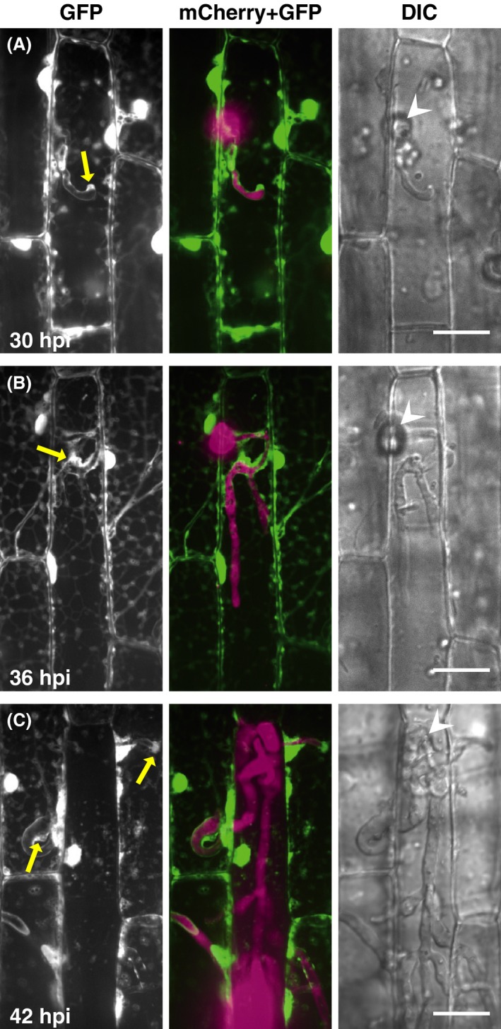
Figure 1.

Visualization of host cytosol during Magnaporthe oryzae infections. Leaf sheaths of transgenic rice plants constitutively expressing AcGFP1 (cyto‐GFP line) were inoculated with a conidial suspension of a compatible strain (Ina86‐137) transformed with tefp::mCherry (TmC1 line) and observed using a laser confocal microscope. Representative data of the 8 (A), 15 (B), and 12 (C) similar images are shown. Cyto‐GFP signals are detected along the primary invasive hyphae both in the first‐invaded (A and B) and second‐invaded cells (C), in addition to the predicted positions of the BIC (yellow arrows). GFP, stacked z‐series confocal fluorescence images of GFP signals corresponding roughly to the surface half of rice epidermal cells. mCherry + GFP, mergers of the GFP image, and stacked z‐series confocal fluorescence images of mCherry signals. Wedge, appressorial penetration site. Size bar = 20 μm. BIC, biotrophic interfacial complex; GFP, green fluorescent protein; DIC, differential interference contrast images.
