Abstract
Aim: The aim of this study is to highlights possible differences in the volume of shaping and canal surface area after the using of common endodontic devices ProTaper Universal and WaveOne systems. Methods: Forty ISO 15, 0.02 taper, S-shaped endo-training Blocks (Dentsply, Maillefer) were assigned in two groups (n = 20 for each group). For each block the initial working length (WL) was evaluated with a 10 K-files (Dentsply Maillefer), so the glide path was created with PathFile 1, 2 and 3 (Dentsply Maillefer) at the WL. After that, simulated canals in the group 1 were shaped with S1, S2, F1 and F2 at WL; while in group 2 it was used single-file WaveOne primary in reciprocating motion. After shaping, the resin blocks were analysed with Skyscan 1172 scanner (Skyscan, Kontich, Belgium) and then volumetrically at a source voltage of 65 kV and a source current of 153 uA. Results: No statistically differences (P > 0.05) have been found in terms of volume and surface area after the use of ProTaper Universal and WaveOne systems. Conclusions: Although, results from micro-CT analysis revealed that Wave One result in a decrease of volume and surface area of shaping than ProTaper Universal, differences are not statistically significant.
Keywords: Reciprocating system, simulated root canals, WaveOne, ProTaper universal, micro-CT
Introduction
Endodontic treatment consists in the shaping, cleaning and closure of the root canal system, in order to maintain the tooth in the oral cavity for aesthetics and occlusal function.
Shaping concepts of the root canals are based on those described by Schilder [1], which included mechanical requirements and biological targets. Although, today the advent of the Ni-Ti alloys has partially facilitated this protocols such concepts are still valid. The super elasticity of this alloy has allowed the realization of tools with high dimensions and taper, which can be used in rotational or alternating movements, with controlled torque and speed using specific dedicated motors [2].
The Ni-Ti instruments easily glide and work in large and regular curvatures while are particularly stressed by tight turns and multiple. It is therefore sought to eliminate these problems with the use of methods involving the use of a single instrument contouring, used with reciprocating motion. In fact, has been demonstrated that reciprocating motion increase cyclic fatigue resistance and decreases apical debris extrusion [3]. The first instrument used as single file was ProTaper F2, since then single-file reciprocating systematic have been developed in order to reduce cost and time of treatment [4]. WaveOne system is a single-file reciprocating tool made from M-Wire with an apically concave triangular cross section and reverse cutting blades [5]. According to the manufacturer, the instrument should rotate at approximately 350 rpm with 30° of clockwise (CW) and 150° of counterclockwise (CCW) rotation angles [6,7]. Since today poor study have evaluated a comparison of a single-file reciprocating system vs multi-files rotational system about tridimensional parameters. The aim of this study was to compare ProTaper and WaveOne systems on simulated root canals by micro-CT evaluations. The null hypothesis tested was that there weren’t statistically significant differences between ProTaper and WaveOne system about volume and surface area of shaping and structure model index (SMI).
Materials and methods
Preparation of specimens
A total of forty ISO 15, 0.02 taper S-shaped Endo Training Blocks (Dentsply Maillefer) were assigned at two groups (n = 20 for each group). In the group 1, two different operators shaped simulated root canals with ProTaper device. The first operator had over 5 years experience in the field of endodontics, the second is a dental student in the last year of university study. For each block the initial working length (WL) was evaluated with a 10 K-files (Dentsply Maillefer), so the glide path was created with PathFile 1, 2 and 3 (Dentsply Maillefer) at the WL. Glyde (Dents ply Maillefer) was used as lubricating agent, so the channel was shaped using the ProTaper sequence S1 S2 F1 F2 at WL, in order to have an apical width of 0.25. In the group 2, the same two operators of Group 1 shaped the simulated canals with WaveOne primary only. The determination of WL and the glide path was performed with the same method of the Group 1. Subsequently, canals were shaped using Wave One Primary reciprocating files till WL with the reciprocating motor X-Smart Plus used with the dedicated manufacturer configuration setup.
Micro-CT measurement
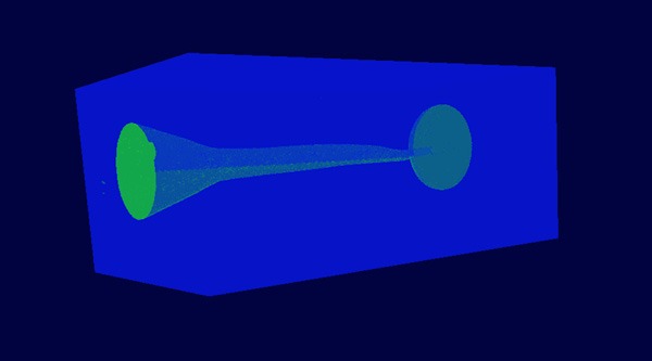
Each resin blocks was placed horizontally on the staging platform of a Skyscan 1172 scanner (Skyscan, Kontich, Belgium) and analysed volumetrically at a source voltage of 65 kV and a source current of 153 uA. Datasets were acquired with a 0.2-degree rotation step and a voxel size of 11.99 um. Scanning of each section generated a total of 1802 slices per block; 3D reconstruction was performed using NRecon software Version 1.6.1.3 (Skyscan) with the following parameters: beam hardening reduction 35%, ring artefact correction 20 and Gaussian low-pass for noise reduction 50%. The CTAn Version 1.9.3 software (Skyscan) was used to calculate the volume of the prepared canal in each block (Figures 1, 2).
Figure 1.

Channel after shaping in resin block. It’s possible to see the plastic material (Blue) and the volume of shaping (green).
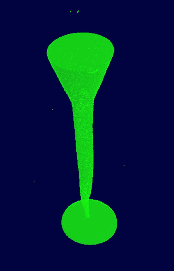
Figure 2.

Form of the channel after shaping with WaveOne system.
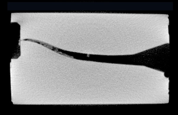
The volume of interest was determined using a standardized rectangular area selection on the binary images. Similar thresholding levels were applied to separate resin blocks from the empty canals. This resulted in binary images of the canals that permitted precise measurements; the software then calculated the respective volumes. A specialist operator that was blinded for this study performed the micro-CT measurements. As results of these evaluations it was possible to calculate: volume of shaping, surface area and the structure model index (SMI) (Figure 3).
Figure 3.

Sample of the CT evaluated block and one of the endodontic system used in the study.
Statistical analysis
Data were analysed by using GraphPad Prism software 6.00 (GraphPad Prism Software, San Diego, CA, USA) version for Windows by an expert in statistical analysis. Statistical significance between different groups was determined by one-way ANOVA and test T. A P-value < 0.05 was taken as threshold to reject the null hypothesis of difference’s absence between groups.
Results
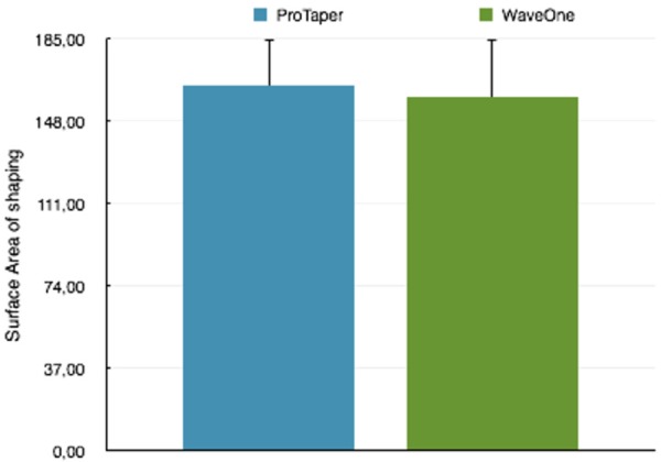
Table 1 reported no differences in terms of quantitative and qualitative three-dimensional parameters, with the use of a multi-files rotational or single-file reciprocating system. Results for volume of shaping and surface area are reported respectively in Figures 4 and 5. These results take us to accept the null hypothesis of no differences between these two methods.
Table 1.
It is reported no differences in terms of quantitative and qualitative three-dimensional parameters, with the use of a multi-files rotational or single-file reciprocating system
| Micro-CT three dimensional parameters | |||
|---|---|---|---|
|
| |||
| ProTaper | WaveOne | P-value | |
| Canal volume (mm3) | 30.10 ± 2.08 | 29.59 ± 2.44 | 0.47 |
| Surface Area (mm2) | 163.62 ± 21.09 | 158.53 ± 25.79 | 0.49 |
| Structurel Model Index | 1.62 ± 0.19 | 1.73 ± 0.33 | 0.21 |
Figure 4.

Comparison between results for volume of shaping with the two used devices.
Figure 5.

Comparison between results for volume of the surface area with the two used devices.
Discussion
The purpose of this study was to evaluate if there are differences in the use of ProTaper or WaveOne system by investigating three-dimensional parameters. To understand this, we performed a shaping comparison on simulated root canals with micro-CT measurements. Although, resin blocks presenting qualitative differences with teeth they represent a valid tool for in vitro evaluations because they permit an easier standardization and comparison of different shaping methods. In addition, it has been demonstrated that micro-CT measurements allows a more accurate analysis than conventional photographic methods since it allows evaluating both 2D and 3D parameters. In past, micro-CT measurements had lower precision caused by a large spatial resolutions [8,9]. Tachibana and Matsumoto were the firsts to evaluate the applicability of computed tomography (CT) imaging in endodontics [10]. Over the years, micro-CT enhanced the spatial resolution analysis, however this led to an increment of scanning and reconstruction time with voxel sizes decreasing to nearly 30 um. Since it has been shown that a repeated use of the instruments may lead to a decrease in the volume shaping, we used a new instruments set for the shaping of each simulated root canal [11]. In previously studies, single-file reciprocating system strongly decrease the mean preparation time in comparison with multi-file rotational system [12]. Other studies focusing on resistance of cyclic fatigue found a higher resistance to fatigue stress of WaveOne than ProTaper (used both in rotational that in reciprocating motion) [13]. However, ProTaper demonstrated to product lower postoperative pain after canal instrumentation than WaveOne single-file system [14]. Moreover, these two systematics seem to be comparable in terms of bacterial removal when used during endodontic retreatment [15]. Our data revealed no differences about volume and surface area of shaping with the use of these two systematics. In addition, thanks to micro-CT has been possible to perform a qualitative shaping evaluation with the structural model index (SMI). Hildebrand and Ruiegsegger [16] firstly introduced by the SMI for the evaluation of bone microarchitecture. Peters et al. [17] were the firsts to use it for endodontic evaluation, this value can vary between 0 (= parallel flat planes) and 4 (= an ideal ball) and characterizes a structure as: “being ribbon-shaped versus cylindrical and is Expressed in arbitrary units” [18]. Also with regard to this parameter our data showed no differences with the use of both the systematics (P-value = 0.21).
Conclusions
Results from this study revealed no statistically differences (P-value > 0.05) between ProTaper and WaveOne groups related to Surfaces Area and Volume of interesting. Same results were obtained in terms of qualitative evaluation by structural model index, confirming absence of differences in the three dimensional parameters with use of multi-file rotational or single-file reciprocating systems.
Disclosure of conflict of interest
None.
References
- 1.Schilder H. Cleaning and shaping the root canal. Dent Clin North Am. 1974;18:269–296. [PubMed] [Google Scholar]
- 2.Peters OA. Current challenges and concepts in the preparation of root canal systems: a review. J Endod. 2004;30:559–567. doi: 10.1097/01.don.0000129039.59003.9d. [DOI] [PubMed] [Google Scholar]
- 3.De-Deus G, Brandao MC, Barino B, Di Giorgi K, Fidel RA, Luna AS. Assessment of apically extruded debris produced by the single-file ProTaper F2 technique under reciprocating movement. Oral Surg Oral Med Oral Pathol Oral Radiol Endod. 2010;110:390–394. doi: 10.1016/j.tripleo.2010.04.020. [DOI] [PubMed] [Google Scholar]
- 4.Yared G. Canal preparation using only one Ni-Ti rotary instrument: preliminary observations. Int Endod J. 2008;41:339–344. doi: 10.1111/j.1365-2591.2007.01351.x. [DOI] [PubMed] [Google Scholar]
- 5.Berutti E, Chiandussi G, Paolino DS, Scotti N, Cantatore G, Castellucci A, Pasqualini D. Canal shaping with WaveOne Primary reciprocating files and ProTaper system: a comparative study. J Endod. 2012;38:505–509. doi: 10.1016/j.joen.2011.12.040. [DOI] [PubMed] [Google Scholar]
- 6.Franco V, Fabiani C, Taschieri S, Malentacca A, Bortolin M, Del Fabbro M. Investigation on the shaping ability of nickel-titanium files when used with a reciprocating motion. J Endod. 2011;37:1398–1401. doi: 10.1016/j.joen.2011.06.030. [DOI] [PubMed] [Google Scholar]
- 7.Berutti E, Paolino DS, Chiandussi G, Alovisi M, Cantatore G, Castellucci A, Pasqualini D. Root canal anatomy preservation of WaveOne reciprocating files with or without glide path. J Endod. 2012;38:101–104. doi: 10.1016/j.joen.2011.09.030. [DOI] [PubMed] [Google Scholar]
- 8.Gambill JM, Alder M, del Rio CE. Comparison of nickel-titanium and stainless steel hand-file instrumentation using computed tomography. J Endod. 1996;22:369–375. doi: 10.1016/S0099-2399(96)80221-4. [DOI] [PubMed] [Google Scholar]
- 9.Dowker SE, Davis GR, Elliott JC. X-ray microtomography: nondestructive three-dimensional imaging for in vitro endodontic studies. Oral Surg Oral Med Oral Pathol Oral Radiol Endod. 1997;83:510–516. doi: 10.1016/s1079-2104(97)90155-4. [DOI] [PubMed] [Google Scholar]
- 10.Tachibana H, Matsumoto K. Applicability of X-ray computerized tomography in endodontics. Endod Dent Traumatol. 1990;6:16–20. doi: 10.1111/j.1600-9657.1990.tb00381.x. [DOI] [PubMed] [Google Scholar]
- 11.Ounsi HF, Franciosi G, Paragliola R, Al-Hezaimi K, Salameh Z, Tay FR, Ferrari M, Grandini S. Comparison of two techniques for assessing the shaping efficacy of repeatedly used nickel-titanium rotary instruments. J Endod. 2011;37:847–850. doi: 10.1016/j.joen.2011.02.030. [DOI] [PubMed] [Google Scholar]
- 12.Burklein S, Hinschitza K, Dammaschke T, Schafer E. Shaping ability and cleaning effectiveness of two single-file systems in severely curved root canals of extracted teeth: Reciproc and WaveOne versus Mtwo and ProTaper. Int Endod J. 2012;45:449–461. doi: 10.1111/j.1365-2591.2011.01996.x. [DOI] [PubMed] [Google Scholar]
- 13.Pirani C, Ruggeri O, Cirulli PP, Pelliccioni GA, Gandolfi MG, Prati C. Metallurgical analysis and fatigue resistance of WaveOne and ProTaper nickel-titanium instruments. Odontology. 2014;102:211–216. doi: 10.1007/s10266-013-0113-6. [DOI] [PubMed] [Google Scholar]
- 14.Nekoofar MH, Sheykhrezae MS, Meraji N, Jamee A, Shirvani A, Jamee J, Dummer PM. Comparison of the Effect of Root Canal Preparation by Using WaveOne and ProTaper on Postoperative Pain: A Randomized Clinical Trial. J Endod. 2015;41:575–8. doi: 10.1016/j.joen.2014.12.026. [DOI] [PubMed] [Google Scholar]
- 15.Martinho FC, Freitas LF, Nascimento GG, Fernandes AM, Leite FR, Gomes AP, Camões IC. Endodontic retreatment: clinical comparison of reciprocating systems versus rotary system in disinfecting root canals. Clin Oral Investig. 2015;19:1411–7. doi: 10.1007/s00784-014-1360-9. [DOI] [PubMed] [Google Scholar]
- 16.Hildebrand T, Ruegsegger P. Quantification of Bone Microarchitecture with the Structure Model Index. Comput Methods Biomech Biomed Engin. 1997;1:15–23. doi: 10.1080/01495739708936692. [DOI] [PubMed] [Google Scholar]
- 17.Peters OA, Laib A, Ruegsegger P, Barbakow F. Three-dimensional analysis of root canal geometry by high-resolution computed tomography. J Dent Res. 2000;79:1405–1409. doi: 10.1177/00220345000790060901. [DOI] [PubMed] [Google Scholar]
- 18.Peters OA, Schonenberger K, Laib A. Effects of four Ni-Ti preparation techniques on root canal geometry assessed by micro computed tomography. Int Endod J. 2001;34:221–230. doi: 10.1046/j.1365-2591.2001.00373.x. [DOI] [PubMed] [Google Scholar]


