Abstract
This study is to investigate the clinical characteristics of patients with nonarteritic anterior ischemic optic neuropathy (NAION). Totally 133 patients (156 eyes) were included in this study. At the first visit and follow-up visits, the patients were subjected to the ophthalmic evaluations, including the fundus photography, visual field (VF) test, fluorescein fundus angiography, and optical coherence tomography (OCT). The visual acuity (VA) of 156 eyes and the VF of 148 eyes were evaluated. For the VA assessment, 59 (38%), 67 (43%), and 30 (19%) cases presented with an initial VA ≥ 20/40, between 20/40 and 20/400, and ≤ 20/400, respectively. VA was improved in 44% and deteriorated in 8% of the patients with VA < 20/20 after NAION onset. Inferior or superior altitudinal defects, constricted fields, and nasal steps were the most common VF defects. In the eyes with VF defects, 32 cases (22%) were improved, and 25 cases (17%) were worsened. In the 61 cases (39%) with VA ≤ 20/200 at NAION onset, neuroepithelial detachment in the fovea was found in 37 eyes (61%). For the optic disc assessment, retinal nerve fiber layer (RNFL) thickening was the most common symptom of NAION. Out of the 36 eyes with ONA or DR, 72% showed VA improvement after the NAION occurrence in the contralateral eye. Poor microcirculation perfusion in the bilateral optic nerve hypoplasia (ONH) might be the underlying mechanism for NAION, which could be relieved by compromising the blood supply to the one side.
Keywords: Nonarteritic anterior ischemic optic neuropathy, short posterior ciliary arteries, optical coherence tomography
Introduction
Nonarteritic anterior ischemic optic neuropathy (NAION) is one of the most common clinical presentations of acute ischemic damage to the optic nerve [1]. NAION is prevalent in the middle-aged and elderly people, affecting increasing population worldwide [2]. Currently, clinical treatments for NAION are mainly empirical, involving a wide range of agents, most of which have not been adequately studied.
Circulatory insufficiency in the optic nerve head is the widely accepted cause of NAION, which is associated with the dysfunction of short posterior ciliary arteries. However, the mechanisms for the vasculopathy and ischemia are still unclear [3]. Epidemiological, clinical and animal studies concerning the disease pathogenesis have identified several systemic and vascular risk factors, including diabetes, hypertension, hypercholesterolemia [4,5], and small cup-to-disk (C/D) ratio [6]. Moreover, the circulatory insufficiency has been found more often in the unaffected eye [7,8], and there is a lower rate of recurrence in the affected eye [9]. Furthermore, ischemia-related cell death of retinal ganglion cells has also been observed in the disease development [10,11]. Therefore, it is of great importance to investigate the intrinsic characteristics of NAION. In this study, the clinical characteristics of NAION patients and their visual outcomes were assessed and analyzed.
Materials and methods
Patients and methods
We had systematically investigated various aspects of NAION in the Fundus Disease Clinic at Nankai University of Tianjin Hospital since 2007. Totally 133 NAION patients (110 patients with data for one eye and 23 patients with data for both; thus, a total of 156 eyes) fulfilled the inclusion criteria and were included in this study, who were first seen at our clinic from 2007 to 2013. Prior written and informed consent were obtained from every patient and the study was approved by the ethics review board of First Hospital of Jilin University.
Criteria for the diagnosis of NAION and inclusion had been previously described by Hayreh et al. [12]: (1) a history of sudden visual loss, without other ocular, systemic, or neurologic diseases that might cause the visual symptoms; (2) optic disc edema (ODE) at onset, which was confirmed by at least three ophthalmology experts; (3) cranial CT, carotid artery Doppler and cranial MRI (only when necessary) excluded diseases that could cause an insufficiency in ophthalmic arterial blood supply, such as brain tumor compression and carotid artery stenosis; (4) optic disc-related visual field (VF) defects in the eye; and (5) no previous corticosteroid therapy or any other treatment for NAION.
Patients who had retinal or optic nerve lesions, or cataracts that could influence their visual status, were excluded. NAION patients with only background diabetic retinopathy were included; however, those with active neovascularization, vitreous hemorrhages, tractional detachment, or other complications influencing their visual acuity (VA) or VF were excluded. Patients with an elevated intraocular pressure (> 21 mmHg) and a shallow anterior chamber, without glaucomatous visual lesions, were included.
Study performed
A detailed ophthalmic and medical history for each patient was obtained at the first visit to our clinic. For the systematic diseases, a history of all previous or current diseases were elicited, particularly concerning the arterial hypertension, diabetes mellitus, ischemic heart disease, stroke, transient ischemic attacks, and carotid artery disease, as well as previous or current drug use. A comprehensive ophthalmic evaluation was performed then by at least two specialists, including: (1) VA measurement according to the Snellen chart; (2) VF measurement with automated perimetry; (3) relative afferent papillary defect assessment; (4) intraocular pressure measurement; (5) slit-lamp examination of the anterior segment, lens, and vitreous humor; (6) direct and indirect ophthalmoscopy; (7) stereoscopic color fundus photography and fluorescein fundus angiography. Moreover, regular blood test and blood pressure measurement (between 8:00 AM and 8:30 AM) were performed. In addition, at the first visit, a systemic evaluation was performed by a cardiologist, internist, or physician. Other systemic or neurologic investigation was also performed to rule out the related causes of visual loss.
Follow-up protocol
Follow-up was initially performed every week, until the ODE was alleviated (lasting for approximately 5-8 w). Thereafter, the patients were examined at 3 m and 6 m, and then yearly.
Visual status evaluation
VA was tested using the Snellen chart and VF defects were evaluated according to a previous method from Hayreh et al. [12]. In this study, automated perimetry (Octopus 101) was used to measure the 30° VF. Mean sensitivity (MS), mean defect (MD), and corrected loss variance (CLV) were documented. Patients with VA ≤ 20/200 underwent fundus OCT (Cirrus HD-OCT ZEISS). Macular and optic disc color pictures, average retinal nerve fiber layer (RNFL) thickness, and average C/D ratio were recorded.
For the eyes with recurrence of NAION, the VA and VF defects were evaluated independently by three ophthalmology experts. Only the evaluation data before the recurrence were used for this study. A change of ≥ 3 lines in the Snellen chart was considered significant (improvement or worsening), which was equivalent to a change of ≥ 0.3 in the logMAR chart.
Statistical analysis
Descriptive statistics (means, standard deviations, and percentages) were computed for the demographic and clinical variables, VA and VF defects. The F test was performed to compare variances, and one-way ANOVA Kruskal-Wallis test was used for the group comparison.
Results
Basic characteristics of the patients in this study
Totally 133 patients (156 eyes) were included in this study, and the basic characteristics of these patients were shown in Table 1. In these patients, 6 cases (4.5%) had simultaneous bilateral NAION, for which only the data from the more severe eye were recorded. Moreover, 23 patients (17%) had successive bilateral NAION. Furthermore, 3 eyes (2%) had NAION recurrence during the follow-up period, for which the data from the last follow-up visit before recurrence were used. In addition, the VF data of 8 eyes (5.1%) were lost during the follow-up period. The median follow-up period was 1.2 y (interquartile range [IQR], 0.7-2.1 y).
Table 1.
Clinical characteristics of patients with nonarteritic anterior ischemic optic neuropathy (NAION)
| Clinical variable | 133 patients (156 eyes) |
|---|---|
| Male patients [n (%)] | 74 (51%) |
| Age at first visit (y) | 55 ± 9.8 |
| NAION eye involvement [n (%)] | |
| Right eye | 54 (32%) |
| Left eye | 56 (33%) |
| Both eyes | 29 (35%)a |
| Follow-up period | n = 148 eyesb |
| Median (25-75%) | 1.2 (0.7-2.1) y |
| Min-max | 1.7 m-5.3 y |
| Systemic conditions [n (%)] | |
| Arterial hypertension | 54 (37.2%) |
| Diabetes mellitus | 37 (25.5%) |
| Ischemic heart disease | 16 (11.0%) |
| Other disease | 8 (0.06%) |
| Cholesterol (mmol/L) | 5.4 ± 1.0 |
| Triglyceride (mmol/L) | 1.9 ± 1.0 |
| Min blood shear rate | 20.6 ± 4.0 |
| Diabetic retinopathy | 18 (11.5%) eyes |
| Initial IOP (mmHg) | 15.2 ± 3.5 |
| Initial blood pressure (mmHg) | 129 ± 17.3/80.3 ± 9.4 |
In these cases, 6 patients had bilateral NAION simultaneously, and 17 patients developed NAION successively;
8 eyes lost visual field (VF) data during follow-up.
Evaluation of visual acuity (VA) and visual field (VF) defects
For the VA assessment, only data from the first and last follow-up visits prior to the occurrence of new ophthalmic diseases were presented. As shown in Table 2, at the first visit, 33% of the eyes had VA ≥ 20/40. Within 3 w after the symptom onset, the VA of 30% of the eyes was between 20/40 and 20/400, and the VA ≤ 20/400 was observed in 19% of the eyes. Out of the 156 eyes with NAION, 59 cases (38%) presented with an initial VA ≥ 20/40, in which 24 cases (41%) were improved and 3 cases (5%) were worsened. Moreover, 23 eyes with NAION (15%) presented with an initial VA ≥ 20/20. Furthermore, 67 eyes with NAION (43%) presented with an initial VA between 20/40 and 20/400, in which 26 cases (39%) were improved and 8 cases (12%) were worsened. In addition, 30 eyes with NAION (19%) presented with an initial VA ≤ 20/400, in which 9 cases (30%) were improved. For patients with VA < 20/20 after the NAION onset, 44% were improved and 8% were worsened. Importantly, the percentage of VA improvement during follow-up in the patients who were first visited at > 3 w after visual loss (22%) were dramatically lower than those who were first visited within 3 w (43%).
Table 2.
Visual acuity (VA) assessment of NAION patients
| Time from the visual loss onset to the first visit | |||||||||
|---|---|---|---|---|---|---|---|---|---|
|
|
|||||||||
| Within 1 w (n = 54 eyes) | 1-3 w (n = 65 eyes) | > 3 w (n = 37 eyes) | |||||||
|
|
|||||||||
| VA | No. | Improved n (%) | Worsened n (%) | No. | Improved n (%) | Worsened n (%) | No. | Improved n (%) | Worsened n (%) |
| 20/15-20/40a | 26 | 12 (46%) | 1 (4%) | 25 | 11 (44%) | 2 (8%) | 8 | 1 (13%) | / |
| 20/40-20/400 | 18 | 7 (39%) | 1 (6%) | 28 | 14 (50%) | 5 (18%) | 21 | 5 (24%) | 2 (10%) |
| < 20/400 | 10 | 3 (30%) | / | 12 | 4 (33%) | / | 8 | 2 (25%) | / |
| In total | 54 | 22 (41%) | 2 (4%) | 65 | 29 (45%) | 7 (11%) | 37 | 8 (22%) | 2 (6%) |
Including 23 eyes with VA ≥ 20/20.
Totally 125 patients (148 eyes) were subjected to the automated perimetry to measure 30° VF at the NAION onset and every follow-up visit. As shown in Table 3, the inferior or superior altitudinal defects, constricted fields, and nasal steps were the most commonly VF defects in these NAION patients. The VF assessment was performed at the first visit and the last follow-up visit before NAION recurrence. Data for 148 eyes with NAION were analyzed (Table 4). The patients had a median follow-up period of 1.5 y (IQR, 0.5-2.2 y). Out of the eyes with VF defects at the first visit, 32 cases (22%) were improved, 25 cases (17%) were worsened, and 91 cases (61%) showed no changes. Moreover, 6 eyes (4%) showed no evidence of 30° VF defect (with the MD value between -2 and +2 dB at the first visit), which was increased to 15 eyes (10%) at the last follow-up visit. Taken together, these results indicate the natural recovery of VA and VF defects in the NAION patients.
Table 3.
Visual field (VF) defects in the NAION patients
| VF defect | Case [n (%)] |
|---|---|
| Inferior altitudinal defect | 25 (16.9%) |
| Superior altitudinal defect | 17 (11.5%) |
| Constricted field | 31 (20.1%) |
| Nasal step | 24 (16.2%) |
| Central scotoma | 2 (1.3%) |
| Central island field only | 8 (5.2%) |
| Peripheral island field only | 11 (7.4%) |
| Irregular field defect | 7 (4.7%) |
| Total visual field defect | 8 (5.2%) |
| Hemiscotosis | 9 (6.1%) |
| Normal | 6 (4.1%) |
Table 4.
VF assessment of the NAION patients
| Case n (%) | VF parameter | |||
|---|---|---|---|---|
|
|
||||
| MS | MD | CLV | ||
| First visit | 14.8 ± 2.2 | 12.6 ± 2.2 | 40.8 ± 6.6 | |
| Improvement | 32 (22%) | 20.6 ± 1.6* | 6.4 ± 1.5** | 37.2 ± 8.9 |
| Deterioration | 25 (17%) | 7.32 ± 3.15* | 22.0 ± 3.14* | 51.6 ± 14.1 |
Abbreviation: MS, mean sensitivity; MD, mean defect; CLV, corrected loss variance. Note: Compared with the first visit;
P < 0.05;
P < 0.01.
VA assessment of NAION eyes with macular lesions
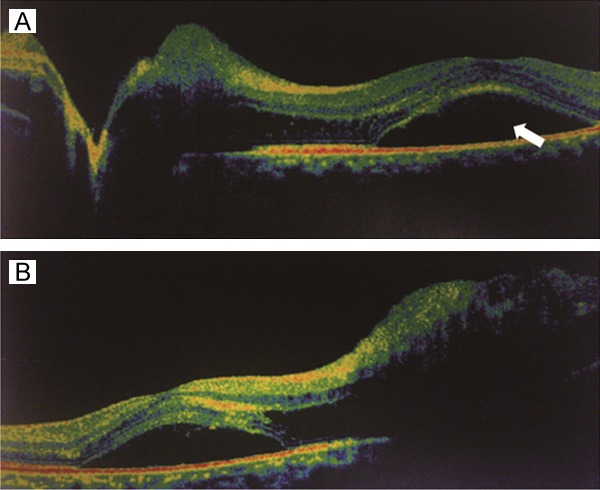
Out of the 156 eyes, 61 cases (39%) with VA ≤ 20/200 were examined by the optical coherence tomography (OCT) at the NAION onset. As shown in Table 5, neuroepithelial detachment in the fovea (Figure 1) was found in 37 eyes (61%), which was the leading cause of low VA in eyes with NAION. For the patients who were first seen at < 1 week after the visual loss, a higher percentage of VA improvement (14/19, 74%) was observed, compared with those who were first visited at > 1 week after the visual loss (6/18, 33%). Moreover, other types of macular lesions were also observed, including macular exudation (10 eyes), and macular nerve fiber layer thickness (7 eyes) and thinness (2 eyes). A significantly higher prevalence of diabetes mellitus was noted in patients with macular exudation (80%) than those with VA ≤ 20/200 (33%) (P < 0.01). Furthermore, macular exudation was not induced by diabetic retinopathy, because no significant difference in the frequency of diabetic retinopathy was observed between the subgroups of patients with or without macular exudation (11.5% v.s. 20%). Taken together, these results suggest that macular lesion is a main cause of low VA in the NAION patients.
Table 5.
VA assessment of NAION eyes with macular changes
| Macular lesion | No. | Improved [n (%)] | No change [n (%)] |
|---|---|---|---|
| Neuroepithelial detachment in the fovea | 37 | 20 (54%) | 17 (46%) |
| Macular exudation | 10 | 6 (60%) | 4 (40%) |
| Macular layer thickness | 7 | 0 (/) | 7 (100%) |
| Macular layer thinness | 2 | 0 (/) | 2 (100%) |
Figure 1.

Neuroepithelial detachment in the fovea. A. Neuroepithelial detachment in the fovea (white arrow) was found in NAION patients with VA ≤ 20/200. B. Neuroepithelial detachment induced by optic disc retinal nerve fiber layer (RNFL) thickening.
Optic disc changes in NAION patients
For the optic disc assessment of the NAION eyes, 78 cases (53%) were examined by OCT at the onset of NAION. In these patients, 22 cases (28%) of the contralateral eye with cup-to-disk (C/D) ratio ≤ 0.1 and 44 cases (56%) with C/D ratio ≤ 0.3 were found. At the onset of NAION, optic disc edema (ODE) usually disrupted the C/D measurement of the affected eye. Thus, the C/D measurement of the contralateral eye was performed for the analysis. ODE increased the optic disc retinal nerve fiber layer (RNFL) thickness and the neuroepithelial detachment. The mean optic disc RNFL thickness in the affected eye was 1.5-4 times that in the contralateral eye (data not shown).
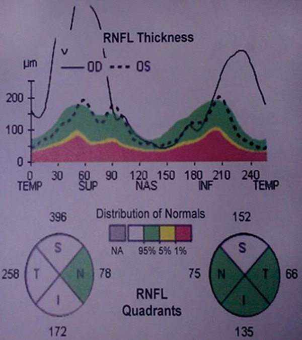
Inferior and superior RNFL thickening of the optic disc was the most common symptom of NAION, appearing in a camel-hump shape (Figure 2). Moreover, optic disc RNFL thickening was prone to induce RNFL detachment (Figure 1). These results suggest that the unique physiologic changes in optic disc might be helpful in predicting and diagnosing NAION.
Figure 2.

Camel-hump shape of optic disc edema (ODE) in NAION patients.
Contralateral visual improvement in NAION patients
To assess the contralateral visual outcomes in the NAION patients, the ophthalmologic information of the contralateral eye was obtained at the disease onset. As shown in Tables 6 and 7, at the NAION onset, 30, 6, 3, and 2 eyes out of the 41 eyes were diagnosed as optic atrophy, diabetic retinopathy (DR), macular hole, and dry-AMD, respectively. Moreover, 36 eyes had VF defects with varying severities. The period between the disease onset and the first visit ranged from 3 to 34 d, with a mean time of 13.2 d. The follow-up period ranged from 3 m to 2.5 y (IQR, 0.5-1.7 y). Patients developed ONA at 4 m to 2 y after the occurrence NAION in the contralateral eye. The VA improvement of ≥ 3 lines was observed in 21 eyes (70%) with optic never atrophy (ONA) and 5 eyes (83%) with DR; however, no VA improvement was noted in patients with macular hole or dry-AMD. The patients with ONA or DR in the contralateral eye had a higher incidence of VA improvement (72%), compared with other NAION patients (44%) (P < 0.01).
Table 6.
VA assessment of the contralateral eye in NAION patients
| Macular lesion | No. | Improved [n (%)] | No change [n (%)] |
|---|---|---|---|
| ONA | 30 | 21 (70%) | 9 (30%) |
| DR | 6 | 5 (83%) | 1 (17%) |
| Others | 5 | 0 (0%) | 5 (100%) |
Abbreviation: ONA, optic never atrophy; DR, diabetic retinopathy.
Table 7.
VF assessment of the contralateral eye in NAION patients
| VF parameter | |||
|---|---|---|---|
|
|
|||
| MS | MD | CLV | |
| First visit | 17.93 ± 2.07 | 10.23 ± 2.06 | 27.7 ± 5.6 |
| Last follow-up | 20.73 ± 2.33 | 6.71 ± 2.22 | 21.35 ± 7.04 |
Abbreviation: MS, mean sensitivity; MD, mean defect; CLV, corrected loss variance.
To determine whether the results of the VF recovery were the same as the VA recovery, the VF data of 36 eyes before and after the NAION occurrence in the contralateral eye were analyzed. In these cases with VF defects, no improvement was found between the first visit and the last follow-up visit. However, the MD values between -2 and +2 dB were observed in 10% of the patients with classical NAION. Moreover, the MD values between -2 and +2 dB were found in 28% of the contralateral eye at the first visit, which was increased to 61% at the last follow-up visit. Taken together, these results suggest that contralateral VA and VF defects could be alleviated in the NAION patients during the follow-up period.
Discussion
Vascular factors involved in glaucomatous optic neuropathy have been attracting increasing attention over the past few years [13]. Unlike intraocular pressure, poor vascular perfusion could be an independent factor in the pathogenesis of glaucomatous optic neuropathy. A great amount of evidence indicates that patients with glaucomatous optic neuropathy have poor vascular circulation in the posterior ciliary, central retinal, and ophthalmic arteries [14]. Clinical studies have shown that ischemic disorders could lead to retinal nerve fiber layer edema, VF defects and macular lesions, in patients with non-glaucomatous optic nerve atrophy (such as atrophy caused by central retinal artery occlusion, proliferative diabetic retinopathy, atherosclerosis with arterial hypertension, and compressive optic neuropathy) [15-18]. Therefore, investigation on NAION may help to increase our understanding of the vascular causes of glaucomatous optic neuropathy.
In this study, we investigated NAION patients in regards of the systemic characteristics, the disease course, and the natural recovery of VA and VF, and similar results were obtained as the previous studies [6,19-21]. Importantly, this study discovered new features of NAION, including the camel-hump shape of the optic disc, macular lesions, and contralateral visual improvement in bilateral sequential NAION. Combined with our clinical experience, these findings might help us to improve the diagnosis and prognosis of the ischemic optic neuropathy.
In this study, most NAION patients had at least one vasculopathy risk factor. Our results showed that hypertension, diabetes and ischemic heart disease were present in 37%, 25.5%, and 11%, respectively, of all the NAION patients. Moreover, higher mean blood cholesterol (5.4 mmol/L) and triglyceride (1.9 mmol/L) levels were commonly seen in the NAION patients, which might increase the blood viscosity. The anatomic optic disc risk factors were also determined in this study. In normal eyes, there is ample room for the optic nerve to exit the eye. The optic cup or disc provides a narrow space for the short posterior ciliary artery (SPCA) to pass through. In our study, over half of the patients had a C/D ratio ≤ 0.3 and 28% of the eyes had a C/D ratio ≤ 0.1. This crowded structure might compress the capillaries and small vessels among the nerve fibers, resulting in poor microvascular perfusion. Therefore, patients with small C/D ratios are more predisposed to NAION [22,23]. In addition, the asymmetric characteristic of NAION was implied in this study. Our results indicated low incidence of NAION in both eyes (4.5% of the patients had simultaneous bilateral NAION), low monocular relapse rate (2%), and relatively higher incidence involving both eyes (17%), which was in line with a previous retrospective study [24]. In the case of bilateral NAION, VA and VF results were significantly inconsistent between the two eyes, which was line with the findings from Hayreh et al. [25]. Based on these results, we speculate that poor microvascular perfusion persists in both eyes in the NAION patients, resulting in poor blood supply. The short posterior ciliary artery occlusion could randomly shut down the blood supply to the unilateral eye, which would swell the ischemic optic nerve axons and aggravate the vicious cycle. That is why monocular occurrence has been commonly seen in NAION patients. Optic atrophy could lead to poor microvascular perfusion at months after the NAION onset, which might result in a poor blood supply to the contralateral eye, making it susceptible to NAION. However, it is still unclear how short posterior ciliary artery occlusion occurs before the NAION onset, and whether there is an inconsistent blood supply to the two eyes after the disease onset [6].
In this study, our results showed that, for patients with successive bilateral NAION or DR with VA ≤ 20/30 (17% of all the patients), when the contralateral eye developed NAION, the eye that initially had NAION showed better VA improvement than the cases with unilateral NAION. In clinic, when one eye has a better VA and then suffers from disease or trauma, the other eye shows spontaneous VA improvement. In our study, the spontaneous VA improvement did not occur in the cases of structural lesions in the fovea, such as macular holes or AMD. This phenomenon might be explained by the following hypotheses: 1) optic nerve cells have nutritional blood dependence; 2) optic nerve cells depend on neurotrophic factors; 3) the visual cortex shows plasticity. The first two hypotheses state that the restoration of blood supply and neurotrophic factors are indispensable for optic neural cells, while the third hypothesis emphasizes that the potential for visual restoration remains in the visual cortex in adults (like the occlusion therapy for amblyopia). According to our results, we believe that the blood supply restoration could lead to VA rehabilitation, because before the NAION onset, patients might have persistent microcirculatory ischemia. After the occurrence of NAION in one eye, the ischemia could induce retinal ganglia cell death, leading to the permanent VF defect in the area of the short posterior ciliary artery. Impaired central vision is caused by the reduced blood supply, which makes the retinal ganglia cells dormant. Once the ischemia is relieved (when the contralateral eye develops NAION, as discussed above), the retinal ganglia cells wake up from the dormant status, which could contribute to the central vision improvement. Of course, further in-depth studies are still needed to confirm the hypothesis.
This is the first study, in China, focusing on the natural history of NAION with a large sample size, and similar results were obtained as the previously published study in the prognoses of VA and VF [19,20]. For the measurement of VF, the 30° automated perimetry was used herein, generating a comparable error of non-VF defect (4.1%) with the other study (6.4%) [12]. Moreover, automated perimetry had the advantage of generating quantitative data that could be used for comparison. However, it is really important to use 30° as the cut-off for the measurement of VF.
In conclusion, our results showed some new characteristics of NAION, including the camel-hump shape of the optic disc, macular lesions, and spontaneous VA improvement in the contralateral eye. Moreover, poor microcirculation perfusion in the bilateral optic nerve hypoplasia (ONH) might be the underlying mechanism for NAION, which could be relieved by compromising the blood supply to the one side. Together with previous studies, these findings support the ischemia/reperfusion theory in the NAION pathogenesis. These findings might improve the understanding of the pathogenesis of NAION and help in the development of treatment for the disease.
Acknowledgements
We are grateful to Dr. Mei Han, Dr. Chunxia Cong, and Xin Wang from Tianjin Eye Hospital for their kind assistance in the data collection and analysis and the manuscript preparation.
Disclosure of conflict of interest
None.
References
- 1.Miller GR, Smith JL. Ischemic optic neuropathy. Am J Ophthalmol. 1966;62:103–15. doi: 10.1016/0002-9394(66)91685-0. [DOI] [PubMed] [Google Scholar]
- 2.Hayreh SS. Anterior ischemic optic neuropathy. III. Treatment, prophylaxis, and differential diagnosis. Br J Ophthalmol. 1974;58:981–9. doi: 10.1136/bjo.58.12.981. [DOI] [PMC free article] [PubMed] [Google Scholar]
- 3.Arnold AC. Pathogenesis of nonarteritic anterior ischemic optic neuropathy. J Neuroophthalmol. 2003;23:157–63. doi: 10.1097/00041327-200306000-00012. [DOI] [PubMed] [Google Scholar]
- 4.Hayreh SS, Joos KM, Podhajsky PA, Long CR. Systemic diseases associated with nonarteritic anterior ischemic optic neuropathy. Am J Ophthalmol. 1994;118:766–80. doi: 10.1016/s0002-9394(14)72557-7. [DOI] [PubMed] [Google Scholar]
- 5.Ischemic Optic Neuropathy Decompression Trial Research Group. Optic nerve decompression surgery for nonarteritic anterior ischemic optic neuropathy (NAION) is not effective and may be harmful. The Ischemic Optic Neuropathy Decompression Trial Research Group. JAMA. 1995;273:625–32. [PubMed] [Google Scholar]
- 6.Hayreh SS. Ischemic optic neuropathy. Prog Retin Eye Res. 2009;28:34–62. doi: 10.1016/j.preteyeres.2008.11.002. [DOI] [PubMed] [Google Scholar]
- 7.Newman NJ, Scherer R, Langenberg P, Kelman S, Feldon S, Kaufman D, Dickersin K Ischemic Optic Neuropathy Decompression Trial Research Group. The follow eye in NAION: report from the ischemic optic neuropathy decompression trial follow-up study. Am J Ophthalmol. 2002;134:317–28. doi: 10.1016/s0002-9394(02)01639-2. [DOI] [PubMed] [Google Scholar]
- 8.Preechawat P, Beau BB, Newman NJ, Biousse V. Anterior ischemic optic neuropathy in patients younger than 50 years. Am J Ophthalmol. 2007;144:653–60. doi: 10.1016/j.ajo.2007.07.031. [DOI] [PubMed] [Google Scholar]
- 9.Hayreh SS. Ipsilateral recurrence of non-arteritic anterior ischemic optic neuropathy. Am J Ophthalmol. 2001;132:734–42. doi: 10.1016/s0002-9394(01)01192-8. [DOI] [PubMed] [Google Scholar]
- 10.Hayreh SS. Animal model for nonarteritic anterior ischemic optic neuropathy. J Neuroophthalmol. 2008;28:79–80. doi: 10.1097/WNO.0b013e318167828e. author reply 80-1. [DOI] [PubMed] [Google Scholar]
- 11.Slater BJ, Mehrabian Z, Guo Y, Hunter A, Bernstein SL. Rodent anterior ischemic optic neuropathy (AION) induces regional retinal ganglion cell apoptosis with a unique temporal pattern. Invest Ophthalmol Vis Sci. 2008;49:3671–6. doi: 10.1167/iovs.07-0504. [DOI] [PMC free article] [PubMed] [Google Scholar]
- 12.Hayreh SS, Zimmerman B. Visual field abnormalities in nonarteritic anterior ischemic optic neuropathy. Arch Ophthalmol. 2005;123:1554–62. doi: 10.1001/archopht.123.11.1554. [DOI] [PubMed] [Google Scholar]
- 13.Hata M, Miyamoto K, Oishi A, Makiyama Y, Gotoh N, Kimura Y, Akagi T, Yoshimura N. Comparison of optic disc morphology of optic nerve atrophy between compressive optic neuropathy and glaucomatous optic neuropathy. PLoS One. 2014;9:e112403. doi: 10.1371/journal.pone.0112403. [DOI] [PMC free article] [PubMed] [Google Scholar]
- 14.Hayreh SS. Evaluation of optic nerve head circulation: review of the methods used. J Glaucoma. 1997;6:319–30. [PubMed] [Google Scholar]
- 15.Hayreh SS, Zimmerman MB. Central retinal artery occlusion: visual outcome. Am J Opthalmol. 2005;140:376–91. doi: 10.1016/j.ajo.2005.03.038. [DOI] [PubMed] [Google Scholar]
- 16.Simó R, Hernández C European Consortium for the Early Treatment of Diabetic Retinopathy (EUROCONDOR) Neurodegeneration in the diabetic eye: new insights and therapeutic perspectives. Trends Endocrinal Metab. 2014;25:23–33. doi: 10.1016/j.tem.2013.09.005. [DOI] [PubMed] [Google Scholar]
- 17.Göbel W, Matlach J. [Hypertensive changes of the funfus] . Ophthalmologe. 2013;110:995–1005. doi: 10.1007/s00347-013-2953-4. [DOI] [PubMed] [Google Scholar]
- 18.Shams PN, Ma R, Pickles T, Rootman J, Dolman PJ. Reduced risk of compressive optic neuropathy using orbital radiotherapy in patients with active thyroid eye disease. Am J Ophthalmol. 2014;157:1299–305. doi: 10.1016/j.ajo.2014.02.044. [DOI] [PubMed] [Google Scholar]
- 19.Hayreh SS, Zimmerman MB. Nonarteritic anterior ischemic optic neuropathy Natural history of visual outcome. Ophthalmology. 2008;115:298–305. e2. doi: 10.1016/j.ophtha.2007.05.027. [DOI] [PMC free article] [PubMed] [Google Scholar]
- 20.Sawle GV, James CB, Russall RW. The natural history of nonarteritic anterior ischemic optic neuropathy. J Neurol Neurosurg Psychiatry. 1990;53:830–3. doi: 10.1136/jnnp.53.10.830. [DOI] [PMC free article] [PubMed] [Google Scholar]
- 21.Salomon O, Huna-Baron R, Steinberg DM, Kurtz S, Seligsohn U. Role of aspirin in reducing the frequency of second eye involvement in patients with non-arteritic anterior ischemic optic neuropathy. Eye. 1999;13:357–9. doi: 10.1038/eye.1999.90. [DOI] [PubMed] [Google Scholar]
- 22.Purvin V, King R, Kawasaki A, Yee R. Anterior ischemic optic neuropathy in eyes with optic disc drusen. Arch Ophthalmol. 2004;122:48–53. doi: 10.1001/archopht.122.1.48. [DOI] [PubMed] [Google Scholar]
- 23.Hayreh SS, Zimmerman B. Nonarteritic anterior ischemic optic neuropathy: fefractive error and its relationship to cup/disc ratio. Am J Opthalmol. 2008;115:2275–81. doi: 10.1016/j.ophtha.2008.08.007. [DOI] [PubMed] [Google Scholar]
- 24.Atkins EJ, Bruce BB, Newman NJ, Biousse V. Treatment of nonarteritic anterior ischemic optic neuropathy. Surv Ophthalmol. 2010;55:47–63. doi: 10.1016/j.survophthal.2009.06.008. [DOI] [PMC free article] [PubMed] [Google Scholar]
- 25.Hayreh SS, Zimmerman B. Bilateral nonarteritic anterior ischemic optic neuropathy: comparison of visual outcome in the two eyes. J Neuroophthalmol. 2013;33:338–43. doi: 10.1097/WNO.0b013e31829b5d03. [DOI] [PubMed] [Google Scholar]


