Abstract
Histamine is an important chemical messenger that regulates multiple physiological processes in both vertebrate and invertebrate animals. Even so, how glial cells and neurons recycle histamine remains to be elucidated. Drosophila photoreceptor neurons use histamine as a neurotransmitter, and the released histamine is recycled through neighboring glia, where it is conjugated to β-alanine to form carcinine. However, how carcinine is then returned to the photoreceptor remains unclear. In an mRNA-seq screen for photoreceptor cell-enriched transporters, we identified CG9317, an SLC22 transporter family protein, and named it CarT (Carcinine Transporter). S2 cells that express CarT are able to take up carcinine in vitro. In the compound eye, CarT is exclusively localized to photoreceptor terminals. Null mutations of cart alter the content of histamine and its metabolites. Moreover, null cart mutants are defective in photoreceptor synaptic transmission and lack phototaxis. These findings reveal that CarT is required for histamine recycling at histaminergic photoreceptors and provide evidence for a CarT-dependent neurotransmitter trafficking pathway between glial cells and photoreceptor terminals.
Author Summary
Neurotransmitter transporters that remove neurotransmitters and recycle them after their release have particular importance at visual synapses, which must signal at high frequencies and therefore required rapid clearance of neurotransmitters from the synaptic cleft. In this study, we identified a SLC22 family transporter, CarT, in the visual system of Drosophila, which is exclusively located to photoreceptor terminals in the lamina neuropil and is responsible for taking up carcinine, an inactive histamine metabolite, from surrounding glia. Loss of CarT disrupts the regeneration of histamine and blocks neurotransmission at photoreceptor cell synapses. Our work provides direct evidence for a local histamine recycling pathway between glial cells and photoreceptor terminals, and shows that a CarT-dependent histamine/carcinine shuttle pathway is critical for maintaining the normal histamine content of neurons.
Introduction
Histamine is an important chemical messenger known to be involved in a broad spectrum of biological processes such as inflammation and gastric acid secretion. It is also recognized as an important neurotransmitter [1]. Recycling histamine at histaminergic synapses is a key event both in maintaining synaptic transmission and in terminating histamine’s action on postsynaptic neurons. The Drosophila visual system uses histamine as the neurotransmitter at photoreceptor synapses, and provides a good genetic model for studying histamine, its metabolism and recycling [2]. The compound eye of Drosophila is composed of ~800 ommatidia, each of which contains eight photoreceptor cells. Of the latter, R1-R6 photoreceptors in each ommatidium project axons from the retina to the underlying lamina neuropil, where they are organized into synaptic modules called cartridges. R7/R8 photoreceptors project axons to the second neuropil, the medulla [3–6]. In lamina cartridges, three epithelial glial cells normally envelop six photoreceptor terminals [7].
Although the synthesis of histamine from histidine occurs de novo under the action of histidine decarboxylase (Hdc) in photoreceptor cells, recycling of histamine is reported to be the dominant pathway for maintaining the histamine content in photoreceptors [8,9]. Both pathways, de novo synthesis and recycling, are required to maintain an adequate content of histamine in photoreceptor cells. Disrupting either pathway affects visual synaptic transmission in Drosophila in the long term [8,10]. Upon light stimulation, photoreceptor terminals release histamine as a neurotransmitter, which activates histamine-gated chloride channels (HisClA) on large monopolar cells (LMCs) in the lamina and hyperpolarizes these postsynaptic neurons [2,4,11]. After its release, histamine is taken up by lamina glia and conjugated to β-alanine, converting it to carcinine by the N-β-alanyl-dopamine synthase, Ebony, which is expressed in epithelial glia [10,12,13]. The metabolized histamine conjugate, carcinine, is then transported back into the photoreceptors and hydrolyzed back to histamine by Tan, an N-β-alanyl-dopamine hydrolase[10,14]. Despite knowledge of these pathways, little is known about the critical step by which carcinine is transported back to the photoreceptors. It has been proposed that the gene inebriated (ine) might encode a carcinine neurotransmitter transporter in photoreceptor cells to take up carcinine from synaptic cleft [15]. However, in this study, we show that Ine fails to function in any clear way in photoreceptor cells. In addition, the cellular location for carcinine uptake, the trafficking route by which it is returned to the photoreceptor cells where the Tan enzyme has to act, and the transporters responsible for carcinine uptake, all remain controversial. Recently, it has been suggested that metabolites of histamine are transported between glia and the cell bodies of photoreceptors through networks of intercellular gap junctions [9].
We identified a photoreceptor cell-enriched neurotransmitter transporter, CarT, which is able to transport carcinine across the membranes of photoreceptors. CarT is predominantly localized to photoreceptor terminals. The cart mutant flies are defective in photoreceptor synaptic transmission, and as a result lack phototaxis. In addition, we found that a human homologue of CarT, Organic Cation Transporter (OCT2), can also transport carcinine in vitro and is thus able to reverse synaptic transmission defects in cart mutant flies. We therefore propose the presence of a novel pathway for histamine recycling, in which the carcinine transporter CarT efficiently takes up carcinine that is released locally from glial cells lying in close vicinity to photoreceptor terminals.
Results
CG9317 encodes a photoreceptor cell-enriched transporter
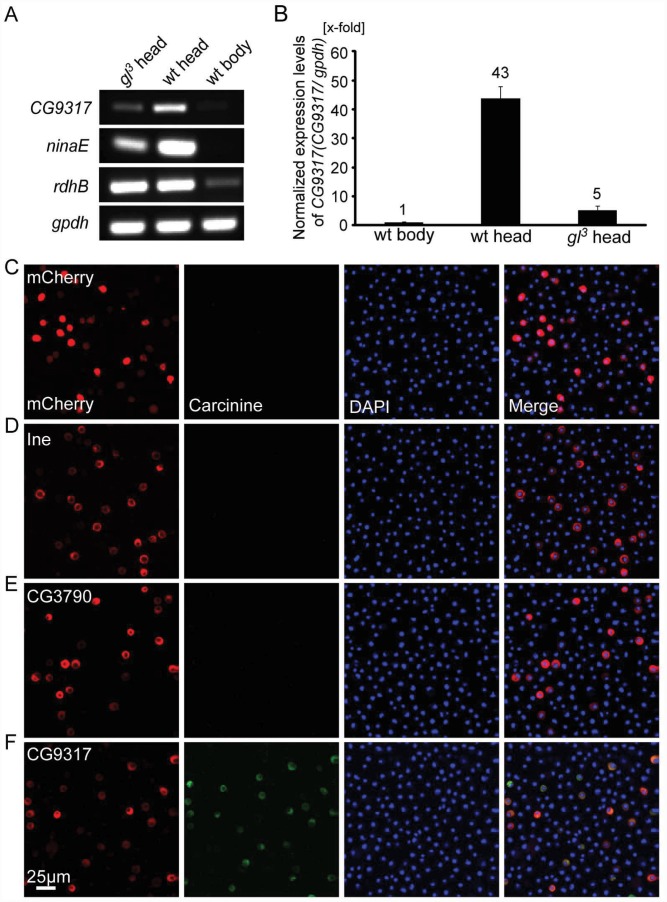
Given that the histamine/carcinine shuttle in the visual system occurs between photoreceptors and surrounding glia cells [7], and that the enzyme Tan responsible for hydrolyzing carcinine to release histamine is exclusively expressed in photoreceptor cells, we assumed that the neurotransmitter transporter responsible for taking up carcinine must be enriched in photoreceptor cells. The gene glass (gl) gene encodes a zinc finger transcription factor, and glass mutations specifically remove photoreceptor cells, but leave other cell types intact. Mutations of glass specifically remove photoreceptor cells, and thus largely abolish the expression of mRNA transcripts of photoreceptor-enriched genes, such as the gene encoding major rhodopsin neither inactivation nor afterpotential E (ninaE). Expression of ninaE is greatly reduced in the heads of gl 3 flies relative to wild-type (w 1118) heads (Fig 1A).
Fig 1. CG9317 is photoreceptor cell-enriched carcinine transporter.
(A-B) Photoreceptor cells express CG9317 at a high level. (A) qPCR experiments show that CG9317 expression is enriched in wild-type (wt: w 1118) heads compared with gl 3 heads or wild-type bodies. (B) The ratio of CG9317 transcript levels versus gpdh transcript levels was determined using quantitative PCR. The mRNA level was normalized to the wild-type body, relative to which the CG9317 transcript levels were increased about 43 fold and 5 fold in the heads of wild-type and gl 3 mutant flies respectively. Error bars indicate the SD. (C-F) S2 cells transiently expressed (C) mCherry, (D) Ine-mCherry, (E) CG3790-mCherry or (F) CG9317-mCherry. Carcinine was added to the culture medium at a final concentration of 20μm. Cells were labeled with rabbit anti-carcinine (green) and DAPI (blue). The mCherry (red) signal was observed directly. Scale bar, 25μm.
By comparing mRNAs isolated from wild-type heads with gl 3 heads or wild-type bodies, we identified a list of genes that are expressed predominantly in photoreceptor cells. We examined both this RNA-seq data and a DNA microarray data set, which screened for genes expressed predominantly in photoreceptor cells and the compound eyes respectively [16]. This enabled us identify candidate genes that might encode the carcinine transporters. CG9317 and CG3790 are both candidate genes for eye-enriched neurotransmitter transporters. Both proteins share significant amino acid identities with the mammalian solute carrier family 22 (SLC22) family proteins, including the mouse OCT2 and OCT3 (S1 Fig). The expression of CG9317 mRNA was greatly reduced in gl 3 fly heads, indicating that CG9317 is expressed predominantly in photoreceptor cells (Fig 1A and 1B). In contrast, the expression levels of the retinal pigment cell marker gene retinol dehydrogenase B (rdhB) remain unchanged for both gl 3 flies and wild-type flies (Fig 1A) [17].
CG9317 is a carcinine transporter in vitro
We next conducted in vitro experiments to examine whether CG9317 and CG3790 can transport carcinine. We expressed mCherry-tagged proteins in S2 cells, and used immunolabeling to examine the intracellular signals for histamine or carcinine. Carcinine or histamine was added to the medium to yield final concentrations of 20μM. After three-hour incubations, the intracellular carcinine or histamine signal was examined. No transporter activity for either carcinine or histamine was detected in S2 cells expressing mCherry alone (Fig 1C and S2A Fig). There was no immunosignal for either carcinine or histamine after expressing Ine, which indicates the probability that Ine does not transport either carcinine or histamine under the conditions tested (Fig 1D and S2B Fig). We next examined the candidate carcinine transporters that are highly expressed in eyes, including CG9317 and CG3790 [16]. CG3790 failed to transport either carcinine or histamine (Fig 1E and S2C Fig). We confirmed these results by using a specific rat anti-carcinine antibody from a different source [18] (S3 Fig). In contrast, a clear immunosignal for carcinine but not histamine was detected in cells expressing CG9317 (Fig 1F and S2D Fig). When we expressed histidine decarboxylase (Hdc) in S2 cells, immunosignal for histamine was observed, which served as a positive control, validating our in vitro histamine immunolabeling method (S2E Fig). These findings suggest that CG9317 encodes a carcinine transporter, which we therefore named CarT (Carcinine Transporter).
Visual synaptic transmission is defective in cart mutants
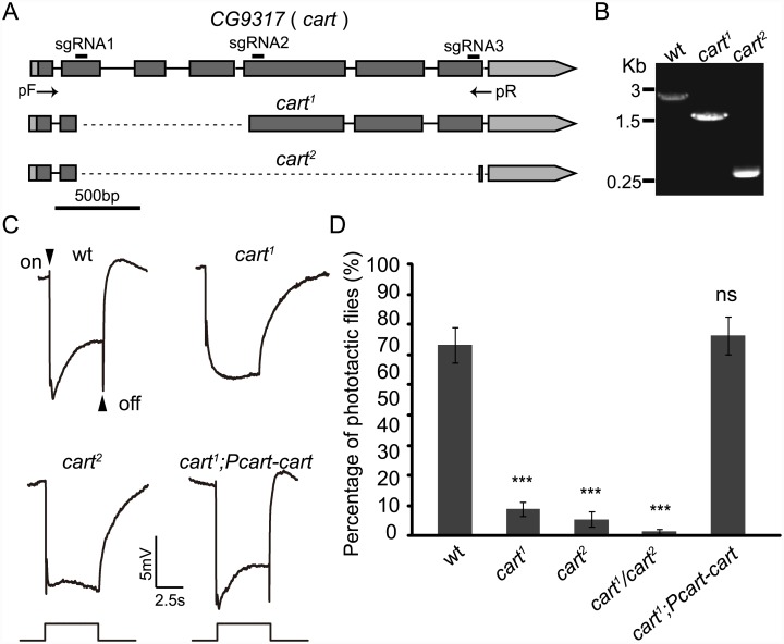
To characterize the requirement for CarT in transmitting visual signals, we generated two different null mutations in the cart gene using the CRISPR-associated single-guide RNA system (Cas9)(Fig 2A)[19]. We identified fly lines containing these cart 1 and cart 2 mutations by PCR using genomic primers outside of the deleted regions (Fig 2A). Full-length PCR products were detected in wild-type flies, whereas shorter PCR products were detected in the cart 1 and cart 2 mutant lines, indicating the disruption of the cart locus in cart 1 and cart 2 flies (Fig 2B). The cart genomic region in both mutations was sequenced, and 1112 and 2344 bp fragments were deleted in cart 1 and cart 2 mutants respectively (S4 Fig).
Fig 2. Mutations in cart eliminate “on” and “off” transients in ERG recordings.
(A) Schemes for cart deletion by sgRNA targeting. The organization of the cart locus and the expected structure of the deletion alleles (cart 1 and cart 2) are shown. Boxes represent exons, and deep gray boxes represent the coding region. The sgRNA1 and sgRNA2 primer pair was used to generate the cart 1 mutation. The sgRNA1 and sgRNA3 primer pair was used to generate the cart 2 mutation. The positions of the DNA primers used for PCR (arrows) are indicated. (B) PCR products obtained from successful cart deletion mutants. The agarose gel electrophoresis of PCR products using the primers indicated in (A) (pF and pR) is shown and genomic DNA templates prepared from wt (w 1118), cart 1 and cart 2 flies. (C) ERG recordings from wt (w 1118), cart 1, cart 2 and cart 1; Pcart-cart flies. Flies (~1 d after eclosion) were dark adapted for 1 min and subsequently exposed to a 5s pulse of orange light. (D) Phototaxis assays revealed a difference in behavior between wt and cart mutant flies. Error bars: SD; significant differences between mutant and wt flies were determined using unpaired t-tests (*p < 0.05; **p < 0.01; ***p < 0.001; ns, not significant).
As cart mutants were not lethal, so we undertook electroretinogram (ERG) recordings directly. ERG recordings are extracellular recordings that measure the summed responses of all retinal cells in response to light. Upon exposure to light, an ERG recording from a wild-type fly contains a sustained depolarizing response from the photoreceptors, and “on” and “off” transients originating from synaptic transmission to the lamina [20] (Fig 2C). Mutations with defective synaptic transmission have obvious reductions in their “on” and “off” transients [6]. As in mutants of genes involved in histamine recycling, ERG transients were not observed in cart 1, cart 2, or cart 1/ cart 2 mutant flies (Fig 2C). Phototaxis is a visual behavior that requires the integrity of the neuron circuits of the visual system [21], and defective synaptic transmission of visual signals results in poor phototaxis [22]. Significantly reduced phototaxis was associated with all the cart 1, cart 2, and cart 1/cart 2 mutations (Fig 2D). To further confirm that the loss of visual synaptic transmission resulted from mutations of the cart locus, we generated a Pcart-cart transgenic fly line expressing the cart cDNA under the control of the cart promoter. The Pcart-cart transgene reversed the loss of “on” and “off” transients and restore phototaxis in the cart 1 mutant flies (Fig 2C and 2D).
CarT functions in photoreceptor cells
Tan, the hydrolase that deconjugates carcinine and releases histamine, localizes to photoreceptor cells and functions downstream of the transport of carcinine. A carcinine transporter coupled with Tan’s action should therefore be expressed and should function in photoreceptor cells. It has been suggested that ine encodes a putative carcinine neurotransmitter transporter in photoreceptor cells [15]. We used the eyeless-GAL4 UAS-FLP (EGUF)/hid technique to generate genetically mosaic flies [23]. The compound eyes of these mosaic flies comprise cells homozygous for a selected mutation, but forming part of an entire mosaic fly that is elsewhere heterozygous for the mutation. Therefore, if Ine functions in the compound eye of Drosophila, eye-specific mutations of ine in ine mosaic flies should mirror at least the same ocular defects in synaptic transmission as those present in the ine mutants.
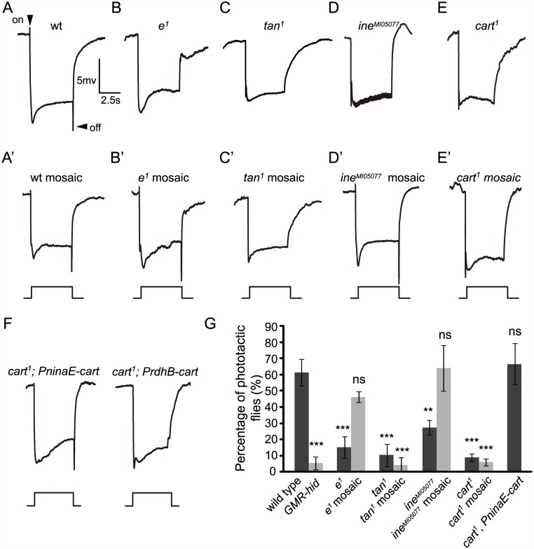
We observed that ERG recordings from wild-type eyes have normal “on” and “off” transients (Fig 3A and 3A’). Mutations in both the ebony (e 1) and the tan (tan 1) genes disrupt histamine recycling and this results in the loss of “on” and “off” transients in their ERG recordings (Fig 3B and 3C) [10]. As expected, e 1 mosaic flies in which all photoreceptors were homozygous mutant for ebony had wild-type “on” and “off” transients. This is because Ebony is not required in the photoreceptors but is required in glial cells lying outside them (Fig 3B’). As Tan functions in the photoreceptor cells of the compound eye, the tan 1 mosaic which lacks tan expression in the photoreceptors displayed reduced “on” and “off” transients (Fig 3C’).
Fig 3. CarT is required for synaptic transmission in photoreceptor cells.
(A-E) ERG paradigm that elicits “on” and “off” transients (arrows) in (A) wt (w 1118) flies but not in (B) e 1, (C) tan 1, (D) ine MI05077 or (E) cart 1 mutant flies. Flies (~1 d after eclosion) were dark adapted for 1 min and subsequently exposed to a 5s pulse of orange light. (A’-E’) “Off” transients were observed in the wt mosaic, e 1 and ine MI05077 mosaic eyes but not in tan 1 or cart 1 mosaic eyes. The genotypes are as follows: (A’: wt mosaic) ey-flp; FRT40A/GMR-hid CL FRT40A. (B’: e 1 mosaic) ey-flp; FRT82B e 1 /FRT82B GMR-hid CL. (C’: tan 1 mosaic) tan 1 FRT19A/GMR-hid FRT19A; ey-flp. (D’: ine MI05077 mosaic) ey-flp; ine MI05077 FRT40A /GMR-hid CL FRT40A. (E’: cart 1 mosaic mosaic) ey-flp; cart 1 FRT40A /GMR-hid CL FRT40A. (F) Expression of CarT in photoreceptor cells (cart 1 ; PninaE-cart), but not in pigment cells (cart 1 ; PrdhB-cart), restored the “on” and “off” transients in cart 1 flies. (G) Quantification of phototaxis of flies with the indicated genotypes. Error bars represent SD. Significant differences between mutant and wt flies were determined using unpaired t-tests (*p < 0.05; **p < 0.01; ***p < 0.001; ns, not significant).
The ERG responses of ine mutants (ine MI05077) contain prominent oscillations superimposed on the sustained depolarizating response and they also have reduced “on” and “off” transients (Fig 3D) [15]. The latter phenotype indicates impaired photoreceptor synaptic transmission. However, as with the ebony mutants, heterozygous flies with homozygous ine mutant compound eyes (ine mosaic flies) had wild-type ERG responses with normal “on” and “off” transients (Fig 3D’), indicating that Ine does not function obligatorily in photoreceptor cells. Therefore, it is unlikely that Ine is directly or necessarily responsible for carcinine uptake at the photoreceptor cell membrane, as previously suggested. Its possible role as a transporter elsewhere is not addressed by these experiments.
Given that expression of the cart gene is enriched in photoreceptor cells, we assumed that CarT is required in photoreceptor cells for synaptic transmission. As expected, homozygous cart 1 mutant eyes lacked “on” and “off” transients despite the heterozygous background elsewhere (Fig 3E and 3E’). This finding indicates that CarT functions in the compound eyes. Photoreceptor cells and retinal pigment cells are the two major cell types in the compound eye. To confirm the retinal cell type in which CarT functions, we expressed CarT specifically in photoreceptor cells using the ninaE promoter or in retinal pigment cells using the rdhB promoter [17] [24]. Photoreceptor-enriched expression of CarT by PninaE-cart restored both the “on” and “off” transients and phototaxis in cart 1 mutant flies, whereas expression of CarT in pigment cells through PrdhB-cart did not (Fig 3F and 3G). These results strongly support the interpretation that CarT functions in photoreceptor cells to maintain synaptic transmission.
In addition, we extended these ERG results by phototaxis assays. Wild-type flies displayed positive phototactic behavior, whereas flies that were homozygous mutant for ebony, tan, ine, or cart all displayed poor phototaxis, indicating that these genes are required for visual synaptic transmission (Fig 3G). Consistent with the ERG results, phototaxis was significantly reduced in the mosaic eyes of tan 1 and cart 1 compared with wild-type flies, whereas phototaxis of both the e 1 and the ine MI05077 mosaic flies did not differ from that in wild-type flies (Fig 3G). These results suggest the possibility that CarT rather than Ine functions as a carcinine transporter in photoreceptor cells.
CarT is predominantly localized to photoreceptor terminals
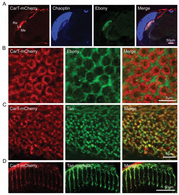
Trafficking of carcinine into photoreceptors is a key step in histamine recycling in Drosophila. As we have proposed here that CarT functions as a carcinine transporter acting at the photoreceptor cell membrane, we examined the localization of CarT to photoreceptor cells to evaluate the cellular location of carcinine transport. Since multiple attempts to generate an anti-CarT antibody failed, we eventually generated transgenic flies that expressed mCherry-tagged CarT driven by the cart promoter. Importantly, the Pcart-cart-mcherry transgene completely reversed the loss of “on” and “off” transients in cart mutant flies (Fig 2C). Although CarT was expressed throughout the photoreceptor neurons, the CarT signal was predominantly detected in the lamina layer where it was marked by the Ebony immunosignal, and not appreciably in the region of the retina (Fig 4A). In cross sections at high magnification we observed that CarT was not co-localized with Ebony to the epithelial glial cells (Fig 4B), but rather co-localized with the photoreceptor cell axon marker Tan to both the lamina and medulla neuropils, to which the R1-R6 and R7/R8 photoreceptors project their axons respectively (Fig 4C and 4D). The finding that CarT expression is enriched in photoreceptor terminals is consistent with the assumption that photoreceptor cells take up carcinine mainly from the local synaptic cleft in the lamina, rather than by a long-distance histamine recycling pathway which is mediated by lamina glia and a retinal pigment cell network [9]. However, we cannot exclude the existence of a long-term trafficking pathway for carcinine.
Fig 4. CarT is localized to terminals of photoreceptor neurons.
(A) CarT expression was enriched in the lamina and medulla neuropils by a mCherry-tagged transgene labeled with anti-mCherry. Pcart-cart-mcherry flies expressing mCherry-tagged CarT driven by the cart promoter were used. Cryosections of fly heads were labeled with anti-mCherry (red), anti-Chaoptin (24B10, expressed in photoreceptors) (blue) and anti-Ebony (green, expressed in lamina epithelial glia). La, lamina; Me, medulla; Re, retina. (B) Cross sections of the lamina of Pcart-cart-mcherry flies immunolabeled with anti-mCherry (red) and anti-Ebony (green) antibodies showed a complementary pattern. (C) Cross sections of the lamina of Pcart-cart-mcherry flies labeled with anti-mCherry (red) and anti-Tan (green) showed an overlapping pattern. (D) Longitudinal sections of the medulla of Pcart-cart-mcherry flies labeled with anti-mCherry (red) and anti-Tan (green) showed an overlap in the pattern of R7/R8 labeling.
Mutant cart 1 shows decreased histamine immunolabeling in photoreceptor terminals
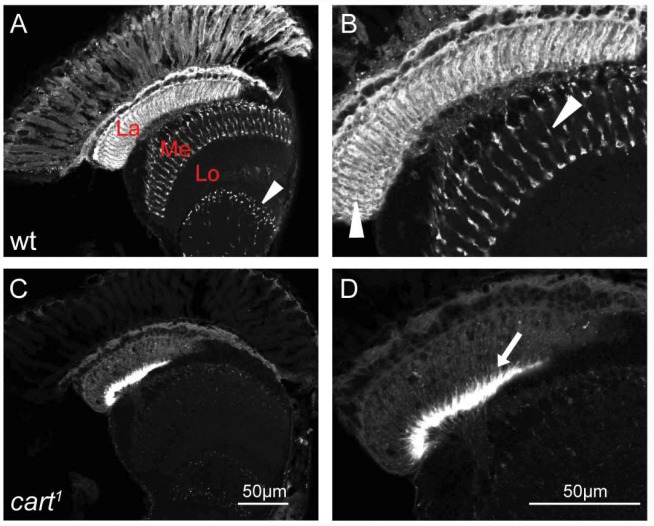
Given that the evidence so far suggests that cart acts to transport carcinine into the photoreceptor, where tan then acts to hydrolyze it and release histamine, we next sought to examine whether loss of cart would decrease histamine labeling. We labeled head cross sections from the cart 1 mutant and from the w 1118 control with anti-histamine antibody. The distribution of histamine signal in cart 1 mutant flies relative to their w 1118 controls reveals a clear loss of photoreceptor signal (Fig 5A and 5B), compatible with the mutant’s inability to take up carcinine and so liberate histamine. In the enlarged images, it is clear that cart 1 mutants showed a dramatic decrease in labeling for histamine in R1-R6 photoreceptor terminals in the lamina, and in R7/R8 photoreceptor terminals in the medulla (Fig 5C and 5D). In contrast to the weak label in R1-R6 photoreceptor terminals in the lamina, a strong label was seen in the underlying marginal glia at the proximal lamina in the cart 1 mutant (Fig 5C and 5D) [3,9,25]. The labeling of this region suggests that histamine might be accumulated at an ectopic site in the cart mutant.
Fig 5. Histamine is reduced in cart 1 mutant photoreceptor neurons.
Histamine was immunolabeled in horizontal sections of heads from (A-B) wild type (wt: w 1118) and (C-D) cart 1 mutant flies. (A) Strong signals in the lamina neuropile (La) and R7/R8 terminals in the distal medulla (Me) were detected in control w 1118. Additional immunolabel also appeared from cells in the lobula (Lo) (arrowhead). (B) The enlarged image of the wt head section in (A). Arrowheads in the lamina and medulla identified histamine positive photoreceptor terminals. (C) Loss of photoreceptor histamine signals and strong signals in lamina marginal glia were detected in cart 1 mutant. (D) The enlarged image of the cart 1 head section in (C) showing labeled marginal glia (arrow) but no photoreceptor signals. Scale bars: 50μm
Expression of human OCT2 in photoreceptor cells rescues the defective visual synaptic transmission in cart 1 flies
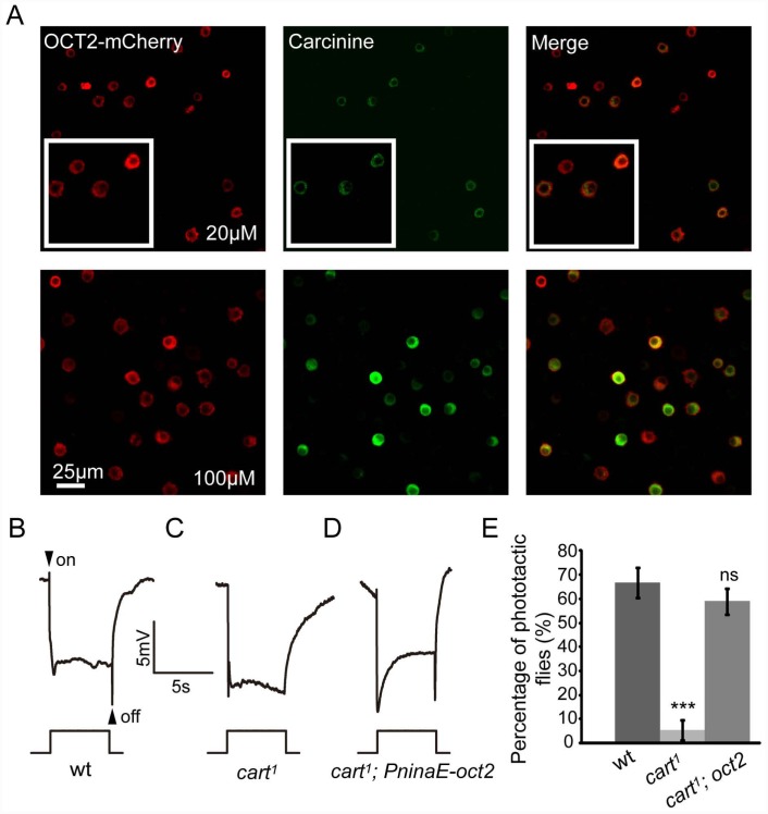
CarT belongs to the SLC22 protein family and is highly homologous to the mammalian OCT2 protein. We therefore wondered whether heterologous expression of OCT2 in cart mutant flies would restore the synaptic transmission of photoreceptors. OCT2 is known to mediate low affinity transport of some monoamine neurotransmitters [26]. However, it is not known whether OCT2 is able to transport carcinine. We performed in vitro assays to determine whether OCT2 can transport carcinine. After expressing OCT2 in S2 cells, carcinine was taken up by the OCT2-positive cells (Fig 6A). These results indicated that OCT2 can indeed transport carcinine. We next generated a PninaE-oct2 transgene to express OCT2 in photoreceptor cells only, and introduced this transgene into the cart 1 mutant background. We found that the expression of human OCT2 in cart 1 mutant fly photoreceptor cells fully restored both the “on” and “off” transients and phototaxis in cart 1 flies (Fig 6B–6E). These results demonstrated a conserved function for OCTs in both a mammal and Drosophila.
Fig 6. Rescue of cart 1 phenotype by expressing human OCT2 in photoreceptors.
(A) OCT2 was able to transport carcinine. S2 cells transiently expressed OCT2-mCherry in a culture medium to which carcinine was added at a final concentration of 20μM or 100 μM. Cells were immunolabeled with anti-carcinine (green), and the mCherry (red) signal was observed directly. The carcinine signal is stronger in the presence of 100μM carcinine. (B-D) ERG recordings from (B) wt: w 1118, (C) cart 1, and (D) cart 1; PninaE-oct2 flies showed restored “on” and “off” transients after photoreceptor cell-specific expression of human OCT2 in cart 1 flies. (E) Quantification of phototactic behaviors of wt, cart 1, and cart 1; PninaE-oct2 (cart 1; oct2) flies. Error bars represent SD. Significant differences between mutant and wt flies were determined using unpaired t-tests (*p < 0.05; **p < 0.01; ***p < 0.001; ns, not significant).
Histamine metabolite levels are altered in cart 1 mutant flies
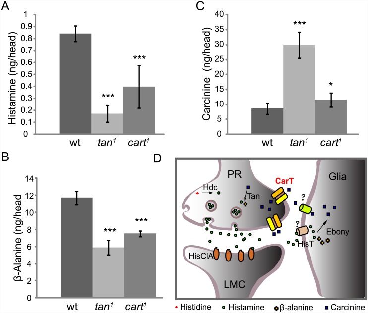
We used high-performance liquid chromatography (HPLC) to examine the in vivo contents of histamine as well as carcinine and β-alanine, the major metabolites in histamine recycling [18,27]. As expected, in the heads of the tan 1 mutant flies, which are defective in their capacity to hydrolyze carcinine into histamine and β-alanine, the head contents of both histamine and β-alanine were significantly reduced (Fig 7A and 7B). The lack of carcinine uptake by photoreceptor cells in cart 1 mutant flies ultimately depletes carcinine in these cells, which reduces the production of histamine and β-alanine mediated by Tan (Fig 7A and 7B). The reduced head contents of histamine and β-alanine are therefore in agreement with the hypothesis that CarT transports carcinine.
Fig 7. Loss of CarT affects the histamine, β-alanine, and carcinine contents in vivo.
(A-C) Head histamine, β-alanine, and carcinine contents in the three genotypes indicated. (A-B) The tan 1 and cart 1 mutants had significantly less histamine and β-alanine than wild-type flies (w 1118). (C) The tan 1 mutants had nearly three times as much carcinine as wild-type flies, and cart 1 flies only showed a 35% increase in carcinine content. Error bars indicate SD; significant differences between mutant and wt flies were determined using unpaired t-tests (*p < 0.05; ***p < 0.001). (D) Model of the pathway for histamine recycling. After a light stimulus, the photoreceptor cells (PR) release histamine, synthesized by histidine decarboxylase (Hdc), into the synaptic cleft to activate histamine-gated chloride channels (HisClA) on postsynaptic neurons (LMC). The released histamine is quickly removed by an unknown histamine transporter in epithelial glial cells that express Ebony, and is then deactivated by conjugation to β-alanine. The histamine metabolite carcinine is then transported out of epithelial glial cells (Glia) by a second unknown transporter, and back to photoreceptors by means of the CarT transporter at the photoreceptor cell terminals, where carcinine is then hydrolyzed back into histamine by Tan ready to be pumped into synaptic vesicles in preparation for further release.
In contrast to histamine and β-alanine, the content of carcinine in the tan 1 mutant heads was approximately three fold higher than the content in wild-type heads, which we interpret to result from diminished hydrolysis of carcinine in photoreceptor cells (Fig 7C). If the flies were not able to transport carcinine into photoreceptor cells for hydrolysis, there should be a greater amount of carcinine in fly heads. As expected, in cart 1 mutants, the head content of carcinine was significantly increased. However, the content of carcinine in the cart 1 mutant was not increased to the same extent as in the tan 1 mutant flies.
Discussion
Although histamine is an important neurotransmitter known to regulate multiple physiological processes, the mechanism by which histamine content is regulated in the nervous system still remains to be elucidated. Our study identifies a mechanism and pathway for the uptake of a primary metabolite of histamine, which has hitherto defied analysis in any nervous system.
Insofar as histamine is the primary neurotransmitter released by photoreceptors in flies [28], the ease with which photoreceptor function and anatomy can be assayed has made the compound eye the preferred system to study histamine recycling. In particular the eye lends itself readily to the identification of genes that regulate neurotransmission, by enabling comprehensive genetic screens [23,29]. Studies in flies have previously identified a histamine/carcinine recycling pathway that involves two enzymes, Ebony, expressed in the epithelial glia, and Tan, expressed in the photoreceptor cells [13,30]. However, the key neurotransmitter transporters required for the histamine/carcinine shuttle pathway have not been identified. Conceptually, the putative carcinine transporter should be functionally coupled to Tan for the uptake of carcinine into photoreceptor cells and its subsequent hydrolysis. For this, both should colocalize to photoreceptors as we have shown in this study.
In this study, we identified a new SLC22A family protein CarT and provided evidence that it is functionally coupled with Tan as a photoreceptor cell-enriched carcinine transporter. CarT is predominantly localized to photoreceptor terminals and is able to transport carcinine in vitro. The decrease in head histamine and β-alanine and the increase in head carcinine in cart 1 support this hypothesis. The reduction in histamine content in cart 1 mutants is ~60%. This amount corresponds rather closely to the reduction in head histamine seen in the mutant sine oculis, which lacks compound eyes and has 28% of the histamine found in the wild-type [27]. The reduction in sine oculis suggests that residual head histamine is not located in the compound eye visual system. In the same way, the reduction in cart 1 is not accessible to photoreceptor synaptic transmission. Moreover, mutant ebony, in which head histamine content is reduced by 50%, has an abnormal ERG and phototaxis, corresponding to the strong ERG and phototaxis defects seen in the cart 1 mutant. Consistent with these HPLC data, we also found a clear difference in the immunosignal for histamine between cart 1 and control fly photoreceptors. The cart head increase in carcinine is not as high as that observed in the head of tan mutants, which raises the question of why the increases in carcinine in tan and cart mutant flies are not similar. This may be because carcinine released in the synaptic cleft in cart 1 mutants is removed by other cells, or alternatively it may enter the hemolymph and be excreted. Consistent with the latter, the carcinine content in the abdomen is increased by 43% in cart 1 compared with control w 1118 flies. We cannot address the carcinine transport by other cells in the lamina, in particular the epithelial and marginal glia, which surround the photoreceptor terminals and which contain carcinine [18], which we propose must therefore express other carcinine transporters.
All of the known plasma membrane neurotransmitter transporters are members of the Solute Carrier (SLC) family of proteins [31]. The most extensively studied of these transporters are members of the SLC6 subfamily, a group of Na+/Cl-—dependent transporters for serotonin, dopamine, norepinephrine, GABA and glycine [32]. OCTs, which belong to the SLC22 subfamily, are known to mediate sodium-independent transport of positively charged organic compounds [33]. The expression of OCT2 in neurons has been evaluated previously, but the neuronal function of OCT2 has not been explored sufficiently [26,33,34]. Carcinine has been identified as a native metabolite related to histamine in multiple tissues in mammals, where it may serve as an antioxidant for scavenging toxic active oxygen species, especially in retinal photoreceptors [35,36]. Our findings that OCT2 can transport the inactive histamine metabolite carcinine both in vitro and in vivo suggests a possible new mechanism for OCTs to function in neurotransmitter recycling and cell protection.
The histamine/carcinine shuttle pathway plays a dominant role in maintaining an adequate level of histamine in photoreceptors. Evidence for the direct uptake of histamine into photoreceptor cells is lacking, insofar as Ebony is necessary to rescue ERG transients in histamine-fed hdc mutant flies [37]. In addition, in our model S2 cells expressing CarT fails to transport histamine, providing further support for the hypothesis that direct uptake of histamine into the photoreceptor terminals may not occur. Although the enzymatic deconjugation of carcinine to yield histamine has been well established, the route through which carcinine is then trafficked back to the photoreceptor has not been established. It has been suggested that recycling of carcinine to photoreceptor cells involves a long-distance pathway mediated by a gap-junction dependent network of lamina and retinal pigment cells [9]. In our study, we observed that CarT is predominantly localized to the terminals of photoreceptor neurons, rather than to their cell bodies in the retina layer, which suggests that carcinine is transported back to photoreceptor cells mainly from the synaptic cleft in the lamina (Fig 7D). It is also possible that this local pathway works in parallel with the long-distance neurotransmitter recycling pathway. Finally, data from the current study together with previous reports now provide evidence for a more complete histamine/carcinine recycling pathway, one which is critical for maintaining the normal histamine content of neurons (Fig 7D).
To complete the model of how histamine is recycled in the fly’s eye (Fig 7D), the remaining question concerns how histamine is transported into the epithelial glia and how carcinine is then transported out of the glia. No specific histamine transporter has been found, in either insects or vertebrates. In insects a mechanism for the fast removal of histamine from the synaptic cleft is essential to maintain the rapid signaling required for insect vision. One transporter may be White [38], but the problem is that in eukaryotes all known ABC transporters move substrates in the opposite direction i.e. out of the cell. To complete the return path for the carcinine will require us to identify how carcinine is exported out of the epithelial glia. To identify the transporter for this function it will be necessary to identify genes, for example from the expression of mRNA transcripts of genes, such as ebony [12, 13], that are enriched in the epithelial glia, in an approach that parallels the one we have adopted here to identify CarT. The transport of β-alanine, the other substrate needed for carcinine synthesis, seems to be of minor importance because this amino acid is present in the head in concentrations greatly exceeding those needed for histamine recycling and it can be also easily synthesized on demand from aspartate or uracil. Answering these questions is necessary to complete the current scheme (Fig 7D) for the recycling of histamine, to which our findings now identify CarT as the photoreceptor uptake transporter.
Methods
Fly stocks
The following stocks were obtained from the Bloomington Stock Center: (1) 122, e 1; (2) 130, tan 1; (3) 38094, ine MI05077; (4) 3605, w 1118; and (5) 24749, M(vas-int.Dm)ZH-2A;M(3xP3-RFP.attP)ZH-86Fb. The (nos-Cas9)attP2 flies were obtained from the lab of Dr. J. Ni at Tsinghua University, Beijing, China. The ey-flp;GMR-hid CL FRT40A/Cyo, ey-flp;FRT42D GMR-hid CL/Cyo, GMR-hid CL FRT19A/FM7;ey-flp, and ey-flp;FRT82B GMR-hid CL /TM3 flies were maintained in the lab of Dr. T. Wang at the National Institute of Biological Sciences, Beijing, China.
Generation of plasmid constructs and transgenic flies
The cart, CG3790, ine, and Hdc cDNA sequences were amplified from GH05908, GH20501, LP16156, and LD44381 cDNA clones obtained from DGRC (Drosophila Genomics Resource Center, Bloomington, IN, USA). The oct2 cDNA sequences were amplified from IOH56335 cDNA clones obtained from Ultimate™ ORF clones (Thermo Fisher Scientific, Waltham, USA). Their entire CDS sequences, excluding the stop codon, were subcloned into the pIB-cmcherry vector (Invitrogen, Carlsbad, USA) for expression in S2 cells. To construct PninaE-cart, PrdhB-cart, and PninaE-oct2, the entire coding region of cart and oct2 was amplified from cDNA clones and cloned into the pninaE-attB and prdhB vectors (both gifts from the lab of Dr. C. Montell at the University of California, Santa Barbara, USA)[17,24,39]. To construct Pcart-cart-mcherry, the promoter region (-2579 to +11 base pairs 5' to the transcription start site) of the cart gene was amplified from genomic DNA, and cart-mcherry was amplified from pIB-cart-mcherry. These constructs were injected into M(vas-int.Dm)ZH-2A;M(3xP3-RFP.attP)ZH-86Fb embryos, and transformants were identified on the basis of eye color. The (3xP3-RFP.attP) locus was removed by crossing with P(Crey) flies.
Generation of the cart mutant flies
The cart 1 and cart 2 mutations were generated using the Cas9/sgRNA system as described previously [19]. Three recognition sequences of guiding RNA to the cart locus were designed with tools available at the following website http://www.flyrnai.org/crispr2/ (sgRNA1: AAAACCGCACGGTATGCAGG, sgRNA2: CCTGTCCGGCGTCACTTATC, sgRNA3: TGAGCGTCATGGACACCCAG). These were cloned into the U6b-sgRNA-short vector. The pU6-sgRNA1 and pU6-sgRNA2 plasmids were used to generate the cart 1 mutant flies, while pU6-sgRNA1 and pU6-sgRNA3 were used to generate the cart 2 mutant flies. Plasmids were injected into the embryos of (nos-Cas9)attP2 flies. The F1 progeny were screened by PCR to identify the cart 1 and cart 2 deletions, using the following primers:
pF: 5’-TGTCGCTACAAATCTTAGATCCAA-3'
pR: 5’-CCATGTCAGATATTGAGGACAACG-3’
Electroretinogram recordings
Two glass microelectrodes filled with Ringer’s solution were inserted into small drops of electrode cream (Sigma, New Jersey, USA) placed on the surfaces of the compound eye and the thorax. A Newport light projector (model 765) was used for stimulation. The source light intensity was 2000lux, and the light color was orange (the source light was filtered by FSR-OG550 filter). ERG signals were amplified with a Warner electrometer IE-210 and recorded with a MacLab/4 s A/D converter and the clampelx 10.2 program (Warner Instruments, Hamden, USA). All recordings were carried out at 23°C.
Carcinine/histamine transport assay
S2 cells were grown in Schneider’s Drosophila medium with 10% Fetal Bovine Serum (Gibco, Carlsbad,USA), and transfected with vigofect reagent (Vigorous Biotechnology, Beijing, China). Carcinine or histamine was added to the medium to yield final concentrations as indicated in the Figure legends. After incubation for 3h, S2 cells were transferred to poly-L-lysine-coated slices, fixed with 4% paraformaldehyde(for carcinine immunolabeling) or 4% 1-ethyl-3-(3-dimethylaminopropyl) carbodiimide (EDAC)(for histamine immunolabeling) for 30min at 25°C, and incubated with rabbit anti-carcinine/histamine (1:100, ImmunoStar, USA)[9] or rat anti-carcinine antibodies (1:100, raised by Dr. Gabrielle Boulianne, from the lab of Dr. I. A Meinertzhagen) [18]. Goat anti-rabbit lgG conjugated to Alexa 488 (1:500, Invitrogen, CA) and goat anti-rat lgG conjugated to Alexa 488 (1:500, Invitrogen, CA) were used as secondary antibodies, and images were recorded with a Nikon A1-R confocal microscope.
Immunohistochemistry
Fly heads were fixed with 4% paraformaldehyde for 2h at 4°C or 4% EDAC (for histamine staining), and immersed in 12% glucose overnight at 4°C. The heads were embedded in O.C.T™ compound (Tissue-Tek, Torrance, USA), and 10μm thick cryosections were cut. Immunolabeling was performed on cryosections sections with mouse anti-24B10 (1:100, DSHB, http://dshb.biology.uiowa.edu/), rat anti-RFP (1:200, Chromotek, Martinsried, Germany), rabbit anti-Ebony (1:200, lab of Dr. S. Carroll, University of Wisconsin, Madison, USA), and anti-Tan (1:200, lab of Dr. B. Hovemann, Ruhr Universität Bochum, Germany) [30] as primary antibodies. For histamine staining, rabbit anti- histamine (1:100, ImmunoStar, USA) was used as a primary antibody. The antibody was preadsorbed with carcinine as previously reported [9]. Goat anti-rabbit lgG conjugated to Alexa 488 (1:500, Invitrogen, USA), goat anti-rat lgG conjugated to Alexa 568 (1:500, Invitrogen, USA) and goat anti-mouse lgG conjugated to Alexa 647 (1:500, Jackson ImmunoResearch, USA) were used as secondary antibodies. The images were recorded with a Nikon A1-R confocal microscope.
The phototaxis assay
A transparent glass tube of 20 cm long and 2.5 cm in diameter was used in this assay. A white light source (with a light intensity of 6000lux) was put at one end of the glass tube, and dark-adapted flies were collected and gently tapped into the other end of the tube. The tube was placed horizontally in the dark, and we counted the number of flies that walked past an 11-cm mark on the tube within 90s after turning the light on. Phototaxis was calculated by dividing the number of flies that walked past the mark as a proportion of the total number of flies. These assays were performed under dark conditions. To quantify the phototactic behaviors of each genotype, three groups of flies were collected for each genotype and three repeats made for each group. Each group contained ≥ 20 flies. Results were expressed as the mean of the mean values for the three groups.
RNA extraction and qPCR
Total RNA was prepared from the heads of three-day-old flies using Trizol reagent (Invitrogen, Carlsbad, USA), followed by TURBO DNA-free DNase treatment (Ambion, Austin, USA). Total cDNA was synthesized using an iScript cDNA synthesis kit (Bio-Rad Laboratories, USA). iQ SYBR green supermix was used for the real-time PCR (Bio-Rad Laboratories, USA). Three different samples were collected from each genotype. The primers used for qPCR were as follows:
ninaE-fwd, 5’-ACCTGACCTCGTGCGGTATTG-3’
ninaE-rev, 5’-GGAGCGGAGGGACTTGACATT-3’
gpdh-fwd, 5’-GCGTCACCTGAAGATCCCATG-3’
gpdh-rev, 5’-CTTGCCATACTTCTTGTCCGT-3’
rdhB-fwd, 5’-TTGAGGCACTCAGGGATCAAG-3’
rdhB-rev, 5’-CACCACATTCGTGTCGAACAG-3’
cart-fwd, 5’-TACAGCACAAGGGTCTCATCC-3’
cart-rev, 5’-AGACCATCCTAATCACGCTGAG-3’
High-performance liquid chromatography (HPLC)
To measurement the total head contents of histamine, β-alanine, and carcinine, flies were decapitated and their heads collected as previously reported [10]. The heads were then processed and analyzed using HPLC with electrochemical detection, all as previously reported [18,27]. Each sample contained ~50 Drosophila heads, and the mean values from five samples were calculated.
Supporting Information
Alignment of the Drosophila CG9317 amino acid sequence with Drosophila CG3790, mouse OCT2, and mouse OCT3. Identical residues, found in at least two proteins, are enclosed in black boxes. CG9317 is 32% identical to OCT2 and 31% identical to OCT3, whereas CG3790 is 30% identical to OCT2 and 29% identical to OCT3. The transmembrane domains are indicated by solid lines above the corresponding sequences. The running tally of amino acids is indicated to the right.
(EPS)
S2 cells transiently expressed (A) mCherry, (B) Ine-mCherry, (C) CG3790-mCherry, (D) CG9317-mCherry or (E) mCherry and Hdc. Histamine was added to the culture medium at a final concentration of 20μM. Cells labeled with anti-histamine (green) and DAPI (blue), or mCherry (red) were observed directly. Scale bar, 25μm
(EPS)
S2 cells transiently expressed (A) mCherry, (B) Ine-mCherry, (C) CG3790-mCherry, or (D) CG9317-mCherry. Carcinine was added to the S2 cells culture medium at a final concentration of 50μM. Cells labeled with rat anti-carcinine (green) and DAPI (blue), and the mCherry (red) signal was observed directly. Scale bar, 20μm.
(EPS)
DNA sequencing results of the Cas9-mediated break points in (A) cart 1 and (B) cart 2 flies. The representative DNA sequence of the wild-type locus (lower trace) shows the break points of the deletions generated. Upstream sequences of the 5’ breakpoints are marked in black, and downstream sequences of 3’ breakpoint are marked in red. The genomic positions corresponding to the break points are indicated.
(EPS)
Acknowledgments
We would like to thank Dr. W. Pak, Dr. S. Carroll, Dr. J Ni, Dr. B. Hovemann, the Bloomington Stock Center and Developmental Studies Hybridoma Bank for stocks and reagents. We thank Y. Wang and X. Liu for assistance on fly injections. We would also like to thank Cao Yang and the National Protein Science Facility at Tsinghua University, Beijing, China for technical assistance.
Data Availability
All relevant data are within the paper and its Supporting Information files.
Funding Statement
This work was supported by a ‘973’ grant (2014CB849700) to TW from the Chinese Ministry of Science (http://www.most.gov.cn/), and by grant DIS000065 to IAM from NSERC (http://www.nserc-crsng.gc.ca/). The funders had no role in study design, data collection and analysis, decision to publish, or preparation of the manuscript.
References
- 1. Haas HL, Sergeeva OA, Selbach O (2008) Histamine in the nervous system. Physiol Rev 88: 1183–1241. 10.1152/physrev.00043.2007 [DOI] [PubMed] [Google Scholar]
- 2. Hardie RC (1989) A histamine-activated chloride channel involved in neurotransmission at a photoreceptor synapse. Nature 339: 704–706. [DOI] [PubMed] [Google Scholar]
- 3. Edwards TN, Meinertzhagen IA (2010) The functional organisation of glia in the adult brain of Drosophila and other insects. Prog Neurobiol 90: 471–497. 10.1016/j.pneurobio.2010.01.001 [DOI] [PMC free article] [PubMed] [Google Scholar]
- 4. Gengs C, Leung HT, Skingsley DR, Iovchev MI, Yin Z, et al. (2002) The target of Drosophila photoreceptor synaptic transmission is a histamine-gated chloride channel encoded by ort (hclA). J Biol Chem 277: 42113–42120. [DOI] [PubMed] [Google Scholar]
- 5. Morante J, Desplan C (2004) Building a projection map for photoreceptor neurons in the Drosophila optic lobes. Semin Cell Dev Biol 15: 137–143. [DOI] [PubMed] [Google Scholar]
- 6. Wang T, Montell C (2007) Phototransduction and retinal degeneration in Drosophila. Pflügers Arch 454: 821–847. [DOI] [PubMed] [Google Scholar]
- 7. Stuart AE, Borycz J, Meinertzhagen IA (2007) The dynamics of signaling at the histaminergic photoreceptor synapse of arthropods. Prog Neurobiol 82: 202–227. [DOI] [PubMed] [Google Scholar]
- 8. Burg MG, Sarthy PV, Koliantz G, Pak WL (1993) Genetic and molecular identification of a Drosophila histidine decarboxylase gene required in photoreceptor transmitter synthesis. EMBO J 12: 911–919. [DOI] [PMC free article] [PubMed] [Google Scholar]
- 9. Chaturvedi R, Reddig K, Li HS (2014) Long-distance mechanism of neurotransmitter recycling mediated by glial network facilitates visual function in Drosophila . Proc Natl Acad Sci U S A 111: 2812–2817. 10.1073/pnas.1323714111 [DOI] [PMC free article] [PubMed] [Google Scholar]
- 10. Borycz J, Borycz JA, Loubani M, Meinertzhagen IA (2002) tan and ebony genes regulate a novel pathway for transmitter metabolism at fly photoreceptor terminals. J Neurosci 22: 10549–10557. [DOI] [PMC free article] [PubMed] [Google Scholar]
- 11. Gisselmann G, Pusch H, Hovemann BT, Hatt H (2002) Two cDNAs coding for histamine-gated ion channels in D. melanogaster . Nat Neurosci 5: 11–12. [DOI] [PubMed] [Google Scholar]
- 12. Richardt A, Kemme T, Wagner S, Schwarzer D, Marahiel MA, et al. (2003) Ebony, a novel nonribosomal peptide synthetase for beta-alanine conjugation with biogenic amines in Drosophila . J Biol Chem 278: 41160–41166. [DOI] [PubMed] [Google Scholar]
- 13. Richardt A, Rybak J, Stortkuhl KF, Meinertzhagen IA, Hovemann BT (2002) Ebony protein in the Drosophila nervous system: optic neuropile expression in glial cells. J Comp Neurol 452: 93–102. [DOI] [PubMed] [Google Scholar]
- 14. True JR, Yeh SD, Hovemann BT, Kemme T, Meinertzhagen IA, et al. (2005) Drosophila tan encodes a novel hydrolase required in pigmentation and vision. PLoS Genet 1: e63 [DOI] [PMC free article] [PubMed] [Google Scholar]
- 15. Gavin BA, Arruda SE, Dolph PJ (2007) The role of carcinine in signaling at the Drosophila photoreceptor synapse. PLoS Genet 3: e206 [DOI] [PMC free article] [PubMed] [Google Scholar]
- 16. Xu H, Lee SJ, Suzuki E, Dugan KD, Stoddard A, et al. (2004) A lysosomal tetraspanin associated with retinal degeneration identified via a genome-wide screen. EMBO J 23: 811–822. [DOI] [PMC free article] [PubMed] [Google Scholar]
- 17. Wang X, Wang T, Ni JD, von Lintig J, Montell C (2012) The Drosophila visual cycle and de novo chromophore synthesis depends on rdhB . J Neurosci 32: 3485–3491. 10.1523/JNEUROSCI.5350-11.2012 [DOI] [PMC free article] [PubMed] [Google Scholar]
- 18. Borycz J, Borycz JA, Edwards TN, Boulianne GL, Meinertzhagen IA (2012) The metabolism of histamine in the Drosophila optic lobe involves an ommatidial pathway: beta-alanine recycles through the retina. J Exp Biol 215: 1399–1411. 10.1242/jeb.060699 [DOI] [PMC free article] [PubMed] [Google Scholar]
- 19. Ren X, Sun J, Housden BE, Hu Y, Roesel C, et al. (2013) Optimized gene editing technology for Drosophila melanogaster using germ line-specific Cas9. Proc Natl Acad Sci U S A 110: 19012–19017. 10.1073/pnas.1318481110 [DOI] [PMC free article] [PubMed] [Google Scholar]
- 20. Heisenberg M (1971) Separation of receptor and lamina potentials in the electroretinogram of normal and mutant Drosophila . J Exp Biol 55: 85–100. [DOI] [PubMed] [Google Scholar]
- 21. Behnia R, Desplan C (2015) Visual circuits in flies: beginning to see the whole picture. Curr Opin Neurobiol 34: 125–132. 10.1016/j.conb.2015.03.010 [DOI] [PMC free article] [PubMed] [Google Scholar]
- 22. Benzer S (1967) Behavioral mutants of Drosophila isolated by countercurrent distribution. Proc Natl Acad Sci U S A 58: 1112–1119. [DOI] [PMC free article] [PubMed] [Google Scholar]
- 23. Stowers RS, Schwarz TL (1999) A genetic method for generating Drosophila eyes composed exclusively of mitotic clones of a single genotype. Genetics 152: 1631–1639. [DOI] [PMC free article] [PubMed] [Google Scholar]
- 24. Mismer D, Rubin GM (1989) Definition of cis-acting elements regulating expression of the Drosophila melanogaster ninaE opsin gene by oligonucleotide-directed mutagenesis. Genetics 121: 77–87. [DOI] [PMC free article] [PubMed] [Google Scholar]
- 25. Saint Marie RL, Carlson SD (1983) The fine structure of neuroglia in the lamina ganglionaris of the housefly, Musca domestica L. J Neurocytol 12: 213–241. [DOI] [PubMed] [Google Scholar]
- 26. Busch AE, Karbach U, Miska D, Gorboulev V, Akhoundova A, et al. (1998) Human neurons express the polyspecific cation transporter hOCT2, which translocates monoamine neurotransmitters, amantadine, and memantine. Mol Pharmacol 54: 342–352. [DOI] [PubMed] [Google Scholar]
- 27. Borycz J, Vohra M, Tokarczyk G, Meinertzhagen IA (2000) The determination of histamine in the Drosophila head. J Neurosci Methods 101: 141–148. [DOI] [PubMed] [Google Scholar]
- 28. Hardie RC (1987) Is histamine a neurotransmitter in insect photoreceptors? J Comp Physiol A 161: 201–213. [DOI] [PubMed] [Google Scholar]
- 29. Johnston D (2002) The art and design of genetic screens: Drosophila melanogaster. Nat Rev Genet 3: 176–188. [DOI] [PubMed] [Google Scholar]
- 30. Wagner S, Heseding C, Szlachta K, True JR, Prinz H, et al. (2007) Drosophila photoreceptors express cysteine peptidase tan. J Comp Neurol 500: 601–611. [DOI] [PubMed] [Google Scholar]
- 31. Blakely RD, Edwards RH (2012) Vesicular and plasma membrane transporters for neurotransmitters. Cold Spring Harb Perspect Biol 4. [DOI] [PMC free article] [PubMed] [Google Scholar]
- 32. Rudnick G, Kramer R, Blakely RD, Murphy DL, Verrey F (2014) The SLC6 transporters: perspectives on structure, functions, regulation, and models for transporter dysfunction. Pflügers Arch 466: 25–42. 10.1007/s00424-013-1410-1 [DOI] [PMC free article] [PubMed] [Google Scholar]
- 33. Roth M, Obaidat A, Hagenbuch B (2012) OATPs, OATs and OCTs: the organic anion and cation transporters of the SLCO and SLC22A gene superfamilies. Br J Pharmacol 165: 1260–1287. 10.1111/j.1476-5381.2011.01724.x [DOI] [PMC free article] [PubMed] [Google Scholar]
- 34. Gorboulev V, Ulzheimer JC, Akhoundova A, Ulzheimer-Teuber I, Karbach U, et al. (1997) Cloning and characterization of two human polyspecific organic cation transporters. DNA Cell Biol 16: 871–881. [DOI] [PubMed] [Google Scholar]
- 35. Flancbaum L, Brotman DN, Fitzpatrick JC, Van Es T, Kasziba E, et al. (1990) Existence of carcinine, a histamine-related compound, in mammalian tissues. Life Sci 47: 1587–1593. [DOI] [PubMed] [Google Scholar]
- 36. Marchette LD, Wang H, Li F, Babizhayev MA, Kasus-Jacobi A (2012) Carcinine has 4-hydroxynonenal scavenging property and neuroprotective effect in mouse retina. Invest Ophthalmol Vis Sci 53: 3572–3583. 10.1167/iovs.11-9042 [DOI] [PMC free article] [PubMed] [Google Scholar]
- 37. Ziegler AB, Brüsselbach F, Hovemann BT (2013) Activity and coexpression of Drosophila black with ebony in fly optic lobes reveals putative cooperative tasks in vision that evade electroretinographic detection. J Comp Neurol 521: 1207–1224. 10.1002/cne.23247 [DOI] [PubMed] [Google Scholar]
- 38. Borycz J, Borycz JA, Kubow A, Lloyd V, Meinertzhagen IA (2008) Drosophila ABC transporter mutants white, brown and scarlet have altered contents and distribution of biogenic amines in the brain. J Exp Biol 211: 3454–3466. 10.1242/jeb.021162 [DOI] [PubMed] [Google Scholar]
- 39. Wang T, Xu H, Oberwinkler J, Gu Y, Hardie RC, et al. (2005) Light activation, adaptation, and cell survival functions of the Na+/Ca2+ exchanger CalX. Neuron 45: 367–378. [DOI] [PubMed] [Google Scholar]
Associated Data
This section collects any data citations, data availability statements, or supplementary materials included in this article.
Supplementary Materials
Alignment of the Drosophila CG9317 amino acid sequence with Drosophila CG3790, mouse OCT2, and mouse OCT3. Identical residues, found in at least two proteins, are enclosed in black boxes. CG9317 is 32% identical to OCT2 and 31% identical to OCT3, whereas CG3790 is 30% identical to OCT2 and 29% identical to OCT3. The transmembrane domains are indicated by solid lines above the corresponding sequences. The running tally of amino acids is indicated to the right.
(EPS)
S2 cells transiently expressed (A) mCherry, (B) Ine-mCherry, (C) CG3790-mCherry, (D) CG9317-mCherry or (E) mCherry and Hdc. Histamine was added to the culture medium at a final concentration of 20μM. Cells labeled with anti-histamine (green) and DAPI (blue), or mCherry (red) were observed directly. Scale bar, 25μm
(EPS)
S2 cells transiently expressed (A) mCherry, (B) Ine-mCherry, (C) CG3790-mCherry, or (D) CG9317-mCherry. Carcinine was added to the S2 cells culture medium at a final concentration of 50μM. Cells labeled with rat anti-carcinine (green) and DAPI (blue), and the mCherry (red) signal was observed directly. Scale bar, 20μm.
(EPS)
DNA sequencing results of the Cas9-mediated break points in (A) cart 1 and (B) cart 2 flies. The representative DNA sequence of the wild-type locus (lower trace) shows the break points of the deletions generated. Upstream sequences of the 5’ breakpoints are marked in black, and downstream sequences of 3’ breakpoint are marked in red. The genomic positions corresponding to the break points are indicated.
(EPS)
Data Availability Statement
All relevant data are within the paper and its Supporting Information files.


