
Figure 4. UBE2T induces EMT of prostate cancer cells.
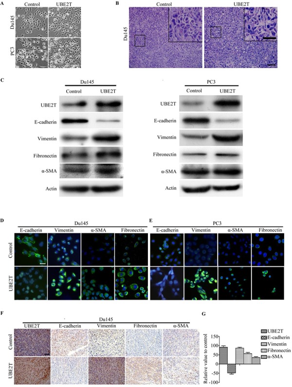
A. Micrographs showing the morphology of Du145, PC3 with overexpression of UBE2T and their corresponding control cells. B. H&E staining of xenograft tumor sections. C. Western blot analysis of the expression of the epithelial cell marker E-cadherin and the mesenchymal cell markers vimentin, fibronectin and alpha-smooth muscle actin in PC3 and Du145 cells overexpressing UBE2T. D. and E. Immunofluorescence images of EMT markers in Du145 and PC3 overexpressing UBE2T. F. Images of immunochemistry staining for UBE2T and EMT markers in sections of xenograft tumors from Du145 cells with or without overexpression of UBE2T. G. Positive cells in (F) were counted and plotted in histogram. Scale bars: 50 μm (A), 100 μm (B and F) and 50 μm (inserts in B), 20 μm (D and E).