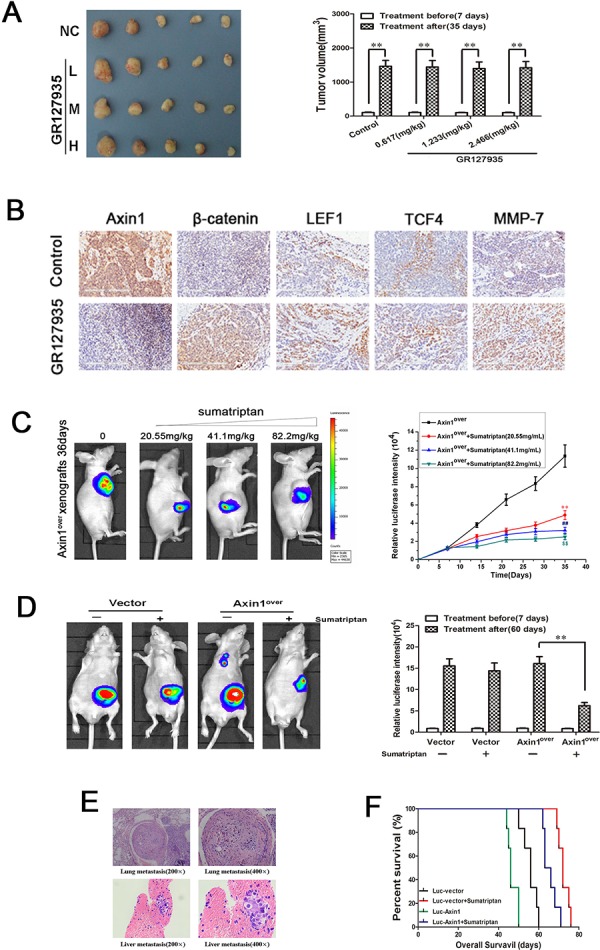
Figure 5. 5-HT1DR modulated colorectal cancer invasion/migration in vivo.

A. Excised tumors on day 28 of treatment with GR127935 (35 days after inoculation). Tumors were weighed (right) and photographed (left). Data are means ± SEM. Significant differences are indicated by **P < 0.01 (representative one in each group has been shown). B. The subcutaneous transplanted tumor tissue in Fig. 5A were subjected to immunohistochemical analysis using Axin1, β-catenin, LEF1, TCF4 and MMP-7 antibody. Images were taken at 200x magnification. Brown staining indicated positive cells. C. Left: Luciferase imaging of the mice with Axin1 overexpression and sumatriptan xenografts on day 36 after tumor cell implantation. Right: the quantification of luciferase intensities in tumors of the four groups. Data are presented as mean ± SD of triplicate experiments. **P < 0.01 sumatriptan (0.617 mg/kg) vs. Control. ##P < 0.01 sumatriptan (1.233 mg/kg) vs. Control. $$P < 0.01 sumatriptan (2.466 mg/kg) vs. Control. D. Luciferase imaging revealed primary tumor growth and its distant metastasis. Right: the quantification of luciferase intensities of the mice treated with sumatriptan for 60 days. Data are means ± SD. significant differences are indicated by **P < 0.01. E. H&E analyses of lung and liver sections in Axin1 overexpressing orthotopic implanted animals by day 60, which was the only group with distant metastasis. F. overall survival of orthotopic model is shown in Fig. 5D.
