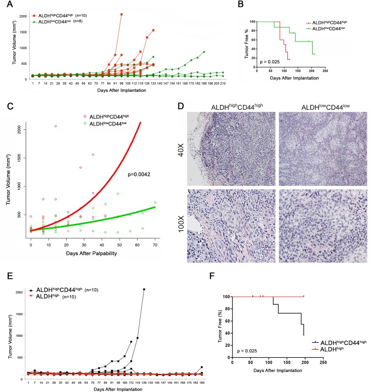
Figure 4. Tumorigenic potential of high passage mucoepidermoid carcinoma cells sorted for ALDH/CD44.
A. Graph depicting the volume of tumors generated by the transplantation of FACS-sorted UM-HMC-3B cells (ALDHhighC44high or ALDHlowCD44low) in immunodeficient mice. 5,000 sorted UM-HMC-3B cells (passage 103) and 900,000 endothelial (HDMEC) cells were seeded on biodegradable scaffolds and transplanted into the subcutaneous space of SCID mice. Tumors were measured weekly and mice were euthanized once the tumors reached 700-1,500 mm3. B. Kaplan-Meyer analysis of time to palpability of tumors generated with ALDHhighCD44high or ALDHlowCD44low cells. Tumors were considered palpable once they reached 200 mm3. C. Regression analysis of growth after palpability (200 mm3) of tumors generated with FACS-sorted ALDHhighCD44high or ALDHlowCD44low cells. D. H&E staining of tumors generated with FACS-sorted ALDHhighCD44high and ALDHlowCD44low cells. Images were taken at 40X and 100X. E. Graph depicting the volume of tumors generated by the transplantation of FACS-sorted UM-HMC-3B cells (ALDHhighC44high or ALDHhigh) in immunodeficient mice. 5,000 sorted UM-HMC-3B cells (passage 104) and 900,000 endothelial (HDMEC) cells were seeded on biodegradable scaffolds and transplanted into the subcutaneous space of SCID mice. Tumors were measured weekly and mice were euthanized once the tumors reached 700-1,500 mm3. F. Kaplan-Meyer analysis of time to palpability of tumors generated with ALDHhighC44high or ALDHhigh cells.

