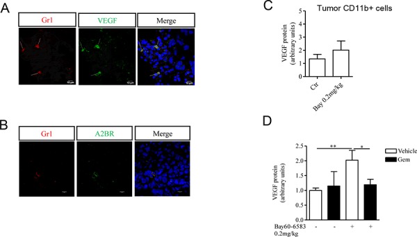
Figure 3. CD11b+Gr1+ cells produce VEGF, that contributes to tumor angiogenesis.

A. Immunofluorescence staining of Gr1+ cells with VEGF in melanoma tissue sections. Magnification 63x. Scale bars represent 10 μm. B. Immunofluorescence staining of Gr1+ cells and A2B receptor in melanoma tissue sections. Magnification 63x. Scale bars represent 10 μm. C. VEGF protein expression determined by Western blotting in CD11b+ cells isolated from tumor tissues of mice treated with Bay60-6583 or vehicle. D. CD11b+Gr1+ cells were depleted by administering gemcitabine (gem, 120mg/kg, i.p.) in melanoma-bearing mice treated with Bay60-6583 or vehicle. Western blotting analysis of VEGF protein expression in melanoma tissue lysates harvested from mice treated as above. Data are from two independent experiments and represent mean ± SEM (n = 5–10 per group). *p < 0.05, **p < 0.01.
